

Abstract
During this study, the in vitro antifungal and antibacterial activities of different extracts (aqueous and organic) obtained from a French propolis batch were evaluated. Antifungal activity was evaluated by broth microdilution on three pathogenic strains: Candida albicans, C. glabrata, and Aspergillus fumigatus. Antibacterial activity was assayed using agar dilution method on 36 Gram-negative and Gram-positive strains including Staphylococcus aureus. Organic extracts showed a significant antifungal activity against C. albicans and C. glabrata (MIC80 between 16 and 31 µg/mL) but only a weak activity towards A. fumigatus (MIC80 = 250 µg/mL). DCM based extracts exhibited a selective Gram-positive antibacterial activity, especially against S. aureus (SA) and several of its methicillin-resistant (MRSA) and methicillin-susceptible (MSSA) strains (MIC100 30–97 µg/mL). A new and active derivative of catechin was also identified whereas a synergistic antimicrobial effect was noticed during this study.
1. Introduction
Propolis is a resinous natural substance collected by honeybees from buds and exudates of various trees and plants, mixed with beeswax and salivary enzymes. Bees generally use it as a sealer, to smooth out the internal walls of the hive, as well as a protective barrier against intruders. Propolis has been used in folk medicine since ancient times due to its pharmacological potential associated with antioxidant [1–3], antifungal [4, 5], antibacterial [6–8], and anti-inflammatory [9] properties.
Propolis is generally composed of 50% of resin and balm (including polyphenolic compounds), 30% of wax and fatty acids, 10% of essential oils, 5% of pollen, and 5% of various organic and inorganic compounds. However, the composition of propolis deeply depends on the vegetation at the site of collection [10]. Indeed, propolis from temperate climatic zones, like in Europe, North America, or nontropical regions of Asia, mainly originates from the bud exudates of Populus species (Salicaceae) and consequently is rich in flavonoids and phenolic acids and their esters [11]; however tropical propolis, originating from regions where neither poplars nor birches grow, is rich in prenylated derivatives of p-coumaric acids, benzophenones, or terpenoids [12, 13].
The antifungal, antibacterial properties and chemical composition of propolis from many countries all over the world have been widely studied [6, 8, 14–20] but few reports were already given for European propolis [21, 22]. In 1990, Grange and Davey [23] highlighted for the first time the bactericidal activity of a French propolis against Gram-positive strains whereas later on, in 2000, Hegazi et al. [22] could associate this antibacterial activity with the presence of benzyl caffeate, pinocembrin, and p-coumaric acid.
During a previous study, we have evaluated the antioxidant and anti-AGEs activities of different solvents extracts [water; 95% EtOH; 70% EtOH; MeOH; dichloromethane (DCM) and DCM/MeOH/H2O (31/19/4)] obtained from a French propolis batch and identified their active constituents [24]. Here we have investigated the in vitro antifungal and antibacterial activities of these extracts. The antifungal activity was studied on three fungal strains (two yeasts, Candida albicans and C. glabrata, and one filamentous fungus, Aspergillus fumigatus). 36 strains of Gram-positive (including Staphylococcus aureus) and Gram-negative (including Escherichia coli) bacteria were used for the antibacterial assays. During this study, a new secondary metabolite was isolated, namely, 8-[(E)-phenylprop-2-en-1-one]-5-methoxy-(±)-catechin.
2. Materials and Methods
2.1. Reagents and Standards
Formic acid, p-coumaric acid, ferulic acid, isoferulic acid, 3,4-dimethoxycinnamic acid, and prenyl caffeate were purchased from Sigma-Aldrich (L'Isle d'Abeau Chesnes, Saint-Quentin-Fallavier, France). Caffeic acid and chrysin were obtained from Acros Organics (Geel, Belgium). Galangin was purchased from Extrasynthese (Genay, France). Pinocembrin and pinobanksin-3-acetate were isolated from the DCM extract of propolis.
2.2. Instrumentation
Optical rotation was measured on a JASCO P-2000 polarimeter. IR spectra were recorded on a Bruker Vertex 70 spectrophotometer. NMR spectra (1D and 2D) were recorded on a Bruker Avance spectrometer at 500 MHz for 1H and 125 MHz for 13C. MS analyses were performed on an ESI/APCI Ion Trap Esquire 3000+ from Bruker. UV absorbances were obtained from a Tecan Infinite M200 microplate spectrophotometer.
2.3. Propolis Samples
In order to analyze a typical French batch, that is, exhibiting an average chemical composition, a mixture of samples (10 g of each), collected in apiaries originating from different regions of France, was used for this study. These samples were provided by “Ballot-Flurin Apiculteurs,” a company specialized in organic beekeeping. Indeed, even collected in the same geographical region, propolis profiles may differ between apiaries and even inside the same apiary from one hive to another one [25]. Keeping in mind any potential economic development, it then appeared more appropriate to study an industrial end-product, that is, a mixture, exhibiting an average chemical composition associated with an average antimicrobial activity, rather than a specific sample. Therefore, 24 batches of propolis collected over two years (2010 and 2011) from different places in France (cf. supporting information 1; see Supplementary Material available online at http://dx.doi.org/10.1155/2015/319240) were homogeneously mixed to undergo this study.
2.4. Extractions
The extraction processes have been already described [24]. Briefly, the propolis batch was homogeneously pulverized in the presence of liquid nitrogen and divided into 1 g samples. Four different extractions were then carried out on 1 g samples with water (E1), 95% EtOH (E2), 70% EtOH (E3), and MeOH (E4). Then, two extractions, preceded by a cyclohexane wax elimination, were independently performed on 1 g samples with DCM (E5) and a mixture of DCM, MeOH, and H2O (31/19/4) (E6). For E1, a decoction of 1 g of propolis powder was boiled in 20 mL H2O at 100°C for 15 min. After cooling, the solidified wax and the residue were removed by filtration, and the filtrate was evaporated to dryness. For other solvents, 1 g of propolis powder (or residue obtained from a previous extraction) was macerated in 3 × 20 mL of solvent. After stirring for 3 × 2 h at room temperature, the mixture was filtered. The filtrates were gathered and evaporated under vacuum. Extraction yields (dried extract/100 g) were as follows: E1 7%; E2 68%; E3 65%; E4 68%; E5 50%; and E6 59%.
2.5. Antifungal Activity
Antifungal activity was assayed on human pathogenic fungi, including two common yeasts (Candida albicans ATCC 66396 and C. glabrata LMA 90-1085) and an opportunistic mould (Aspergillus fumigatus CBS 11326). The strains were obtained from the Parasitology and Mycology Laboratory at the University Hospital Center of Angers, France. Microorganisms were cultivated at 37°C on yeast extract-peptone-dextrose-agar (YPDA) containing 0.5 g/L chloramphenicol for two (C. albicans and C. glabrata) or three (A. fumigatus) days. Tests were performed according to a procedure described by Alomar et al. [27], following the guidelines of the approved reference method of the National Committee for Clinical Laboratory Standards (NCCLS) for yeasts [28] and filamentous fungi [29]. Briefly, the yeast suspensions were prepared in RPMI-1640 culture medium and adjusted spectrophotometrically at 630 nm to reach a final concentration of ca. 0.5 × 10³ to 2.5 × 10³ cells/mL. The tests were performed using sterile 96 flat shaped well microtiter plates. Serial twofold sample dilutions were made in DMSO. Sample solutions were dispensed at a volume of 5 μL in triplicate into the wells to obtain final concentrations from 250 to 1.95 μg/mL. After 48 h at 37°C for C. albicans and C. glabrata and 72 h for A. fumigatus, the spectrophotometric MIC endpoint was calculated from the turbidimetric data as the lowest sample concentration causing a growth inhibition equal to or greater than 80% of the control (MIC80). Amphotericin B was used as a positive control.
2.6. Antibacterial Activity
Antibacterial activity was evaluated on 36 human pathogenic bacterial strains collected by the Laboratory of Bacteriology at the University Hospital Center of Angers, France: seven strains of Acinetobacter baumannii (RCH, SAN008, 12, AYE, CIP7034, 107292, and 5377), two of Escherichia coli (ATCC25922 and a clinical isolate), three of Pseudomonas aeruginosa (ATCC27853 and two clinical isolates), and 4 clinical isolates of Enterobacter cloacae, E. aerogenes, Klebsiella oxytoca, and Salmonella enteritidis (phage type 4) for Gram-negative bacteria; thirteen strains of Staphylococcus aureus (ATCC25923, six methicillin-susceptible clinical isolates, six methicillin-resistant clinical isolates), two clinical isolates of S. epidermidis (methiS and methiR), three clinical isolates of Enterococcus faecalis and one of E. faecium, and one clinical isolate of Corynebacterium striatum for Gram-positive bacteria. Tests were performed using the methodology described by Alomar et al. [30]. Briefly, a stock solution of each sample was prepared in triplicate at 20 mg/mL in DMSO under sterile conditions. Serial dilutions were prepared (sample concentrations: 10, 20, 30, etc., to 100 μg/mL) and 0.1 mL of each dilution was added to 19.9 mL of Mueller-Hinton agar (Merck, Germany) and transferred to Petri plates. Bacterial strains (2 × 10⁴ CFU/mL) were suspended in sterile NaCl aqueous solution (0.15 M) and inoculated on the different Petri plates using the multipoint inoculator (AQS, England). After 24 h of incubation at 37°C, the minimum inhibitory concentration (MIC100, μg/mL) against bacterial strains was defined as the lowest concentration of each sample that inhibited visible growth. A blank was made inoculating the strains on Mueller-Hinton agar without any extract or compound. Oxacillin was used to distinguish the methicillin-resistant from the susceptible staphylococcal strains.
2.7. HPLC-DAD and HPLC-MS Procedures
Dry extracts were dissolved in MeOH (5 mg/mL for the aqueous extract and 10 mg/mL for the organic solvents ones) and centrifuged at 13000 rpm for 10 min prior to injection (10 μL) into the HPLC system. Analytical HPLC was run on a 2695 Waters separation module equipped with a diode array detector 2996 Waters. Separation was achieved on a LiChrospher column 100 RP-18 (125 × 4 mm i.d., 5 μm) protected with a LiChrocart 4-4 guard cartridge (4 × 4 mm i.d.) at a flow rate of 1 mL/min. The mobile phase consisted of 0.1% formic acid in water (solvent A) and MeOH (solvent B) and the separation was performed using the linear gradient: 25–100% B in 40 min. UV detection was achieved at two wavelengths: 254 and 280 nm.
The mass analyses were performed with an ESI interface coupled to an ion trap mass analyzer in both positive and negative modes, with the following conditions: collision gas, He; collision energy amplitude, 1.3 V; nebulizer and drying gas, N2, 7 L/min; pressure of nebulizer gas, 30 psi; dry temperature, 340°C; flow rate, 1.0 mL/min; solvent split ratio 1 : 9; scan range, m/z 100–1000.
2.8. Identification of Propolis Constituents
18 and 22 were directly identified in the DCM extract by HPLC/UV/MS and comparison with the literature data [31, 32], whereas 3, 6, 7, 8, 10, and 32 were compared with authentic standards (Sigma-Aldrich and Acros organics, cf. Section 2.1). A flash chromatography was then carried out in order to identify the other phenolic constituents. 50.0 g of pulverized propolis was firstly extracted with cyclohexane (3 × 200 mL, 2 h, room temperature) to eliminate waxes. After filtration, the residue was extracted with DCM (5 × 200 mL, 2 h, room temperature) to give 25.0 g of dry DCM extract (50% yield). 21.0 g of this extract was fractionated using a CombiFlash Teledyne ISCO apparatus and a prepacked silica gel column (Interchim PF-50SI HC/300 g, 50 μm), at a flow rate of 100 mL/min and with the following gradient elution system: cyclohexane (C6H12) 100% (2.0 L), C6H12 : EtOAc 90 : 10 (1.7 L), C6H12 : EtOAc 90 : 10 to 80 : 20 (2.2 L), 80 : 20 to 70 : 30 (2.5 L), C6H12 : EtOAc 70 : 30 to 60 : 40 (2.2 L), and C6H12 : EtOAc 60 : 40 to 50 : 50 (3.0 L) then DCM : MeOH 96 : 4 (2.2 L). UV detection (λ 254 and 280 nm) and TLC monitoring allowed collecting 21 fractions (F1–21). 48 [33] was identified in F1, 14 [34] and 17 [31] were identified in F11, 1 and 2 [19] were identified in F13, and finally 23 [31] was identified in F15 by HPLC/UV/MS and comparison with the literature data. The remaining constituents were isolated and identified through 1D and 2D NMR analysis (cf. Section 2.2). 200 mg of F1 was chromatographed on a silica gel column (Grace, 24 g) by flash chromatography at a flow rate of 25 mL/min with a mixture of C6H12 and EtOAc (B) [gradient: 1% B (30 min), 2% B (5 min), 2–5% B (2 min), 5% B (2 min), 5–30% B (1 min), and 30% B (5 min)] to give 46 [33, 35] (5 mg), 47 [33, 36] (3 mg), and 49 [37] (5 mg). F2 (1.5 g) gave 43 [38] whereas F4 (696 mg) and F5 (384 mg) allowed us to, respectively, identify 44 [37, 39] and 45 [40]. 500 mg of F6 (1.6 g) was chromatographed on reverse-phase- (RP-) Flash chromatography (Interchim column PF-30C18 HC/6 g, 30 μm) at a flow rate of 15 mL/min with water and MeOH (B) [gradient: 25–30% B (20 min), 30–40% B (2 min), 40% B (8 min), 40–45% B (1 min), 45% B (12 min), 45–50% B (2 min), and 50% B (20 min)] to give 9 [31] (10 mg), 25 [38] (126 mg), and a mixture of 35 and 36 [34] (15 mg). Similarly 500 mg of F7 was fractionated [gradient: 30–50% B (25 min), 50–60% B (25 min), and 60–65% B (20 min)] to give 11 [41] (2 mg), 33 [31, 42] (128 mg), 34 [38] (65 mg), and 42 [31] (41 mg) whilst 500 mg of F8 [gradient: 30–45% B (20 min), 45% B (30 min), 45–48% B (5 min), 48–55% B (5 min), 55% B (8 min), 55–60% B (1 min), and 60% B (12 min)] gave 13 [38] (3 mg), 28 [31, 43] (224 mg), and 41 [34] (28 mg). 500 mg of F9 [gradient: 30–60% B (50 min) and 60–65% B (5 min)] yielded mixture of 4 [34] and 5 [44, 45] (8 mg), 15 [43] (33 mg), 24 [32, 46] (16 mg), 32 [38] (150 mg), and another mixture of 37 [16] and 38 [32] (3 mg). 27 [34] and 29 [31] were identified from F10 (1.3 g). 500 mg of F11 [flow rate of 20 mL/min, gradient: 25–75% B (55 min)] gave 26 [47] (82 mg), 31 [31] (56 mg), and 39 [32] (70 mg). 300 mg of F13 [flow rate of 15 mL/min, gradient: 30–45% B (20 min), 45% B (20 min), 45–50% B (10 min), 50–60% B (5 min), and 60% B (5 min)] gave 10 [31] (2 mg) and 16 [38] (3 mg). 30 [48] was directly identified in F17 (543 mg). 500 mg of F18 [gradient: 40% B (7 min), 40–50% B (2 min), and 50% B (30 min)] gave a mixture of 8 and 12 (cf. F19) together with the new compound 40 (12 mg). 500 mg of F19 was chromatographed [gradient: 25% B (25 min), 25–35% B (1 min), and 35% B (20 min)] to give 8 (8 mg) and 12 (26 mg) [31]. Finally 500 mg of F20 allowed us to isolate [gradient: 30–40% B (20 min), 40–43% B (20 min), and 43–50% B (10 min)] 19 [32] (3 mg) and a mixture of 20 [49] and 21 [50] (7 mg).
3. Results and Discussion
3.1. Antifungal and Antibacterial Activities
Table 1 shows the minimum inhibitory concentration of at least 80% of fungal growth (MIC80) obtained with E1–6 extracts for Candida albicans, C. glabrata, and Aspergillus fumigatus. E1 did not exhibit any interesting antifungal activity (MIC80 > 250 μg/mL for the three strains) whereas E2–6 showed significant antifungal activities (MIC80 between 16 and 31 μg/mL) on both C. albicans and C. glabrata. These results are in agreement with those previously obtained for an Argentinian propolis on several Candida species (MIC100 in a range of 31 to 125 μg/mL) [51] as well as with Greece and Cyprus ones (MIC100 20 μg/mL) [21]. E2–6 also exhibited a weak activity towards A. fumigatus (MIC80 250 μg/mL).
Table 1.
Antifungal activity against Candida albicans, C. glabrata, and Aspergillus fumigatus.
| Extract | Solvent | C. albicans | C. glabrata | A. fumigatus |
|---|---|---|---|---|
| MIC80 (µg/mL) | ||||
| E1 | H2O | >250 | >250 | >250 |
| E2 | 95% EtOH | 31.25 | 15.63 | 250 |
| E3 | 70% EtOH | 31.25 | 31.25 | 250 |
| E4 | MeOH | 31.25 | 31.25 | 250 |
| E5 | DCM | 31.25 | 31.25 | 250 |
| E6 | Mixed solvents | 15.63 | 31.25 | 250 |
| Amphotericin B | 0.125 | 0.125 | 6 | |
According to Ríos and Recio [52] a MIC100 < 100 μg/mL should be considered as a promising value for a crude extract (versus 10 μg/mL for pure compounds). This is the reason why Table 2 gives the results of the antibacterial activity of E1–6 at the concentration of 100 μg/mL for 28 strains of Gram-negative and Gram-positive bacteria.
Table 2.
Antibacterial activity of E1–6 against 28 Gram-negative and Gram-positive strains.
| Number | Bacterial strains | Extracts (100 µg/mL) | |||||
|---|---|---|---|---|---|---|---|
| E1: H2O | E2: 95% EtOH | E3: 70% EtOH | E4: MeOH | E5: DCM | E6: mixed solvents | ||
| Gram-negative: | |||||||
| 1 | Acinetobacter baumannii (RCH) | − | − | − | − | − | − |
| 2 | Acinetobacter baumannii (SAN008) | − | − | − | − | − | − |
| 3 | Acinetobacter baumannii (12) | − | − | − | − | − | − |
| 4 | Acinetobacter baumannii (AYE) | − | − | − | − | − | − |
| 5 | Acinetobacter baumannii (CIP7034) | − | − | − | − | − | − |
| 6 | Acinetobacter baumannii (CIP107292) | − | − | − | − | − | − |
| 7 | Acinetobacter baumannii (CIP5377) | − | − | − | − | − | − |
| 8 | Enterobacter cloacae (0705A1743) | − | − | − | − | − | − |
| 9 | Enterobacter aerogenes (0705A0867) | − | − | − | − | − | − |
| 10 | Escherichia coli (ATCC25922) | − | − | − | − | − | − |
| 11 | Escherichia coli (0705A0434) | − | − | − | − | − | − |
| 12 | Klebsiella oxytoca (0705C0187) | − | − | − | − | − | − |
| 13 | Pseudomonas aeruginosa (ATCC27853) | − | − | − | − | − | − |
| 14 | Pseudomonas aeruginosa (0704C0134) | − | − | − | − | − | − |
| 15 | Pseudomonas aeruginosa (0703C0259) | − | − | − | − | − | − |
| 16 | Salmonella enteritidis (4) | − | − | − | − | − | − |
|
| |||||||
| Gram-positive: | |||||||
| 17 | Corynebacterium striatum (56) | − | + | + | + | + | + |
| 18 | Enterococcus faecalis (11003508001) | − | − | − | − | − | − |
| 19 | Enterococcus faecalis (11003492701) | − | − | − | − | − | − |
| 20 | Enterococcus faecalis (11004774001) | − | − | − | − | − | − |
| 21 | Enterococcus faecium (11502441401) | − | − | − | − | − | − |
| 22 | Staphylococcus aureus (ATCC25923) | − | − | − | + | + | + |
| 23 | MRSA (0706C0025) | − | − | − | + | + | + |
| 24 | MRSA (0702E0196) | − | − | − | − | + | + |
| 25 | MSSA (0703H0036) | − | − | − | − | − | − |
| 26 | MSSA (0701A0095) | − | − | − | − | + | + |
| 27 | S. epidermidis methiS a (12004523201) | − | − | − | − | − | − |
| 28 | S. epidermidis methiR b (12552599902) | − | − | − | − | − | − |
−: no antibacterial activity, +: antibacterial activity, a methicillin-Susceptible, b methicillin-Resistant.
Results showed that Gram-negative bacteria were not susceptible to E1–6 at this concentration. In contrast, organic solvents extracts were active on several Gram-positive bacteria such as Corynebacterium striatum (sometimes involved in pleuropulmonary infections) (E2–5) and especially Staphylococcus aureus, including for the latter several methicillin-resistant (MRSA) and methicillin-susceptible (MSSA) clinical isolates (E5-6). Sometimes called “golden staph,” S. aureus is the most pathogenic species of Staphylococcus genus. It might cause food poisoning, skin infections, abscesses, and diseases like pneumonia, meningitis, and sepsis. S. aureus is additionally one of the major causes of hospital-acquired infections, and the treatment of some multiresistant strains has become quite problematic. Among them, MRSA appears in France as one of the most commonly multiresistant strains encountered in hospitals.
MIC100 of E1–6 were determined on the 6 susceptible Gram-positive strains as well as on 8 other MRSA and MSSA strains. Results are given in Table 3.
Table 3.
MICs of E1–6 against 14 Gram-positive strains including MRSA and MSSA.
| Number | Bacterial strains | MIC100 (µg/mL) | ||||||
|---|---|---|---|---|---|---|---|---|
| E1: H2O | E2: 95% EtOH | E3: 70% EtOH | E4: MeOH | E5: DCM | E6: mixed solvents | Oxacillin | ||
| 17 | Corynebacterium striatum | >100 | 83 ± 6 | 90 ± 0 | 77 ± 12 | 63 ± 15 | 87 ± 21 | — |
| 22 | Staphylococcus aureus (ATCC25923) | >100 | >100 | >100 | 90 ± 0 | 60 ± 10 | 67 ± 15 | ≤0.25 |
| 23 | MRSA (0706C0025) | >100 | >100 | >100 | 90 ± 0 | 57 ± 12 | 30 ± 0 | ≥4 |
| 24 | MRSA (0702E0196) | >100 | >100 | >100 | >100 | 80 ± 10 | 77 ± 23 | ≥4 |
| 25 | MSSA (0703H0036) | >100 | >100 | >100 | >100 | >100 | >100 | ≤0.25 |
| 26 | MSSA (0701A0095) | >100 | >100 | >100 | >100 | 87 ± 6 | 83 ± 29 | ≤0.25 |
| 29 | MRSA (11004533801) | >100 | >100 | >100 | >100 | 80 ± 0 | 87 ± 21 | ≥4 |
| 30 | MRSA (11004691801) | >100 | >100 | >100 | >100 | 77 ± 6 | 67 ± 23 | ≥4 |
| 31 | MRSA (11004787401) | >100 | >100 | >100 | >100 | 97 ± 6 | >100 | ≥4 |
| 32 | MRSA (11006153901) | >100 | >100 | >100 | >100 | 77 ± 6 | 73 ± 29 | ≥4 |
| 33 | MSSA (11004327701) | >100 | >100 | >100 | >100 | 77 ± 6 | 73 ± 12 | 0.25 |
| 34 | MSSA (11004480701) | >100 | >100 | >100 | >100 | 80 ± 0 | 97 ± 12 | 0.5 |
| 35 | MSSA (11004691801) | >100 | >100 | >100 | >100 | 77 ± 6 | 90 ± 17 | 0.5 |
| 36 | MSSA (11004010401) | >100 | >100 | >100 | >100 | 77 ± 6 | 90 ± 0 | ≤0.25 |
E1 did not show any interesting activity on the 14 studied strains (MIC100 > 100 μg/mL). E2–6 showed interesting activities against Corynebacterium striatum with MIC100 ranging from 63 to 90 μg/mL. E5 and E6 exhibited the best antibacterial activities against the Staphylococcus strains with MIC100 up to 57 and 30 μg/mL, respectively. Among the alcoholic extracts, only E4 showed a moderate activity (MIC100 90 μg/mL) against S. aureus and one MRSA whereas E2 and E3 appeared as inactive. These overall activities therefore appeared to be better than those reported by Grange and Davey for the antibacterial activity of a French propolis on S. aureus and MRSA (MIC100 188–375 μg/mL) [23]. Our global antibacterial activity against MRSA and MSSA could be compared with those reported for propolis collected in Solomon Islands, exhibiting MIC100 between 64 and 128 μg/mL [6]. Similarly E4 was more active than a methanolic propolis extract from Jordan (585 μg/mL against S. aureus and 4700 μg/mL against MRSA) [20].
These results suggested that antifungal and antibacterial activities of propolis extracts could be related to their flavonoids contents [24]. Indeed, whereas E1–6 exhibited high total polyphenol contents (239–281 mg GAE/g), only those showing both high flavone/flavonol (FF) and flavanone/dihydroflavonol (FD) contents (i.e., E5-6) were active on the studied strains. In addition the higher the cumulative contents FF+FD were, the stronger the antibacterial activity was, as shown with E5 (254 mg/g) and E6 (236 mg/g) > E2–4 (220–228 mg/g). These results are in agreement with those reported by Velazquez et al. [15] for different Mexican propolis collected in Sonora State where EEP from the areas of Ures (410 mg/g), Caborca (332 mg/g), and Pueblo de Alamos (209 mg/g) showed MIC100 against S. aureus of 100, 200, and >400 μg/mL, respectively.
3.2. Chemical Composition
Figure 1 shows the HPLC chromatogram of the DCM extract E5. 48 compounds were identified by comparison with the literature data (UV/MS) and pure standards or, when needed, through 1H and 13C (1D and 2D) NMR analysis after compound isolation.
Figure 1.

HPLC chromatograms of E5: 1 3,4-dihydroxybenzaldehyde, 2 4-hydroxybenzoic acid, 3 caffeic acid, 4 vanillin, 5 4-hydroxyacetophenone, 6 p-coumaric acid, 7 ferulic acid, 8 isoferulic acid, 9 benzoic acid, 10 3,4-dimethoxycinnamic acid, 11 3-phenylpropanoic acid, 12 pinobanksin-5-methyl ether, 13 cinnamic acid, 14 4-methoxycinnamic acid, 15 pinobanksin, 16 naringenin, 17 quercetin, 18 quercetin-3-methyl ether, 19 pinocembrin-5-methyl ether, 20 1,3-di-p-coumaroylglycerol, 21 1-p-coumaroyl-3-feruloylglycerol, 22 kaempferol, 23 apigenin, 24 cinnamylidene acetic acid, 25 pinocembrin, 26 benzyl caffeate, 27 isopent-3-enyl caffeate, 28 pinobanksin-3-acetate, 29 prenyl caffeate, 30 2-acetyl-1,3-dicoumaroylglycerol, 31 phenylethyl caffeate (CAPE), 32 chrysin, 33 benzyl p-coumarate, 34 galangin, 35 benzyl ferulate, 36 prenyl ferulate, 37 kaempferide, 38 rhamnocitrin, 39 cinnamyl caffeate, 40 8-[(E)-phenylprop-2-en-1-one]-5-methoxy-(±)-catechin (new), 41 cinnamyl isoferulate, 42 cinnamyl p-coumarate, 43 pinostrobin, 44 alpinone-3-acetate, 45 tectochrysin, 46 benzyl cinnamate, 47 cinnamyl benzoate, 48 cinnamyl cinnamate, and 49 cinnamyl cinnamylidene acetate.
Additionally a new flavan-3-ol was identified as the 8-[(E)-phenylprop-2-en-1-one]-5-methoxy-(±)-catechin 40 (Figure 2).
Figure 2.
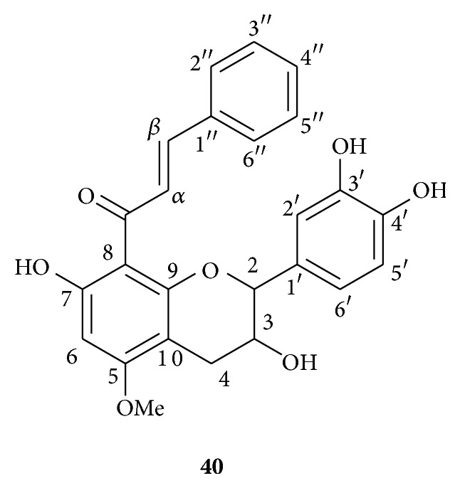
Chemical structure of the new compound 40.
Compound 40 was obtained as a yellow amorphous solid (0.6 μg/g of DCM extract). The molecular formula was determined as C25H22O7 by HRESIMS (found for [M+H]+ 435.1436; calculated 435.1438). The UV spectrum showed an absorption maximum at 350 nm. The IR spectrum indicated the presence of OH (3400 cm−1) as well as conjugated ketone carbonyl (1610 cm−1) groups. The 1H NMR spectrum exhibited signals due to a hydrogen-bonded OH at δ H 14.49, two trans-olefinic protons (δ H 8.06 and 7.63, 2 d, J = 15.7 Hz), aromatic rings (9H, δ H 6.15–7.30), and one methoxyle (δ H 3.92). It also showed the characteristic signals of a flavan-3-ol moiety at δ H 4.68 (1H, d, J = 8.9 Hz, H2), 4.21 (1H, m, H3), 3.07 (1H, dd, J = 16.2, 5.7 Hz, H4a), and 2.53 (1H, dd, J = 16.2, 9.5 Hz, H4b). The 13C NMR and HMQC spectra confirmed the presence of 25 carbons with typical flavan-3-ol signals at δ C 84.2 (C2), 66.8 (C3), and 30.6 (C4). In the 1H NMR spectrum, the signals at δ H 7.11 (1H, d, J = 1.4 Hz), 6.96 (1H, dd, J = 8.4, 1.4 Hz), and 6.91 (1H, d, J = 8.4 Hz) suggested the presence of a 1′,3′,4′-trisubstituted ring B whereas a singlet at δ H 6.15 (1H) indicated a pentasubstituted ring A. Two multiplets at δ H 7.17 (2H) and 7.29 (3H) revealed the presence of a phenyl residue. The HMBC spectrum showed a long-range correlation between the two trans-olefinic protons [δ H 7.63 (1H, d, J = 15.7, Hα) and 8.06 (1H, d, J = 15.7, Hβ)] and the ketone carbon at δ C 193.2. This correlation revealed the presence of an α,β-unsaturated ketone group. The trans-olefinic proton Hβ at δ H 8.06 was also correlated with the phenyl quaternary carbon at δ C 136.2 (C1′′). This correlation implied the presence of a (2E)-4-phenylprop-2-en-1-one moiety. A correlation between the methoxyle protons (δ H 3.92) and the carbon at δ C 165.1 (C5) proved that the OCH3 was attached to C5. The NOESY spectrum showed that this methoxyle was spatially close to the proton at δ H 6.15 (H6), whereas a long-range COSY indicated a correlation between H6 and one of the hydroxyl groups at δ H 14.49 (OH7). Therefore a (2E)-4-phenylprop-2-en-1-one moiety was located at C8 (δ C 105.9). Finally, it appeared that the aromatic ring B was substituted at C3′ and C4′ by two hydroxyl groups (NMR spectra cf. supporting information 2). 1H and 13C NMR data together with 2D NMR correlations for 40 are summarized in Table 4 and Figure 3.
Table 4.
1H and 13C NMR data of the new compound 40 (in acetone-d6).
| Position | 40 | |
|---|---|---|
| δ H, mult. (J in Hz) | δ C, mult. | |
| 2 | 4.68, d (8.9) | 84.2, CH |
| 3 | 4.21, m | 66.8, CH |
| 4 | a 3.07, dd (16.2, 5.7) | 30.6, CH2 |
| b 2.53, dd (16.2, 9.5) | ||
| 5 | 165.1, qC | |
| 6 | 6.15, s | 93.5, CH |
| 7 | 168.1, qC | |
| 8 | 105.9, qC | |
| 9 | 157.6, qC | |
| 10 | 102.4, qC | |
| 1′ | 130.7, qC | |
| 2′ | 7.11, d (1.4) | 116.2, CH |
| 3′ | 146.6, qC | |
| 4′ | 146.2, qC | |
| 5′ | 6.91, d (8.4) | 116.0, CH |
| 6′ | 6.96, dd (8.4, 1.4) | 121.3, CH |
| 1′′ | 136.2, qC | |
| 2′′ | 7.28, m | 129.2, CH |
| 3′′ | 7.16, m | 129.7, CH |
| 4′′ | 7.28, m | 130.8, CH |
| 5′′ | 7.16, m | 129.7, CH |
| 6′′ | 7.28, m | 129.2, CH |
| OH-7 | 14.49, s | |
| α | 7.63, d (15.7) | 143.1, CH |
| β | 8,06, d (15.7) | 128.4, CH |
| C=O | 193.2, qC | |
| OCH3-5 | 3.92, s | 56.5, CH3 |
Figure 3.

2D NMR studies of compound 40: COSY (bold lines), selected HMBC (solid arrows: 1H → 13C), and NOESY (dashed arrows) correlations.
40 had no optical rotation and, thus, was isolated here as a racemate mixture of 8-[(E)-phenylprop-2-en-1-one]-(2R,3S)-5-methoxycatechin (40a) and 8-[(E)-phenylprop-2-en-1-one]-(2S,3R)-5-methoxycatechin (40b). Sha et al. already isolated a similar compound, only differing from 40 by a 1′,3′,5′-trisubstituted aromatic ring B, from a Chinese propolis [26] (Figure 4).
Figure 4.
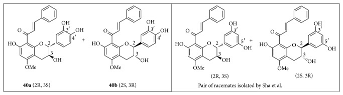
Structures of 8-[(E)-phenylprop-2-en-1-one]-(2R,3S)-5-methoxycatechin (40a) and 8-[(E)-phenylprop-2-en-1-one]-(2S,3R)-5-methoxycatechin (40b) and a similar pair of racemates isolated by Sha et al. 2009 [26].
3.3. Antifungal and Antibacterial Activities of 40
The new flavan-3-ol 40 did not show any antifungal activity on the three strains studied (Table 5). However, though active neither on S. aureus nor on MSSA, its MIC100 on MRSA numbers 23 and 24 were lower or equal to 10 μg/mL (close to oxacillin: ≥4 μg/mL).
Table 5.
Antifungal and antibacterial activities of 40.
| Antifungal activity | Antibacterial activity | |||
|---|---|---|---|---|
| Fungal strains | MIC80 (µg/mL) | Number | Bacterial strains | MIC100 (µg/mL) |
| C. albicans | >250 | 22 | S. aureus (ATCC25923) | >100 |
| C. glabrata | >250 | 23 | MRSA (0706C0025) | ≤10 |
| A. fumigatus | >250 | 24 | MRSA (0702E0196) | ≤10 |
| 25 | MSSA (0703H0036) | >100 | ||
| 26 | MSSA (0701A0095) | >100 | ||
3.4. Major Compounds Activities
Antifungal and antibacterial activities were then individually evaluated for the five major compounds identified in E2–6, namely, pinobanksin-3-acetate (28), pinocembrin (25), chrysin (32), galangin (34), and prenyl caffeate (29) [24]. Their MIC80 towards C. albicans, C. glabrata, and A. fumigatus as well as their MIC100 towards S. aureus, MRSA, and MSSA are given in Table 6.
Table 6.
Antifungal and antibacterial activities of five major compounds.
| Antifungal strains | MIC80 (µg/mL) | ||||||
|---|---|---|---|---|---|---|---|
| Pinobanksin-3-acetate (28) | Pinocembrin (25) | Chrysin (32) | Galangin (34) | Prenyl caffeate (29) | Amphotericin B | ||
| Candida albicans | 250 | 62 | >250 | >250 | 62 | 0.125 | |
| Candida glabrata | 250 | 125 | >250 | >250 | 16 | 0.125 | |
| Aspergillus fumigatus | >250 | 250 | >250 | >250 | 125 | 6 | |
|
| |||||||
| Number | Bacterial strains | MIC100 (µg/mL) | |||||
| Pinobanksin-3-acetate (28) | Pinocembrin (25) | Chrysin (32) | Galangin (34) | Prenyl caffeate (29) | Oxacillin | ||
|
| |||||||
| 22 | Staphylococcus aureus (ATCC25923) | >100 | 100 ± 0 | >100 | >100 | 63 ± 6 | ≤0.25 |
| 23 | MRSA (0706C0025) | >100 | >100 | >100 | >100 | 70 ± 0 | ≥4 |
| 24 | MRSA (0702E0196) | >100 | >100 | >100 | >100 | 70 ± 0 | ≥4 |
| 25 | MSSA (0703H0036) | >100 | >100 | >100 | >100 | 93 ± 6 | ≤0.25 |
| 26 | MSSA (0701A0095) | >100 | >100 | >100 | >100 | 93 ± 6 | ≤0.25 |
Pinobanksin-3-acetate (28), chrysin (32), and galangin (34) appeared as inactive. Pinocembrin (25) showed a moderate activity towards Candida albicans, C. glabrata (MIC80 62–125 μg/mL), and S. aureus (MIC100 100 μg/mL). Overall prenyl caffeate (29) exhibited the best activities (MIC100 up to 16 μg/mL against C. glabrata and up to 63 μg/mL against S. aureus and MRSA). Even so it appeared that these compounds were not individually as active as it could be expected from E5-6 results (MIC100 30–97 μg/mL). As far as S. aureus and MRSA are concerned, this kind of synergistic effects was recently pointed out by Darwish et al. [20] who evaluated the antibacterial activities of pinobanksin-3-acetate, pinocembrin, and chrysin isolated from a Jordanian propolis. Therefore these results are also in agreement with Kujumgiev et al. stating that, in spite of a great chemodiversity, no specific compounds can be associated with the antimicrobial activities of propolis extracts whereas, obviously, different flavonoid combinations are essential for these activities [7]. The antimicrobial of propolis extracts most probably involves a complex mechanism. It can be attributed to the synergistic effects of phenolic compounds such as cinnamic acid and ester derivatives, including caffeic acid and CAPE, as well as flavonoids including quercetin and naringenin [17, 53, 54]. Indeed, each of these compounds would be able to increase membrane permeability and inhibit bacterial mobility [54], thus contributing to the antimicrobial activity of propolis but also to its synergism with other antibiotics [53, 55, 56]. It is the reason why Stepanović et al. could notice the antibacterial and synergistic actions of propolis extracts with ampicillin, ceftriaxone, and doxycycline towards Staphylococcus aureus and with nystatin towards Candida albicans, stating that the bacterial resistance to antibiotics had no influence on the susceptibility to propolis extracts [57]. In vitro studies of synergism carried by Fernandes Jr. et al. also revealed synergistic effects of EEP with chloramphenicol, gentamicin, netilmicin, tetracycline, vancomycin, and clindamycin [58]. Therefore our findings are in total accordance with these results and, now that antibiotic resistance to bacteria has become a major public health concern [59], could bring valuable knowledge to develop new antimicrobial drugs for challenging S. aureus infections.
4. Conclusions
On the basis of these results, it may be concluded that organic solvents extracts of a French poplar type propolis are associated with a good antifungal activity towards Candida albicans and C. glabrata, correlated with high flavonoid contents. However only DCM based extracts (E5-6) showed a significant antibacterial activity against both methicillin-resistant and methicillin-susceptible Staphylococcus aureus strains. Unfortunately these extracts are not compatible with a pharmaceutical use because of their toxicity, whereas EtOH based extracts were not as active as expected. Therefore it would be interesting to develop some alternative extraction of propolis using a nontoxic solvent such as subcritical water. In addition, it should be noticed that, as an intrinsic polytherapy, propolis may also circumvent the development of drug resistance by bacteria [60].
Supplementary Material
The collection sites of the different propolis samples as well as the full NMR data set of new compound 40 (8-[(E)-phenylprop-2-en-1-one]-5-methoxy-(±)-catechin) are available online as Supplementary Material.
Acknowledgments
The authors thank Mrs. Isabelle Péruchès, from Ballot-Flurin Apiculteurs, who provided propolis samples, Benjamin Siegler and Dr. Ingrid Freuze from the “Plateforme d'Ingenierie et Analyses Moléculaires” (PIAM) at the Faculty of Sciences in Angers for NMR and MS analyses.
Abbreviations
- APCI:
Atmospheric pressure chemical ionization
- ATCC:
American Type Culture Collection
- AYE:
Patient code
- CAPE:
Caffeic acid phenylethyl ester
- CBS:
Centraal Bureau voor Schimmelcultures
- CIP:
Collection de l'Institut Pasteur
- COSY:
Correlation spectroscopy
- DCM:
Dichloromethane
- DMSO:
Dimethyl sulfoxide
- EEP:
Ethanolic extract of propolis
- ESI:
Electrospray ionization
- FD:
Flavanone/dihydroflavonol
- FF:
Flavone/flavonol
- GAE:
Gallic acid equivalent
- HMBC:
Heteronuclear multiple bond correlation
- HMQC:
Heteronuclear multiple quantum correlation
- HPLC:
High performance liquid chromatography
- HRESIMS:
High resolution electrospray ionization mass spectrometry
- IR:
Infrared
- LMA:
Laboratoire de Mycologie d'Angers
- MIC:
Minimum inhibitory concentration
- MRSA:
Methicillin-resistant Staphylococcus aureus
- MS:
Mass spectrometry
- MSSA:
Methicillin-susceptible Staphylococcus aureus
- NCCLS:
National Committee for Clinical Laboratory Standards
- NMR:
Nuclear magnetic resonance
- NOESY:
Nuclear Overhauser effect spectroscopy
- RCH:
Patient code
- RP:
Reversed phase
- RPMI:
Roswell Park Memorial Institute
- UV:
Ultraviolet
- YPDA:
Yeast peptone dextrose agar.
Conflict of Interests
This study was financed by Ballot-Flurin Apiculteurs Cie. There are no other competing interests.
References
- 1.Cottica S. M., Sawaya A. C. H. F., Eberlin M. N., Franco S. L., Zeoula L. M., Visentainer J. V. Antioxidant activity and composition of propolis obtained by different methods of extraction. Journal of the Brazilian Chemical Society. 2011;22(5):929–935. doi: 10.1590/S0103-50532011000500016. [DOI] [Google Scholar]
- 2.Miguel M. G., Nunes S., Dandlen S. A., Cavaco A. M., Antunes M. D. Phenols and antioxidant activity of hydro-alcoholic extracts of propolis from Algarve, South of Portugal. Food and Chemical Toxicology. 2010;48(12):3418–3423. doi: 10.1016/j.fct.2010.09.014. [DOI] [PubMed] [Google Scholar]
- 3.Gülçin I., Bursal E., Şehitoĝlu M. H., Bilsel M., Gören A. C. Polyphenol contents and antioxidant activity of lyophilized aqueous extract of propolis from Erzurum, Turkey. Food and Chemical Toxicology. 2010;48(8-9):2227–2238. doi: 10.1016/j.fct.2010.05.053. [DOI] [PubMed] [Google Scholar]
- 4.Ota C., Unterkircher C., Fantinato V., Shimizu M. T. Antifungal activity of propolis on different species of Candida . Mycoses. 2001;44(9-10):375–378. doi: 10.1046/j.1439-0507.2001.00671.x. [DOI] [PubMed] [Google Scholar]
- 5.Sawaya A. C. H. F., Palma A. M., Caetano F. M., et al. Comparative study of in vitro methods used to analyse the activity of propolis extracts with different compositions against species of Candida. Letters in Applied Microbiology. 2002;35(3):203–207. doi: 10.1046/j.1472-765X.2002.01169.x. [DOI] [PubMed] [Google Scholar]
- 6.Raghukumar R., Vali L., Watson D., Fearnley J., Seidel V. Antimethicillin-resistant Staphylococcus aureus (MRSA) activity of ‘pacific propolis’ and isolated prenylflavanones. Phytotherapy Research. 2010;24(8):1181–1187. doi: 10.1002/ptr.3096. [DOI] [PubMed] [Google Scholar]
- 7.Kujumgiev A., Tsvetkova I., Serkedjieva Y., Bankova V., Christov R., Popov S. Antibacterial, antifungal and antiviral activity of propolis of different geographic origin. Journal of Ethnopharmacology. 1999;64(3):235–240. doi: 10.1016/S0378-8741(98)00131-7. [DOI] [PubMed] [Google Scholar]
- 8.Popova M., Silici S., Kaftanoglu O., Bankova V. Antibacterial activity of Turkish propolis and its qualitative and quantitative chemical composition. Phytomedicine. 2005;12(3):221–228. doi: 10.1016/j.phymed.2003.09.007. [DOI] [PubMed] [Google Scholar]
- 9.Castaldo S., Capasso F. Propolis, an old remedy used in modern medicine. Fitoterapia. 2002;73(supplement 1):S1–S6. doi: 10.1016/s0367-326x(02)00185-5. [DOI] [PubMed] [Google Scholar]
- 10.Marcucci M. Propolis: chemical composition, biological properties and therapeutic activity. Apidologie. 1995;26(2):83–99. doi: 10.1051/apido:19950202. [DOI] [Google Scholar]
- 11.Bankova V. S., De Castro S. L., Marcucci M. C. Propolis: recent advances in chemistry and plant origin. Apidologie. 2000;31(1):3–15. doi: 10.1051/apido:2000102. [DOI] [Google Scholar]
- 12.Kumazawa S., Hamasaka T., Nakayama T. Antioxidant activity of propolis of various geographic origins. Food Chemistry. 2004;84(3):329–339. doi: 10.1016/S0308-8146(03)00216-4. [DOI] [Google Scholar]
- 13.Bankova V. Chemical diversity of propolis and the problem of standardization. Journal of Ethnopharmacology. 2005;100(1-2):114–117. doi: 10.1016/j.jep.2005.05.004. [DOI] [PubMed] [Google Scholar]
- 14.Salatino A., Fernandes-Silva C. C., Righi A. A., Salatino M. L. F. Propolis research and the chemistry of plant products. Natural Product Reports. 2011;28(5):925–936. doi: 10.1039/c0np00072h. [DOI] [PubMed] [Google Scholar]
- 15.Velazquez C., Navarro M., Acosta A., et al. Antibacterial and free-radical scavenging activities of Sonoran propolis. Journal of Applied Microbiology. 2007;103(5):1747–1756. doi: 10.1111/j.1365-2672.2007.03409.x. [DOI] [PubMed] [Google Scholar]
- 16.Agüero M. B., Gonzalez M., Lima B., et al. Argentinean propolis from Zuccagnia punctata cav. (Caesalpinieae) exudates: phytochemical characterization and antifungal activity. Journal of Agricultural and Food Chemistry. 2010;58(1):194–201. doi: 10.1021/jf902991t. [DOI] [PubMed] [Google Scholar]
- 17.Santos F. A., Bastos E. M. A., Uzeda M., et al. Antibacterial activity of Brazilian propolis and fractions against oral anaerobic bacteria. Journal of Ethnopharmacology. 2002;80(1):1–7. doi: 10.1016/S0378-8741(02)00003-X. [DOI] [PubMed] [Google Scholar]
- 18.Koo H., Gomes B. P. F. A., Rosalen P. L., Ambrosano G. M. B., Park Y. K., Cury J. A. In vitro antimicrobial activity of propolis and Arnica montana against oral pathogens. Archives of Oral Biology. 2000;45(2):141–148. doi: 10.1016/S0003-9969(99)00117-X. [DOI] [PubMed] [Google Scholar]
- 19.Mohammadzadeh S., Shariatpanahi M., Hamedi M., Ahmadkhaniha R., Samadi N., Ostad S. N. Chemical composition, oral toxicity and antimicrobial activity of Iranian propolis. Food Chemistry. 2007;103(4):1097–1103. doi: 10.1016/j.foodchem.2006.10.006. [DOI] [Google Scholar]
- 20.Darwish R. M., Fares R. J. A., Zarga M. H. A., Nazer I. K. Antibacterial effect of Jordanian propolis and isolated flavonoids against human pathogenic bacteria. African Journal of Biotechnology. 2010;9(36):5966–5974. [Google Scholar]
- 21.Kalogeropoulos N., Konteles S. J., Troullidou E., Mourtzinos I., Karathanos V. T. Chemical composition, antioxidant activity and antimicrobial properties of propolis extracts from Greece and Cyprus. Food Chemistry. 2009;116(2):452–461. doi: 10.1016/j.foodchem.2009.02.060. [DOI] [Google Scholar]
- 22.Hegazi A. G., Abd El Hady F. K., Abd Allah F. A. M. Chemical composition and antimicrobial activity of European propolis. Zeitschrift fur Naturforschung, Section C. 2000;55(1-2):70–75. doi: 10.1515/znc-2000-1-214. [DOI] [PubMed] [Google Scholar]
- 23.Grange J. M., Davey R. W. Antibacterial properties of propolis (bee glue) Journal of the Royal Society of Medicine. 1990;83(3):159–160. doi: 10.1177/014107689008300310. [DOI] [PMC free article] [PubMed] [Google Scholar]
- 24.Boisard S., Le Ray A.-M., Gatto J., et al. Chemical composition, antioxidant and anti-AGEs activities of a French poplar type propolis. Journal of Agricultural and Food Chemistry. 2014;62(6):1344–1351. doi: 10.1021/jf4053397. [DOI] [PubMed] [Google Scholar]
- 25.de Castro Ishida V. F., Negri G., Salatino A., Bandeira M. F. C. L. A new type of Brazilian propolis: prenylated benzophenones in propolis from Amazon and effects against cariogenic bacteria. Food Chemistry. 2011;125(3):966–972. doi: 10.1016/j.foodchem.2010.09.089. [DOI] [Google Scholar]
- 26.Sha N., Guan S.-H., Lu Z.-Q., et al. Cytotoxic constituents of Chinese propolis. Journal of Natural Products. 2009;72(4):799–801. doi: 10.1021/np900118z. [DOI] [PubMed] [Google Scholar]
- 27.Alomar K., Gaumet V., Allain M., Bouet G., Landreau A. Synthesis, crystal structure, characterisation, and antifungal activity of 3-thiophene aldehyde semicarbazone (3STCH), 2,3-thiophene dicarboxaldehyde bis(semicarbazone) (2,3BSTCH2) and their nickel (II) complexes. Journal of Inorganic Biochemistry. 2012;115:36–43. doi: 10.1016/j.jinorgbio.2012.04.022. [DOI] [PubMed] [Google Scholar]
- 28.National Commitee for Clinical Laboratory Standards. Reference Method for Broth Dilution Antifungal Susceptibility Testing of Yeasts. Villanova, Pa, USA: National Commitee for Clinical Laboratory Standards; 1997. [Google Scholar]
- 29.National Commitee for Clinical Laboratory Standards. NCCLS Document. M38-A. Villanova, Pa, USA: Clinical and Laboratory Standards Institute; 2002. Reference method for broth dilution antifungal susceptibility testing of filamentous fungi, approved standard. [Google Scholar]
- 30.Alomar K., Landreau A., Kempf M., Khan M. A., Allain M., Bouet G. Synthesis, crystal structure, characterization of zinc(II), cadmium(II) complexes with 3-thiophene aldehyde thiosemicarbazone (3TTSCH). Biological activities of 3TTSCH and its complexes. Journal of Inorganic Biochemistry. 2010;104(4):397–404. doi: 10.1016/j.jinorgbio.2009.11.012. [DOI] [PubMed] [Google Scholar]
- 31.Pellati F., Orlandini G., Pinetti D., Benvenuti S. HPLC-DAD and HPLC-ESI-MS/MS methods for metabolite profiling of propolis extracts. Journal of Pharmaceutical and Biomedical Analysis. 2011;55(5):934–948. doi: 10.1016/j.jpba.2011.03.024. [DOI] [PubMed] [Google Scholar]
- 32.Falcão S. I., Vale N., Gomes P., et al. Phenolic profiling of Portuguese propolis by LC-MS spectrometry: uncommon propolis rich in flavonoid glycosides. Phytochemical Analysis. 2013;24(4):309–318. doi: 10.1002/pca.2412. [DOI] [PubMed] [Google Scholar]
- 33.Li F., Awale S., Tezuka Y., Esumi H., Kadota S. Study on the constituents of mexican propolis and their cytotoxic activity against PANC-1 human pancreatic cancer cells. Journal of Natural Products. 2010;73(4):623–627. doi: 10.1021/np900772m. [DOI] [PubMed] [Google Scholar]
- 34.Rubiolo P., Casetta C., Cagliero C., Brevard H., Sgorbini B., Bicchi C. Populus nigra L. bud absolute: a case study for a strategy of analysis of natural complex substances. Analytical and Bioanalytical Chemistry. 2013;405(4):1223–1235. doi: 10.1007/s00216-012-6537-y. [DOI] [PubMed] [Google Scholar]
- 35.Sova M., Perdih A., Kotnik M., et al. Flavonoids and cinnamic acid esters as inhibitors of fungal 17β-hydroxysteroid dehydrogenase: a synthesis, QSAR and modelling study. Bioorganic & Medicinal Chemistry. 2006;14(22):7404–7418. doi: 10.1016/j.bmc.2006.07.027. [DOI] [PubMed] [Google Scholar]
- 36.Correia R., DeShong P. Palladium-catalyzed arylation of allylic benzoates using hypervalent siloxane derivatives. The Journal of Organic Chemistry. 2001;66(21):7159–7165. doi: 10.1021/jo010627f. [DOI] [PubMed] [Google Scholar]
- 37.Athikomkulchai S., Awale S., Ruangrungsi N., Ruchirawat S., Kadota S. Chemical constituents of Thai propolis. Fitoterapia. 2013;88:96–100. doi: 10.1016/j.fitote.2013.04.008. [DOI] [PubMed] [Google Scholar]
- 38.Bertelli D., Papotti G., Bortolotti L., Marcazzan G. L., Plessi M. 1H-NMR simultaneous identification of health-relevant compounds in propolis extracts. Phytochemical Analysis. 2012;23(3):260–266. doi: 10.1002/pca.1352. [DOI] [PubMed] [Google Scholar]
- 39.Gripenberg J., Honkanen E., Silander K., Stenhagen E., Thorell B. The structure of alpinone. Acta Chemica Scandinavica. 1956;10:393–396. doi: 10.3891/acta.chem.scand.10-0393. [DOI] [Google Scholar]
- 40.Park Y., Moon B.-H., Yang H., Lee Y., Lee E., Lim Y. Complete assignments of NMR data of 13 hydroxymethoxyflavones. Magnetic Resonance in Chemistry. 2007;45(12):1072–1075. doi: 10.1002/mrc.2063. [DOI] [PubMed] [Google Scholar]
- 41.Markham K. R., Mitchell K. A., Wilkins A. L., Daldy J. A., Lu Y. HPLC and GC-MS identification of the major organic constituents in New Zealand propolis. Phytochemistry. 1996;42(1):205–211. doi: 10.1016/0031-9422(96)83286-9. [DOI] [Google Scholar]
- 42.Bankova V. S. Synthesis of natural esters of substituted cinnamic acids. Journal of Natural Products. 1990;53(4):821–824. doi: 10.1021/np50070a007. [DOI] [PubMed] [Google Scholar]
- 43.Fang J.-M., Su W.-C., Cheng Y.-S. Flavonoids and stilbenes from armand pine. Phytochemistry. 1988;27(5):1395–1397. doi: 10.1016/0031-9422(88)80201-2. [DOI] [Google Scholar]
- 44.Greenaway W., Scaysbrook T., Whatley F. R. Composition of propolis in Oxfordshire, U.K. and its relation to poplar bud exudate. Zeitschrift für Naturforschung C: Journal of Biosciences. 1988;43(3-4):301–304. [Google Scholar]
- 45.Mazille F., Schoettl T., Lopez A., Pulgarin C. Physico-chemical properties and photo-reactivity relationship for para-substituted phenols in photo-assisted Fenton system. Journal of Photochemistry and Photobiology A: Chemistry. 2010;210(2-3):193–199. doi: 10.1016/j.jphotochem.2009.12.015. [DOI] [Google Scholar]
- 46.Sanyal R., Badami B. V. A new Synthesis of 3-arylpropenoic acids and 5-phenyl-2,4- pentadienoic acid from 4-acetyl-3-arylsydnones and arylaldehydes. Organic Communications. 2009;2(2):42–48. [Google Scholar]
- 47.Yamauchi R., Kato K., Oida S., Kanaeda J., Ueno Y. Benzyl caffeate, an antioxidative compound isolated from propolis. Bioscience, Biotechnology and Biochemistry. 2014;56(8):1321–1322. doi: 10.1271/bbb.56.1321. [DOI] [Google Scholar]
- 48.Banskota A. H., Nagaoka T., Sumioka L. Y., et al. Antiproliferative activity of the Netherlands propolis and its active principles in cancer cell lines. Journal of Ethnopharmacology. 2002;80(1):67–73. doi: 10.1016/S0378-8741(02)00022-3. [DOI] [PubMed] [Google Scholar]
- 49.Koshino H., Terada S.-I., Yoshihara T., et al. Three phenolic acid derivatives from stromata of Epichloe typhina on Phleum pratense . Phytochemistry. 1988;27(5):1333–1338. doi: 10.1016/0031-9422(88)80188-2. [DOI] [Google Scholar]
- 50.Cooper R., Gottlieb H. E., Lavie D. New phenolic diglycerides from Aegilops ovata. Phytochemistry. 1978;17(9):1673–1675. doi: 10.1016/S0031-9422(00)94673-9. [DOI] [Google Scholar]
- 51.Agüero M. B., Svetaz L., Sánchez M., et al. Argentinean Andean propolis associated with the medicinal plant Larrea nitida Cav. (Zygophyllaceae). HPLC–MS and GC–MS characterization and antifungal activity. Food and Chemical Toxicology. 2011;49(9):1970–1978. doi: 10.1016/j.fct.2011.05.008. [DOI] [PubMed] [Google Scholar]
- 52.Ríos J. L., Recio M. C. Medicinal plants and antimicrobial activity. Journal of Ethnopharmacology. 2005;100(1-2):80–84. doi: 10.1016/j.jep.2005.04.025. [DOI] [PubMed] [Google Scholar]
- 53.Krol W., Scheller S., Shani J., Pietsz G., Czuba Z. Synergistic effect of ethanolic extract of propolis and antibiotics on the growth of Staphylococcus aureus . Arzneimittel-Forschung. 1993;43(5):607–609. [PubMed] [Google Scholar]
- 54.Mirzoeva O. K., Grishanin R. N., Calder P. C. Antimicrobial action of propolis and some of its components: the effects on growth, membrane potential and motility of bacteria. Microbiological Research. 1997;152(3):239–246. doi: 10.1016/s0944-5013(97)80034-1. [DOI] [PubMed] [Google Scholar]
- 55.Bonvehí J. S., Coll F. V., Jordà R. E. The composition, active components and bacteriostatic activity of propolis in dietetics. Journal of the American Oil Chemists' Society. 1994;71(5):529–532. doi: 10.1007/bf02540666. [DOI] [Google Scholar]
- 56.Wojtyczka R. D., Dziedzic A., Idzik D., et al. Susceptibility of Staphylococcus aureus clinical isolates to propolis extract alone or in combination with antimicrobial drugs. Molecules. 2013;18(8):9623–9640. doi: 10.3390/molecules18089623. [DOI] [PMC free article] [PubMed] [Google Scholar]
- 57.Stepanović S., Antić N., Dakić I., Švabić-Vlahović M. In vitro antimicrobial activity of propolis and synergism between propolis and antimicrobial drugs. Microbiological Research. 2003;158(4):353–357. doi: 10.1078/0944-5013-00215. [DOI] [PubMed] [Google Scholar]
- 58.Fernandes A., Jr., Balestrin E. C., Betoni J. E. C., Orsi R. D. O., da Cunha M. D. L. R. D. S., Montelli A. C. Propolis: anti-Staphylococcus aureus activity and synergism with antimicrobial drugs. Memórias do Instituto Oswaldo Cruz. 2005;100(5):563–566. doi: 10.1590/s0074-02762005000500018. [DOI] [PubMed] [Google Scholar]
- 59.Davies J., Davies D. Origins and evolution of antibiotic resistance. Microbiology and Molecular Biology Reviews. 2010;74(3):417–433. doi: 10.1128/MMBR.00016-10. [DOI] [PMC free article] [PubMed] [Google Scholar]
- 60.Pamplona-Zomenhan L. C., Pamplona B. C., da Silva C. B., Marcucci M. C., Mimica L. M. J. Evaluation of the in vitro antimicrobial activity of an ethanol extract of Brazilian classified propolis on strains of Staphylococcus aureus . Brazilian Journal of Microbiology. 2011;42(4):1259–1264. doi: 10.1590/S1517-83822011000400002. [DOI] [PMC free article] [PubMed] [Google Scholar]
Associated Data
This section collects any data citations, data availability statements, or supplementary materials included in this article.
Supplementary Materials
The collection sites of the different propolis samples as well as the full NMR data set of new compound 40 (8-[(E)-phenylprop-2-en-1-one]-5-methoxy-(±)-catechin) are available online as Supplementary Material.