Abstract
While antegrade nailing for proximal and diaphyseal femur fractures is a commonly utilized fixation method with benefits including early mobilization and high rates of fracture union, both intraoperative and postoperative complications may occur. Intraoperative errors include leg length discrepancy, anterior cortical perforation, malreduction of the fracture, and neurovascular injury, and postoperative complications include nonunion, malunion, infection, and hardware failure. This case series reviews complications affecting the distal femur after intramedullary nailing including fracture surrounding a distal femoral interlocking screw (Case #1), nonunion after dynamization with nail penetration into the knee joint (Case #2), and anterior cortical perforation (Case #3). Prevention of intraoperative and postoperative complications surrounding intramedullary nailing requires careful study of the femoral anatomy and nail design specifications (radius of curvature), consideration of the necessity of distal interlocking screws, the need for close radiographic follow-up after nail placement with X-rays of the entire length of the nail, and awareness of possible nail penetration into the knee joint after dynamization.
Key words: femoral intramedullary nail, femoral cortical perforation, dynamization, fracture nonunion
Introduction
Intramedullary nailing is a commonly utilized method of treatment for proximal femur and femoral shaft fractures. First introduced in the 1940s, this method of fixation provides excellent stability to well-reduced femoral intertrochanteric, subtrochanteric, and diaphyseal fractures.1,2 Intramedullary nailing is associated with early patient mobilization, high rates of fracture union and low rates of intraoperative complications relative to other modalities of fixation.1
Antegrade intramedullary nailing confers the advantage of superior healing rates and fewer knee related complications when compared to retrograde nailing for the treatment of femoral shaft fractures.1 The relative ease of patient positioning (patient supine with a bump, with or without ipsilateral traction) and identifiable operative starting points (piriformis fossa or greater trochanter) make the antegrade approach to femoral nailing the preferred method of fixation among most surgeons.1 However, there is no difference between postoperative femoral length or coronal axis deviation when comparing antegrade to retrograde femoral nails.3,4 The majority of proximal femur and femoral shaft fractures can be treated with antegrade femoral nailing but relative contraindications include morbid obesity, a distal femoral shaft fracture, and ipsilateral pelvic or hip fractures.1
The documented complications specific to antegrade femoral nailing can be broadly categorized into intraoperative and postoperative events. Intraoperative errors include leg length discrepancy, rotational malalignment, anterior cortical perforation, malreduction of the fracture, and neurovascular injury.3-7 The incidence of femoral malalignment post intramedullary nailing is highly associated with the location and pre-operative stability of the fracture. In one series, 30% of proximal femur fractures, 2% of middle third femur fractures, and 10% of distal third femur fractures were associated with malalignment.3 Proximal femoral shaft fractures were most commonly malaligned in a varus deformity.3 Additionally, significant leg length discrepancies (over 1.25 cm) have been reported in 7% of patients treated with intramedullary nails for a femoral shaft fracture, particularly in comminuted fractures with unclear landmarks.5
Additionally, distal femoral cortical breach is a rare but an important complication of antegrade femoral nailing.7,8 Anatomic factors contributing to the occurrence of distal anterior cortical breach include fracture pattern and femoral radius of curvature in the sagittal plane. The anatomic average radius of curvature in the femur is 120 cm, while that of modern intramedullary nails range from 150 to 300 cm.7,9 Straighter implants are associated with higher rates of penetration of the anterior cortex.8 Additionally, the medullary canal of the femur lies slightly anterior within the distal femur and the anterior femoral cortex undergoes significant thinning with age.7 Surgeonrelated factors contributing to the occurrence of cortical breach include improper starting point or technical errors. An anterior starting point along the piriformis fossa facilitates proper anteversion of the cephalomedullary screw within the femoral neck, however it increases the risk of breaching the anterior femoral cortex with the nail.7 A recently published technique for both piriformis and trochanteric entry nails includes using sequential, percutaneous, anteriorly placed Steinmann pins that direct the guidewire posteriorly in the distal femur. This technique is designed to protect the anterior femoral cortex and may prove useful in preventing anterior cortical perforation.9 Postoperative complications include nonunion, malunion, infection, and hardware failure. Reported rates of non-union for antegrade femoral nails range from 0.3% to 7.6%.10-12 Malunion, most frequently rotational, is seen in up to 28% of cases.8 Rates of infection for closed femur fractures treated with antegrade intramedullary nailing range from 0.7% to 2.1%.10 The rate of post-operative implant failure has been reported to be 6.7%, and is usually associated with the cephalomedullary or distal locking screw.10
The aim of this case series is to review the complications specifically affecting the distal femur associated with antegrade femoral intramedullary nailing. We present three cases from our tertiary care center that highlight significant complications associated with intramedullary nailing and tips and techniques to avoid them.
Case Report #1
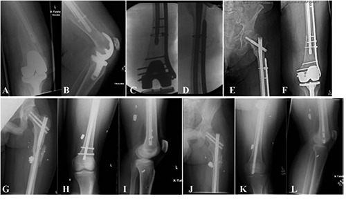
A 95 year-old female with a history of femoral intramedullary nailing for an intertrochanteric femur fracture in addition to an ipsilateral total knee arthroplasty was admitted after sustaining a fall. Radiological examination revealed an oblique periprosthetic distal femur fracture, spiraling around her intramedullary nail, ending just proximal to her total knee arthroplasty (Figure 1A,B). In the operating room, the patient’s distal interlocking screw was removed and reduction was achieved with a clamp and two cerclage wires. A 16-hole distal femoral locking plate was placed after fracture reduction, including screw placement through both the plate as well as the nail (Figure 1C,D). She was allowed to touch-down weight-bear immediately following surgery on the affected extremity. Follow-up X-rays 8 weeks postoperatively demonstrated stable hardware with a well-maintained fracture reduction (Figure 1E,F).
Figure 1.

Anteroposterior (A) and lateral (B) view of the knee demonstrating an oblique periprosthetic distal femur fracture extending from the distal interlocking screw. Anteroposterior view of the knee (C) and femur (D) intraoperatively after removal of the distal interlocking screw, cerclage fixation and a 16-hole distal femur locking plate; 8-week postoperative x-rays (E,F) with maintained fixation. Antero-posterior of the hip (G) and femur (H), and lateral of the knee (I) status post intramedullary fixation of a left subtrochanteric femur fracture with retained radio-opaque fragments. Anteroposterior of the hip (J), femur (K), and lateral of the femur (L) demonstrating subtrochanteric nonunion 3.5 years after initial fixation with retained radio-opaque fragments and distal cortical perforation into the knee joint.
Case Report #2
A 28 year-old male who had previously sustained a gunshot wound leading to a left subtrochanteric femur fracture treated with trochanteric entry recon nail resulted in a fracture nonunion (Figure 1G-I). After 12 months, he underwent nail dynamization with removal of his distal interlocking screw to allow compression of his non-united fracture. He was subsequently lost to follow-up for 3 years. He then presented for follow up with recent onset of left knee and thigh pain. Physical examination revealed knee range of motion from 30-90° and imaging of the entire femur demonstrated continued nonunion at his fracture site as well as penetration of the distal femoral cortex with migration of the nail into the knee joint (Figure 1J-L).
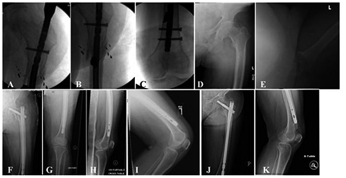
A CT was obtained confirming the nonunion; workup for infection revealed no elevation of inflammatory markers. The patient was taken to the operating room and underwent removal of his previous intramedullary nail with revision intramedullary nailing with dynamic compression across the fracture site (Figure 2A-C). He was made weight-bearing as tolerated postoperatively and X-rays four weeks postoperatively demonstrated stable nail position and fracture reduction. However, he was again lost to follow-up and has not returned to clinic.
Case Report #3
An 85-year-old female presented to our institution after a non-syncopal fall with left hip pain with a basicervical femoral neck fracture (Figure 2D,E). She was taken to the operating room and underwent trochanteric entry intramedullary nail fixation with a size 10 mm, radius 150 cm, 300 mm nail with no distal interlocking screw fixation. Reduction was achieved on a fracture table and intraoperative X-rays were consistent with an acceptable reduction and hardware placement without anterior cortical perforation (Figure 2F-H). She was made weight-bearing as tolerated postoperatively. The remainder of her immediate postoperative course was uncomplicated and patient was discharged to a rehabilitation facility.
While at the rehabilitation facility, she participated in physical therapy and subsequently felt a new pain her distal femur. At her three-week postoperative visit, repeat whole length femur radiographs revealed failure of hardware with nail penetration and fracture of the anterior cortex of her distal femur (Figure 2I). The decision was made to pursue non-operative management and protected weight-bearing. Follow-up X-rays demonstrated healing at both her proximal and distal fracture sites with callus formation (Figure 2J,K).
Figure 2.

Anteroposterior of the femur before (A) and after (B) dynamic compression across the fracture site with final distal fixation (C). Preoperative anteroposterior (D) and lateral (E) of a basicervical femoral neck fracture. Postoperative Anteroposterior of the hip (F), femur (G) and lateral of the femur (H) demonstrating intramedullary fixation with cortical abutment of the distal femur without perforation. Lateral of the knee (I) taken three weeks postoperative with anterior cortical perforation. Repeat imaging 3 months postoperatively with a healed femoral neck fracture (J) and distal femur fracture (K).
Discussion
Intramedullary femoral nailing has become the most common technique to stabilize femur fractures due to its advantages including less dissection and blood loss, faster time to healing, and earlier mobilization in comparison to plating.1 However, this technique is not without complications. This case series highlights three important complications that can affect the distal femur following antegrade intramedullary nailing: periprosthetic fracture through a distal interlocking screw, nail penetration into the knee joint, and anterior perforation of the nail. Prevention and treatment of these complications requires a sound understanding of the anatomy of the femur and the design specifications of femoral intramedullary nails. This case series underscores the necessity of follow-up distal femur radiographs in patients after long antegrade femoral nail placement.
Modern femoral intramedullary nails provide options for distal interlocking screws, which have been shown to provide fixation with an increase in stiffness and rotational load in unstable fracture patterns.13,14 However, recent evidence has demonstrated similar torsional strength in locked and unlocked nails in stable intertrochanteric hip fractures.15 Furthermore, distal interlocking screws provide a stress riser for fracture, and can add both operative time and radiation exposure to the case. This time may be valuable in elderly, medically sick patients with multiple comorbidities and higher perioperative risk of cardiopulmonary complications and also adds to the overall cost. Further research is needed to assess whether unlocked femoral nails provide adequate fixation for fractures while decreasing the rate of distal femoral periprosthetic fracture. Distally-locked fractures may also be vulnerable to spiral fracture distal to the interlocking screw, and frequently require fixation with open reduction and plating. However, we have observed that in unlocked nails the fracture occurs in the shaft of the femur and fixation can be achieved by closed reduction (rotating the fracture around the nail) and placing distal interlocking fixation through the existing nail.
An additional cause of fracture at the distal end of femoral nails is a mismatch of the radius of curvature and anterior bow of the femur, resulting in anterior cortical perforation of the distal femur.7,8,16 Using a starting point anterior to the midline and an implant with a smaller radius of curvature has been recommended in order to avoid this complication.17 Despite the use of a modern nail with a smaller radius of curvature (150 cm), our case demonstrates that anterior cortical perforation may still occur, especially in patients with a radius of curvature that falls outside the normal range. A recent study of 214 antegrade cephalomedullary nails found one anterior cortical perforation (0.47%), while 16% were within 3mm of the anterior cortex and 50% were within 7mm of the cortex.18 Although rare, anterior cortical perforation is a significant complication associated with intramedullary nails requiring altered weight bearing and sometimes revision surgery.
An additional pitfall of femoral intramedullary nail fixation is nonunion. Non-unions after intramedullary nailing are frequently managed with nail dynamization, and the removal of distal interlocking screws permits the nail to migrate distally. Patient 2 in this series demonstrates a case of extreme fracture shortening with subsequent intra-articular nail penetration into the knee. There are no previous case reports of modern nail failure by this mechanism. Surgeons must be aware that this risk exists when dynamizing a nail, as shortening at the fracture site may result in distal cortical penetration if there is not enough remaining intramedullary length and if the fracture shortens substantially.
Conclusions
This case series highlights the distal femoral complications following antegrade femoral nailing, the need for close follow-up with distal femur imaging for patients with intramedullary nails in place, and appropriate surgical planning for both primary and revision surgery with femoral intramedullary nails.
Acknowledgments
the authors would like to thank Dr. Christopher Born and Dr. Roman Hayda for their technical expertise.
References
- 1.Ricci WM, Gallagher B, Haidukewych GJ. Intramedullary nailing of femoral shaft fractures: current concepts. J Am Acad Orthop Surg 2009;17:296-305. [DOI] [PubMed] [Google Scholar]
- 2.Wild M, Gehrmann S, Jungbluth P, et al. Treatment strategies for intramedullary nailing of femoral shaft fractures. Orthopedics 2010;33:726. [DOI] [PubMed] [Google Scholar]
- 3.Ricci WM, Bellabarba C, Lewis R, et al. Angular malalignment after intramedullary nailing of femoral shaft fractures. J Orthop Trauma 2001;15:90-5 [DOI] [PubMed] [Google Scholar]
- 4.Salem KH, Maier D, Keppler P, et al. Limb malalignment and functional outcome after antegrade versus retrograde intramedullary nailing in distal femoral fractures. J Trauma 2006;61:375-81. [DOI] [PubMed] [Google Scholar]
- 5.Herscovici D, Scaduto JM. Assessing leg length after fixation of comminuted femur fractures. Clin Orthop Relat Res 2014;472:2745-50. [DOI] [PMC free article] [PubMed] [Google Scholar]
- 6.Hüfner T, Citak M, Suero EM, et al. Femoral malrotation after unreamed intramedullary nailing: an evaluation of influencing operative factors. J Orthop Trauma 2011;25:224-7. [DOI] [PubMed] [Google Scholar]
- 7.Ostrum RF, Levy MS. Penetration of the distal femoral anterior cortex during intramedullary nailing for subtrochanteric fractures: a report of three cases. J Orthop Trauma 2005;19:656-60. [DOI] [PubMed] [Google Scholar]
- 8.Neumayer F, Löcherbach C, McManus J, Chevalley F. Medial femoral condyle fracture as a complication of antegrade intramedullary nailing. Arch Orthop Trauma Surg 2013;133:1273-9. [DOI] [PubMed] [Google Scholar]
- 9.Scolaro JA, Endress C, Mehta S. Prevention of cortical breach during placement of an antegrade intramedullary femoral nail. Orthopedics 2013;36:688-92. [DOI] [PubMed] [Google Scholar]
- 10.Karadimas EJ, Papadimitriou G, Theodoratos G, et al. The effectiveness of the antegrade reamed technique: the experience and complications from 415 traumatic femoral shaft fractures. Strateg Trauma Limb Reconstr 2009;4:113-21. [DOI] [PMC free article] [PubMed] [Google Scholar]
- 11.Malik MHA, Harwood P, Diggle P, Khan SA. Factors affecting rates of infection and nonunion in intramedullary nailing. J Bone Joint Surg Br 2004;86B:556-60. [PubMed] [Google Scholar]
- 12.El Moumni M, Leenhouts PA, ten Duis HJ, Wendt KW. The incidence of non-union following unreamed intramedullary nailing of femoral shaft fractures. Injury 2009;40:205-8. [DOI] [PubMed] [Google Scholar]
- 13.Gallagher D, Adams B, El-Gendi H, et al. Is distal locking necessary? A biomechanical investigation of intramedullary nailing constructs for intertrochanteric fractures. J Orthop Trauma 2013;27:373-8. [DOI] [PubMed] [Google Scholar]
- 14.Kane P, Vopat B, Paller D, et al. A biomechanical comparison of locked and unlocked long cephalomedullary nails in a stable intertrochanteric fracture model. J Orthop Trauma 2014;28:715-20. [DOI] [PMC free article] [PubMed] [Google Scholar]
- 15.Kane PM, Vopat B, Paller D, et al. Effect of distal interlock fixation in stable intertrochanteric fractures. Orthopedics 2013;36:e859-64. [DOI] [PubMed] [Google Scholar]
- 16.Roberts JW, Libet LA, Wolinsky PR. Who is in danger? Impingement and penetration of the anterior cortex of the distal femur during intramedullary nailing of proximal femur fractures: preoperatively measurable risk factors. J Trauma Acute Care Surg 2012;73:249-54. [DOI] [PubMed] [Google Scholar]
- 17.Kanawati AJ, Jang B, McGee R, Sungaran J. The influence of entry point and radius of curvature on femoral intramedullary nail position in the distal femur. J Orthop 2014;11:68-71. [DOI] [PMC free article] [PubMed] [Google Scholar]
- 18.Bazylewicz DB, Egol KA, Koval KJ. Cortical encroachment after cephalomedullary nailing of the proximal femur: evaluation of a more anatomic radius of curvature. J Orthop Trauma 2013;27:303-7. [DOI] [PubMed] [Google Scholar]


