Abstract
Ever since Hattori et al. had described the mediastinal Mullerian cyst in 2005 there has been several new cases described in the literature. We report a 51-year-old woman with an incidentally found 2 cm × 3 cm mass in her left paravertebral mediastinum. She underwent thoracoscopic removal with the impression of a neurogenic tumor and was unexpectedly found with a ciliated cyst of Mullerian origin.
Keywords: Mediastinal cyst, Mullerian duct, video assisted thoracoscopic surgery
Introduction
The Mullerian cyst was first described in 2005 by Hattori and colleagues occurring in the mediastinum (1). Since then, there have been 14 established cases reported in the English literature (2). We report a case of Mullerian cyst occurring in the mediastinum in a 51-year-old female patient that was resected by video assisted thoracoscopic surgery.
Case report
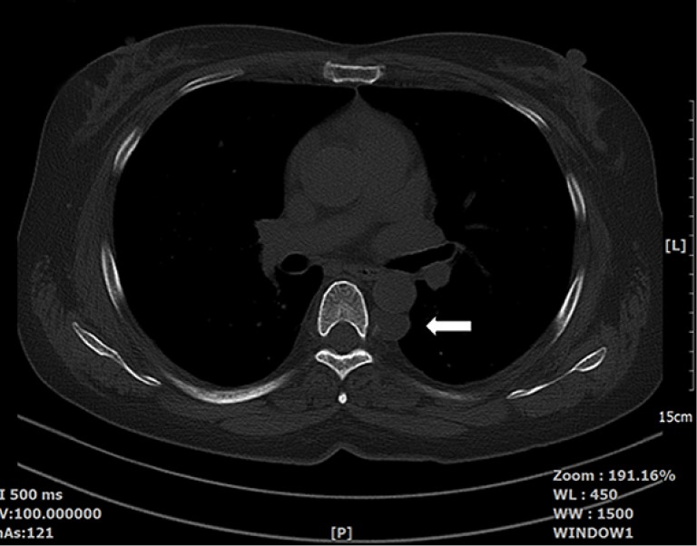
A 51-year-old woman visited our clinic with symptoms of right chest wall pain of one week onset after blunt trauma. The patient had a history of spine operation 6 years ago, but no gynecologic history of any surgical procedure. The mass was not visible by simple X-rays and was found incidentally by computer tomographic scans of her chest. The computer tomographic scan revealed a 2 cm × 3 cm oval shaped mass over her 6th thoracic paravertebral area (Figure 1). One week later thoracoscopic resection of the mass was performed.
Figure 1.

The computer tomographic scan shows a 2 cm × 3 cm mass (arrow) in her posterior mediastinum over her 6th thoracic vertebra.
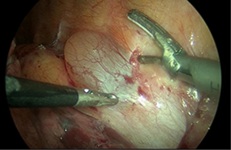
The patient was placed in a lateral position with three ports; the 5 mm camera (5 mm 30 degree, Storz) port in her 8th intercostal space in her mid-clavicular line anteriorly, a second 3 mm port in her 6th intercostal space cranially and posteriorly, and a third 5 mm port caudally in her 9th intercostal space posteriorly. Initially visualization was done with the 5 mm camera and was then switched to a 3 mm camera (Storz) (Figure 2). The tumor was juxtaposed to her descending aorta and was found to be cystic. The cyst was drained and cyst removed in its entirety with the help of Harmonic scalpel. The mass was removed through an endoscopic bag, necessitating extension of the 5 mm camera port to 1 cm to allow passage of the endoscopic bag, and sent for pathologic examination.
Figure 2.

The thoracoscopic operative view showing the mass with a 3 mm camera (Storz) over the patient’s 6th thoracic vertebra abutting her aorta. The 5 mm grasper and 5 mm harmonic scalpel instruments are seen manipulating the cystic mass.
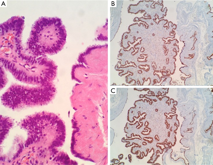
The resected specimen histologically revealed a unilocular cyst with ciliated and flattened epithelium with scant connective tissue and surrounding smooth muscle bundles in the cystic wall. Immunochemistry confirmed presence of both estrogen and progesterone receptors in its epithelium (Figure 3).
Figure 3.
(A) Ciliated columnar cells and flattened epithelium and surrounding smooth muscle bundles are seen in the cystic wall (Hematoxylin and eosin, 400×); (B,C) immunostaining shows positive staining for estrogen and progesterone receptors, respectively (estrogen receptor alpha and progesterone receptor, 100×).
Her postoperative course was uneventful, her drain was removed on her first postoperative day. She was not discharged immediately due to her pain involved with her multiple rib fractures. There are no signs of recurrence 9 months after the operation and she is without pain and more than happy with the cosmesis.
Discussion
Mullerian cysts occurring in the mediastinum have only recently been reported. Retrospective analyses done in two studies have discovered these cysts in 15.8% (3 of 19 cases of mediastinal cysts) and 4.3% (7 established cases of 163 mediastinal cysts) (3,4). These studies show that the Mullerian cysts may not be so rare.
Mullerian cysts generally occur in woman 40 to 60 years of age, are found in paravertebral location between the 3rd to 8th thoracic vertebra (2). They are reported frequently in women in their perimenopausal period with many cases occurring in obese women and women with a gynecologic history (1). All cases were positive for either estrogen or progesterone receptor. Our particular case was also found in a 51-year-old woman over her 6th thoracic vertebra and immunohistochemical stains have confirmed positivity for estrogen and progesterone receptors.
The origin of the Mullerian cyst is unknown and a subject of interest. The origins of Mullerian cysts in the abdomen can be explained by aberrant Mullerian duct remnants, but this occurring in the chest cavity is a mystery. The lesion has been proposed to be derived from primary Mullerian apparatus (5).
The behavior of the cyst appears benign and there are no present reports of recurrence. Mullerian cysts in a woman between her 40 to 60 age group should be considered in differential diagnosis of posterior mediastinal paravertebral cysts.
Acknowledgements
This work was supported by Wonkwang University in 2014.
Disclosure: The authors declare no conflict of interest.
References
- 1.Hattori H.Ciliated cyst of probable mullerian origin arising in the posterior mediastinum. Virchows Arch 2005;446:82-4. [DOI] [PubMed] [Google Scholar]
- 2.Simmons M, Duckworth LV, Scherer K, et al. Mullerian cysts of the posterior mediastinum: report of two cases and review of the literature. J Thorac Dis 2013;5:E8-10. [DOI] [PMC free article] [PubMed] [Google Scholar]
- 3.Hattori H.High prevalence of estrogen and progesterone receptor expression in mediastinal cysts situated in the posterior mediastinum. Chest 2005;128:3388-90. [DOI] [PubMed] [Google Scholar]
- 4.Thomas-de-Montpréville V, Dulmet E.Cysts of the posterior mediastinum showing müllerian differentiation (Hattori's cysts). Ann Diagn Pathol 2007;11:417-20. [DOI] [PubMed] [Google Scholar]
- 5.Batt RE, Mhawech-Fauceglia P, Odunsi K, et al. Pathogenesis of mediastinal paravertebral müllerian cysts of Hattori: developmental endosalpingiosis-müllerianosis. Int J Gynecol Pathol 2010;29:546-51. [DOI] [PubMed] [Google Scholar]


