Abstract
Cytochrome P450 1b1 (Cyp1b1) expression is absent in mouse hepatocytes, but present in liver endothelia and activated stellate cells. Increased expression during adipogenesis suggests a role of Cyp1b1 metabolism in fatty acid homeostasis. Wild-type C57BL/6j (WT) and Cyp1b1-null (Cyp1b1-ko) mice were provided low or high fat diets (LFD and HFD, respectively). Cyp1b1-deletion suppressed HFD-induced obesity, improved glucose tolerance and prevented liver steatosis. Suppression of lipid droplets in sinusoidal hepatocytes, concomitant with enhanced glycogen granules, was a consistent feature of Cyp1b1-ko mice. Cyp1b1 deletion altered the in vivo expression of 560 liver genes, including suppression of PPARγ, stearoyl CoA desaturase 1 (Scd1) and many genes stimulated by PPARα, each consistent with this switch in energy storage mechanism. Ligand activation of PPARα in Cyp1b1-ko mice by WY-14643 was, nevertheless, effective. Seventeen gene changes in Cyp1b1-ko mice correspond to mouse transgenic expression that attenuated diet-induced diabetes. The absence of Cyp1b1 in mouse hepatocytes indicates participation in energy homeostasis through extra-hepatocyte signaling. Extensive sexual dimorphism in hepatic gene expression suggests a developmental impact of estrogen metabolism by Cyp1b1. Suppression of Scd1 and increased leptin turnover support enhanced leptin participation from the hypothalamus. Cyp1b1-mediated effects on vascular cells may underlie these changes.
INTRODUCTION
Cytochrome P450 1b1 (Cyp1b1) is expressed in a spatial and temporal manner during development, notably in rhombomere 4 of the neural crest, the hind brain and the eye [1–3]. Human developmental aberrations in both the eye [3] and the liver [4] arise from Cyp1b1 deficiency. Cyp1b1 is expressed in multi-potential mesenchymal stromal cells [5], liver stellate cells [6–7], endothelium [8–9] and microvessels of the brain [10]. Cyp1b1 is, however, essentially absent from adult mouse hepatocytes [7, 11]. Effects of Cyp1b1 deletion on the vasculature depend on increased oxidative signaling [8–9] and diminished estrogen metabolism [12].
Hepatic fatty acid metabolism is regulated by the inter-play between a set of nuclear receptors. These include peroxisome proliferator-activated receptors gamma and alpha (PPARγ, PPARα, respectively), constitutive androstane receptor and hepatocyte nuclear factor 4 alpha (HNF4α), which are activated by fatty acids or their derivatives, and pregnane X receptor, liver X receptor and farnesyl X receptor, which are activated by cholesterol derivatives [13–14]. Hepatic PPARs play a key role in maintaining energy homeostasis during periods of fasting, including the regulation of the storage of fatty acids as triglyceride droplets [15]. Activation results in increased fatty acid uptake and oxidation, lipogenesis and gluconeogenesis/glycolysis. The microsomal oxidase, stearoyl CoA desaturase 1 (Scd1), which is particularly sensitive to suppression by hypothalamic leptin signaling, enhances this storage while decreasing mitochondrial oxidation [16–17].
Cyp1b1 expression increases in the early phase of in vitro adipogenic differentiation, in parallel with PPARγ expression [5]. The endothelium plays a critical role in the development and maintenance of both liver and adipose tissue. Cyp1b1 expression in endothelial cells and pericytes decreases peroxidation of unsaturated fatty acids, while promoting capillary morphogenesis in vitro, and neovascularization in vivo [8–9, 18–19]. The association of Cyp1b1 activity with adipogenic PPARγ expression and decreased fatty acid peroxidation suggests that Cyp1b1 may play an important role in this aspect of energy homeostasis. To test this hypothesis, we maintained C57BL/6j (WT) and Cyp1b1-null (Cyp1b1-ko) mice on either a low fat/high carbohydrate diet (LFD) or a near iso-caloric high fat/low carbohydrate diet (HFD). We used various time periods post-weaning, which distinguish different phases of the response to high dietary fat. We report that Cyp1b1 deletion lowers adiposity without affecting caloric intake, suggesting altered energy utilization.
The major phenotypic features associated with Cyp1b1 deletion include protection from the deleterious effects of excess dietary fat, including oxidative stress, hepatic steatosis and Type 2 diabetes. Microarray data, from the livers of the Cyp1b1-ko mice that are resistant to diet-induced obesity (DIO), indicates that Cyp1b1 deletion substantially alters the expression of genes associated with fatty acid homeostasis. We identified 17 gene changes that, when introduced singly into transgenic mice, produce protection from these conditions.
Many genes are constitutively activated by PPARα, a protein that is enhanced by mobilization of fatty acids during fasting. Here, analyses of livers from mice fed ad libitum demonstrate an extensive suppression of this constitutive PPAR regulation with Cyp1b1 deletion. Much of this suppression process diverts hepatic fatty acid metabolism from the formation of triglyceride droplets to mitochondrial oxidation and glycogen storage. Substantial differences in gene expression in Cyp1b1-ko mice fed high or low fat diets suggests that Cyp1b1 metabolism of endogenous substrates affects extra-hepatic signaling to the hepatocyte, thereby controlling fatty acid metabolism within these cells. Adipose suppression is similar in male and female mice, yet many gene changes suggest affects on sexually dimorphic control. Estradiol is a Cyp1b1 substrate [20], which is generated by aromatase within the hypothalamus and controls energy homeostasis through activation of estrogen receptor alpha (ER-α) [21–22]. One possibility is that increased estradiol concentrations in the hypothalamus may affect sexually dimorphic responses in Cyp1b1-ko mice.
The suppression effects of Cyp1b1 deletion on adiposity and liver steatosis are retained in both male and female mice from 6 to 36 weeks of age. We have identified likely participants in this novel external modulation of hepatocyte activity by Cyp1b1 metabolism by focusing on similarly conserved gene responses. A small group of genes have been identified in this way, which have each been implicated in fatty acid homeostasis, including Scd1 and a subset of PPARα-regulated genes.
MATERIALS AND METHODS
Ethics statement
Mice were maintained in the AAALAC-accredited University of Wisconsin School of Medicine and Public Health animal care facility. Experimental protocols were approved by the University of Wisconsin School of Medicine and Public Health Animal Care and Use Committee (ACUC; Protocol number: M00682), in strict accordance to the EU Directive 2010/63/EU.
Mouse breeding and maintenance
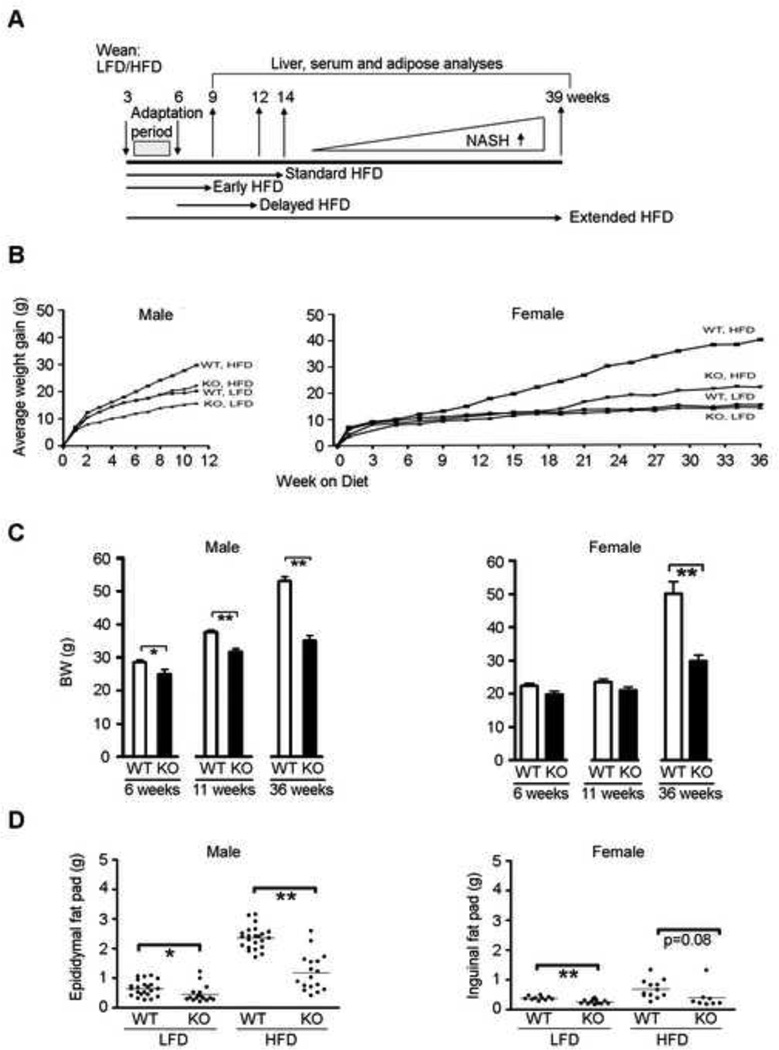
Mice were provided food and water ad libitum and were maintained on a 12 hour light/dark cycle. The mice were fed isocaloric diets containing 10 percent fat/70 percent carbohydrate (D12450B) or 60 percent fat/20 percent carbohydrate (D12492) from Research Diets Inc. (New Brunswick, NJ). The protein content of each of these diets is depleted of iso-flavones, which have major endocrine impacts, and the fat is derived from lard. Diets were administered for 6, 11 or 36 weeks post-weaning, as indicated (Figure 1A). The initial Cyp1b1-ko founders (on C57BL/6 background) were obtained from Dr. Frank Gonzalez [23]. The original colony (1997) was backcrossed to Jackson C57BL/6j (WT) mice through 10 generations, but subsequently inbred for several years. This colony provided consistent obesity responses over the generations used in these studies, with the exception of blood glucose analyses. Treatments were compared between littermates from multiple litters over more than 12 months. Comprehensive data was collected for food consumption (Figure S1A) and body weight (BW) gain over this period of time (Figure 1B). A second phase of the study was started when an increased proportion of Cyp1b1-ko mice demonstrated DIO. Cyp1b1-ko mice that demonstrated DIO, which was characteristic of WT mice, were re-derived and refreshed by a single mating with WT animals. This Cyp1b1-ko colony predominantly retained high obesity with the HFD challenge. Subsequent back-crossing was completed by mating Cyp1b1-heterozygote females to Jackson WT males. The F3-F5 generations of this back-cross demonstrated obesity suppression and were used for glucose measurements. Additional mating examined the effect of breeding the smallest (S1&2) and largest (L1&2) progeny, respectively, from the initial backcross, as well as the effect of mating a heterozygous male or female, respectively, with a Cyp1b1-ko partner. Mice were sacrificed by CO2 asphyxiation and blood was collected by cardiac puncture. Tissues were weighed and flash frozen in liquid nitrogen and stored at −80° C for protein and mRNA analyses. Tissue samples were also frozen in sectioning medium or fixed in 10% formalin solution for histology.
Figure 1. Cyp1b1-ko mice exhibit decreased adiposity.
A. Experimental design to determine effect of Cyp1b1 deletion on diet responses.
B. Average body weight (BW) gain in male and female C57BL/6j (WT) and Cyp1b1-ko (KO) mice for, respectively, 11 and 36 weeks post-weaning.
C. Average BW of male and female WT and KO mice fed the HFD for 6, 11 and 36 weeks post-weaning. The bar graphs indicate the average BW ± SEM. (Male WT: n=20, 6-week; n=23, 11-week; n=11, 36-week. Male KO: n=4, 6-week; n=17, 11-week; n=6, 36-week. Female WT: n=13, 6-week; n=12, 11-week; n=3, 36-week. Female KO: n=7, 6-week; n=10, 11-week; n=3, 36-week.)
D. Epididymal and inguinal fat pad mass in male and female WT and KO mice fed the LFD or HFD for 11 weeks post-weaning. Each dot represents an individual animal. The horizontal line represents the average fat pad mass of each group. (WT males: n=22 LFD, n=23 HFD. KO males: n=16 LFD, n=17 HFD. WT females: n=10 LFD, n=12 HFD. KO females: n=12 LFD, n=8 HFD.)
*p<0.05, ** p<0.01.
Wy-14,643 treatment of mice
Mice were given a single oral gavage treatment of Wy-14,643 (Cayman Chemical Company, Ann Arbor, MI) at 50mg/kg and sacrificed 48 hours thereafter. Wy-14,643 was dissolved in DMSO and added to olive oil (Sigma-Aldrich, St. Louis, MO) to generate a 20% DMSO-WY/olive oil solution. Control mice were similarly treated with the 20% DMSO/olive oil solvent control.
RNA extraction and real-time PCR
Total RNA was isolated from 50 mg of tissue, using TRIzol (Life Technologies, Grand Island, NY). RNA (1.5 µg) was reverse transcribed with Reverse Transcriptase (Promega, Madison, WI) in 20 µL reaction volume. The RT reaction was diluted 5-fold and 5 µL was amplified by real-time PCR (qPCR) in a 25 µL reaction mixture containing Master Sybr Green (Bio-Rad, Hercules, CA). Primers (Table S1) were designed to span the intron-exon borders, using Primer Express software (Applied Biosystems, Grand Island, NY). qPCR was performed on a MyiQ real-time PCR system (Bio-Rad, Hercules, CA): an initial 2 min denaturation step at 95° C, followed by 40 cycles of 15 sec each at 95° C and a 1 min annealing step at 60° C. Gene expression was quantified in replicate samples using the delta ct methodology [24], normalizing to cyclophillin expression.
Magnetic resonance imaging (MRI) analysis
MRI measurements were completed on a representative set of four female mice using a 3.0T clinical imager (MR750, GE Healthcare, Waukesha, WI), equipped with a custom-built quadrature birdcage coil specifically sized for rodents [25–27]. Imaging was performed using an investigational version of a quantitative 3D multi-echo chemical-shift encoded fat-water separation technique that has been previously described and validated in mice [25–27]. Prior to imaging, mice were anesthetized with pentobarbital via intraperitonel (IP) injection (40mg/kg BW). Quantification of adipose volume was completed using OsiriX computer software through segmentation of the fat-only images.
Blood glucose monitoring
To avoid the stress associated with a major fasting intervention, we routinely measured early morning (7:30-9 AM), non-fasted glucose levels in the WT and Cyp1b1-ko mice after 5 and 10 weeks on the HFD. Tail vein blood levels were measured using a standard glucometer (One-touch Ultra 2, Milpitas, CA). Despite the appreciable BW gain between weeks 5 and 10 of HFD exposure, the blood glucose levels for each mouse remained within 10 percent, suggesting that larger differences were not due to stress or feeding variability.
Glucose tolerance testing (GTT)
Mice were fasted for 16 hours. Blood glucose levels were measured before (0) and 15, 30, 60 and 120 min following the IP administration of dextrose (2 g/kg body mass), as described above. GraphPad Prism 5 (La Jolla, CA) was used to determine the area under the curve (AUC) by plotting each individual animal, setting the baseline to the glucose level at the zero time point.
Serum metabolic profile
Blood samples were collected by cardiac puncture at sacrifice and the serum isolated by centrifugation at 14,000 rpm for 10 min at 4° C. Serum was frozen in liquid nitrogen and stored at −80° C until analysis. All analyses were completed as per manufacturer’s instructions. Insulin and leptin levels were measured using a Mercodia (Uppsala, Sweden) and an RnD Systems (Minneapolis, MN) ELISA kit, respectively, on 4-hour fasted animals. Serum triglyceride, free fatty acid and cholesterol levels were measured using the Sigma-Aldrich (St. Louis, MO) serum triglyceride determination, the Zen-Bio (Research Triangle Park, NC) Elisa and the Pointe Scientific (Canton, MI) colorimetric assay kits, respectively, on samples derived from non-fasted animals.
Hepatic triglycerides
Frozen liver tissue (50 mg), collected from mice fed ad libitum, was homogenized in ethanol (1 ml), cleared by centrifugation and the supernatant isolated for analysis. Hepatic triglycerides were measured using the Sigma-Aldrich (St. Louis, MO) serum triglyceride determination kit, as above.
Immunohistochemistry
Fixed white adipose, liver and skin tissues were embedded in paraffin, sectioned and stained with hematoxylin/eosin (H&E) or periodic acid-Schiff (PAS) (± diastase) stain, according to the manufacturer’s protocol. Frozen liver tissues were sectioned and stained with Oil Red O. The relative adipocyte area was determined using NIH ImageJ software. An H&E-stained adipose image was captured from one slide of each of four animals per group (WT and Cyp1b1-ko, LFD and HFD) and the average relative area was calculated by tracing 50 adipocytes per image.
Microarray analysis
Total liver RNA was isolated using RNeasy Mini kits, according to manufacturer’s instructions (Qiagen, Valencia, CA). RNA concentration was quantified and the purity determined by A260/A280 ratio and by visual inspection via denaturing gel electrophoresis. All Agilent mouse microarray (Santa Clara, CA) analyses were performed with three biological replicates from WT and Cyp1b1-ko mice fed either the LFD or HFD. All methods used for microarray preparation were completed as previously described [28]. All arrays were normalized and quality assessed using EDGE software. ANOVA analysis was completed to determine statistical significance.
The Agilent dual color microarray employs a competitive binding of, respectively, a Cy3-labeled treatment sample and a Cy5-labeled mouse liver reference standard (a pool of three WT/LFD samples), which is presented as a Cy3/Cy5 binding ratio. The WT/LFD Cy5 binding provides the reference level of expression of each gene. We have considered genes that exhibit a WT/LFD Cy5 binding >200. This cutoff includes over 5,000 liver genes and greatly reduces false positives that readily appear as expression nears background. We further considered the relevant expression differences between the treatment groups to be two-fold or greater with p<0.05 (Table S2). This is based on the Limma program that tests significance in these array comparisons based on ANOVA [28]. The data discussed in this publication have been deposited in NCBI's Gene Expression Omnibus and are accessible through GEO Series accession number GSE53910 (http://www.ncbi.nmk.nih.gov/geo/query/acc.cgi?acc=GSE53910).
Other statistical analyses
Excluding microarray analyses, all remaining statistical comparisons were assessed by unpaired 2-tailed Student t tests (GraphPad Prism, La Jolla, CA), which provide a numerical p-value for each comparison. Analyses are reported as mean ± SEM. Experiments were designed to include at least 8 mice in each genotype/diet group, representing animals from a minimum of 4 litters.
RESULTS
Cyp1b1 deletion suppresses post-weaning weight gain
The association of Cyp1b1 with adipogenesis and pro-oxidant signaling suggested that Cyp1b1 metabolism may play a significant role in mediating the responsiveness of liver and adipose tissue to dietary fat. Cyp1b1 is appreciably expressed in adipose tissue of C57BL/6j mice, but is expressed at very low levels in the liver [Figure S1B], most of which can be accounted for by expression in endothelial cells (Sheibani et al, unpublished data). To examine this further, we compared the effects of a high fat/low carbohydrate (60 percent fat/20 percent carbohydrate, HFD) and a low fat/high carbohydrate (10 percent fat/70% carbohydrate, LFD) diet on WT and Cyp1b1-ko mice. The two defined diets, which are free of protein-associated isoflavones, are of similar caloric content and are commonly used for studies of DIO [29]. Male and female mice were placed on the respective diet at weaning (postnatal day 21; continued for 6, 11 or 36 weeks) or late in liver maturation (6 weeks of age; 6 week exposure) (Figure 1A). The three periods of HFD intake correspond to different stages of non-alcoholic hepatic steatosis (NASH) in C57BL/6 mice [30].
The HFD significantly increased the BW of male WT mice after 3 weeks, relative to their LFD counterparts, while the WT females needed an additional 6 weeks for an equivalent increase (Figure 1B). The 11 week BW gain for both genders was suppressed by Cyp1b1 deletion (Figure 1B). In females, the BW suppression at 36 weeks reached levels that were comparable to those observed in the 11 week male mice (Figure 1C). This was associated with a suppression of adiposity, as measured by decreased mass of epididymal and inguinal adipose tissue depots in male and female mice (Figure 1D), respectively, which correlated with the BW changes (Figure S2A and S2B). Suppression of adiposity in Cyp1b1-ko mice was also seen on the LFD (Figure 1D).
The adipose response of male Cyp1b1-ko offspring to the HFD divided into three levels of suppression that correlated with the BW changes, often within the same litter: 60–85 percent suppression (8/17), 30–50 percent suppression (7/17), no suppression (2/17) (Figure 1D). The female littermates were consistently more highly suppressed. These data were collected from Cyp1b1-ko mice obtained from multiple litters over three generations, with similar response variability throughout this period. In later studies, which included Cyp1b1-ko mice from other laboratories, the proportion of mice exhibiting resistance to suppression was higher. These resistant mice have been examined further (see below).
Liver and fat pad each undergo extensive development between birth and 3 weeks post-weaning [31–32]. The BW commonly varied by 50–100 percent in the mice at weaning (7- to 14 g), particularly in the Cyp1b1-ko colony. However, weaning weight did not correlate with the 11 week BW (Maguire and Larsen, unpublished data). Notably, differences in weaning weight equalized within this initial three weeks on either diet. This rapid post-weaning correction in BW suggests that the differences at weaning arise from the access or response to maternal milk. Adiposity and BW were similarly suppressed by Cyp1b1 deletion when a 6 week HFD exposure was initiated during (3–9 weeks) or after (6–12 weeks) this adaptation period (Figure S2C).
General suppression in adipose is linked to adipocyte size
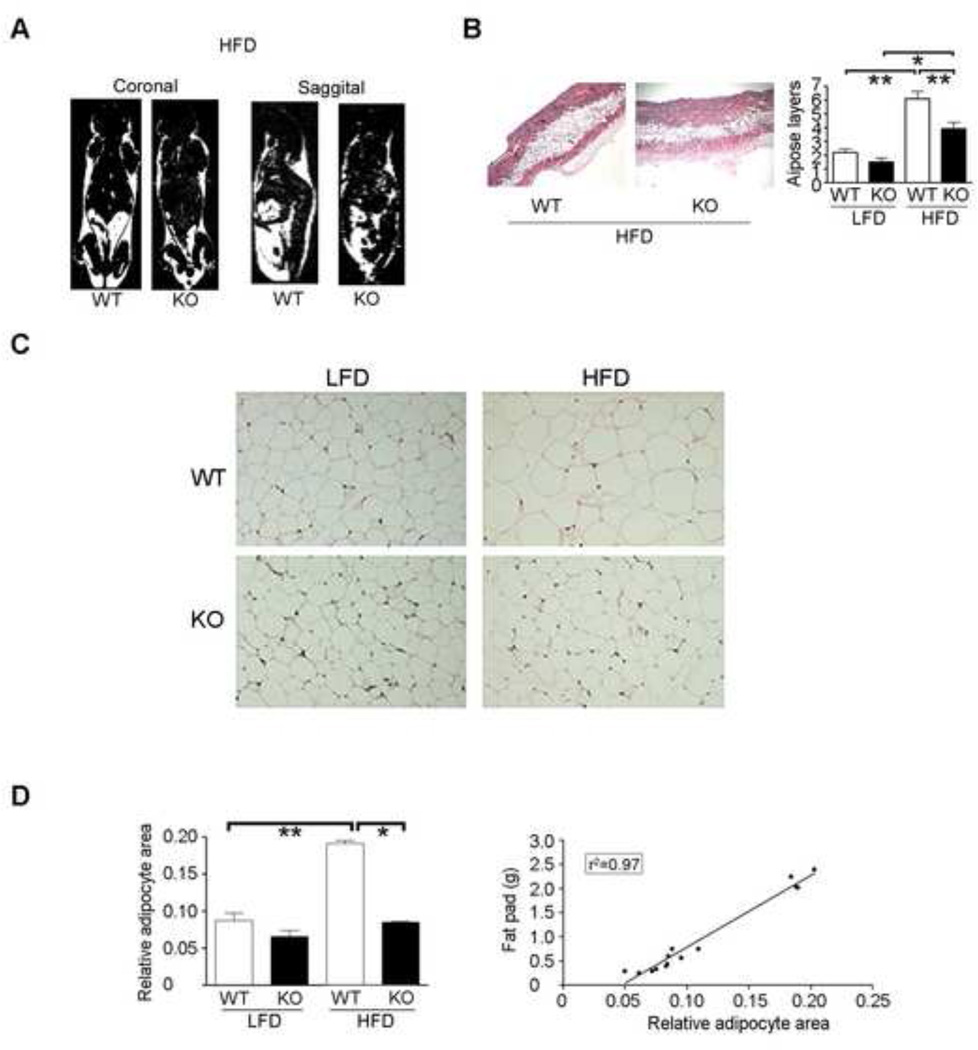
In order to examine whether adipose suppression changes were fat pad selective, MRI analyses, examining coronal and sagittal reformatted images through the mouse midline, were completed on representative mice at 11 weeks post-weaning. We selected Cyp1b1-ko mice exhibiting typical adipose suppression. The fat-only MRI images demonstrated the general suppression of adipose tissue volume in all fat pads within the Cyp1b1-ko mice exposed to the HFD (Figure 2A). Likewise, quantification of the average number of sub-dermal adipose layers showed that Cyp1b1 deletion suppressed diet-mediated adipose deposition (Figure 2B).
Figure 2. Anomalous characteristics of adipose of Cyp1b1-ko mice.
A. Reformatted fat-only MRI images (coronal and sagittal reformats) of representative C57BL/6j (WT) and Cyp1b1-ko (KO) mice after 11 weeks on the HFD.
B. H&E staining (HFD) and quantification of sub-dermal adipose tissue layers (HFD and LFD) from WT and KO mice after 11 weeks on the respective diets. The bar graph represents the average number of adipose layers ± SEM (WT: n=4 LFD and HFD; KO: n=2 LFD, n=4 HFD).
C. H&E stained epididymal fat pad adipocytes of representative male WT and KO mice after 11 weeks on the LFD and HFD.
D. Image J analysis of relative adipocyte area with correlation to fat pad mass. The bar graph represents the average area ± SEM of 50 adipocytes per image from 4 mice per strain/treatment group.
*p<0.05, ** p<0.01.
Adiposity can increase as a consequence of the increased size of the adipocytes, as found in ER-α-ko mice [33], or increased numbers of adipocytes. This alternative, which is found in aromatase-deficient mice [34], derives from either increased proliferation or commitment of progenitor cells. Adipose tissue is comprised of adipocytes, supporting vasculature and macrophages [35]. The cross-sectional area of adipocytes doubles in WT mice fed the HFD for 11 weeks (Figure 2C) [36]. The decreased fat pad mass in the Cyp1b1-ko mice, relative to their WT counterparts, correlated with a decrease in adipocyte size (r2=0.97) (Figure 2D). The effect of Cyp1b1 deficiency is, therefore, targeted to lipid accumulation rather than to an increased number of adipocytes, thus representing a reversal of the ER-α-ko phenotype.
Effect of Cyp1b1-ko on serum lipids
Serum cholesterol ester and triglyceride levels, which are each present in the various lipoprotein fractions (HDL, LDL and VLDL) exhibited different sensitivities to HFD and Cyp1b1 status (Table 1). Serum cholesterol esters paralleled the adiposity changes, in a manner that is typical of DIO [37]. Levels almost doubled in the WT mice challenged with the HFD, but were only minimally affected in the Cyp1b1-ko mice. Serum triglycerides increased by 45 percent in the WT mice switched to HFD. Increased triglyceride levels were also observed in Cyp1b1-ko mice, irrespective of diet. By contrast, serum free-fatty acids were unaffected by either diet or Cyp1b1 status.
Table 1.
Physiological parameters in male mice at 11 weeks of HFD exposure.
| Strain/ Treatment |
Blood glucose (mg/dL)a |
Serum free fatty acids (µM)b |
Serum Insulin (µg/L)c |
Serum triglycerides (µg/µl)b |
Serum cholesterol (mg/dL)b |
Hepatic triglycerides (µg/g)b |
|---|---|---|---|---|---|---|
| WT, LFD | 159 ± 18 (n=4) | 804 ± 131 (n=10) | 1.64 ± 0.31 (n=4) | 0.60 ± 0.06 (n=6) | 89 ± 3.5 (n=4) | 10.8 ± 2.3 (n=2) |
| KO, LFD | 101 ± 8* (n=3) | 902 ± 131 (n=6) | 1.60 ± 0.26 (n=4) | 0.86 ± 0.10* (n=4) | 96 ± 5.5 (n=6) | 12.6 ± 0.3 (n=2) |
| WT, HFD | 210 ± 14 (n=4) | 913 ± 95 (n=12) | 6.34 ± 2.5 (n=4) | 0.81 ± 0.05* (n=6) | 150 ± 10.2* (n=10) | 23.5 ± 2.4* (n=7) |
| KO, HFD | 106 ± 8 ** (n=3) | 1025 ± 27 (n=7) | 1.61 ± 0.3 (n=4) | 0.89 ± 0.12 (n=3) | 104 ± 9.0**,*** (n=7) | 9.8 ± 2.2** (n=6) |
SEM
Significantly different from WT, LFD.
Significantly different from WT, HFD.
Significantly different from KO, LFD.
Mice were fasted for 16 hours.
Mice were fed ad litibum.
Mice were fasted for 4 hours.
Blood samples from these individual mice have also been analyzed by tandem mass spectrometry, examining a set of lysophosphatidylcholine derivatives (LPCs). The detailed findings for mice from the 11 week studies have been recently described [38]. Interestingly, the 18:0 LPC parallels the cholesterol ester/obesity response, whereas the other major LPC, 16:0, is unaffected by these conditions.
Suppression of blood glucose by Cyp1b1 deletion
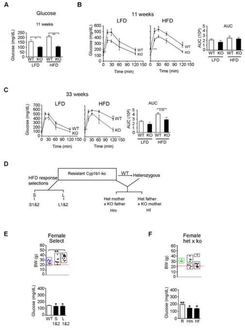
Serum insulin levels increased by approximately 4-fold in WT mice fed the HFD, but were unaffected in the Cyp1b1-ko mice (Table 1). The fasted blood glucose levels were significantly lower in Cyp1b1-ko mice than WT mice on the LFD. The HFD challenge increased levels in WT mice by 30 percent, while the Cyp1b1-ko animals remained unchanged (Figure 3A and Table 1).
Figure 3. Glucose levels and tolerance in Cyp1b1-ko mice: Effects of selective breeding and backcrossing with WT mice on HFD responsiveness.
A. Fasting blood glucose levels in C57BL/6j (WT) and Cyp1b1-ko (KO) male mice fed the LFD or HFD for 11 weeks. The bar graph represents the average fasting glucose level ± SEM of WT (n=4) and KO (n=3) mice under LFD and HFD conditions, respectively.
B. Glucose tolerance testing (GTT) on WT and KO male mice fed the LFD or HFD for 11 weeks. The area under the curve (AUC) provides a measure of glucose sensitivity. The bar graph represents the average fasting glucose level ± SEM for WT mice under LFD (n=4) and HFD (n=3) conditions, respectively.
C. GTT of WT and KO female mice fed the LFD or HFD for 33 weeks. The area under the curve (AUC) provides a measure of glucose sensitivity. The bar graph represents the average fasting glucose level ± SEM of WT (n=4) and KO (n=3) mice under LFD and HFD conditions, respectively.
D. Breeding design used to restore the suppressed obesity phenotype in a colony of KO mice that had become suppression-resistant with prolonged inbreeding. Male and female KO mice from the colony with the lowest and highest BW responses to HFD, respectively, were bred and their progeny (S1&2, L1&2) were compared for HFD responsiveness. Resistant KO mice from the colony were also bred with WT mice to provide heterozygote progeny (Het). These mice were then bred with resistant KO mice. Progeny from het mothers (Hm) and het fathers (Hf) were compared for HFD responses.
E. BW and non-fasting blood glucose levels of female WT, S1&2 and L1&2 selectively bred KO mice. The red dotted line corresponds to the mean BW for fully suppressed KO mice (Figure 1). Each dot represents the BW of an individual animal (WT, n=8; S1&2, n=8; L1&2, n=12). The bar graph represents the average non-fasting glucose level ± SEM for 9 mice in each of the three groups.
F. BW and non-fasting blood glucose levels in female resistant (R) and mated het x KO mice (het mother, Hm; het father, Hf,). The red dotted line corresponds to the mean BW for fully suppressed KO mice (Figure 1). Each dot represents the BW of an individual animal (R, n=4; Hm, n=8; Hf, n=10). The bar graph represents the average non-fasting glucose level ± SEM of the mice presented in the dot plot. *Significantly different from WT, p<0.05; **Significantly different from WT, p<0.01.
Glucose intolerance, associated with DIO, becomes more evident in C57BL/6j mice as obesity increases. In standard glucose tolerance tests (GTT), at 11 weeks, the HFD had little impact on the response. However, the blood glucose, measured in males, was decreased at all time-points in Cyp1b1-ko mice fed the LFD and HFD, relative to their WT counterparts, without significant decreases in the area under the curve (AUC) (Figure 3B). The AUC were, however, significantly decreased at this 11 week time point in our second study [38]. After 33 weeks, the glucose tolerance curves of the females on the LFD were similar to those of the 11 week males. Again, the female Cyp1b1-ko mice on the LFD consistently retained the lowered glucose levels (Figure 3C). This profile is indicative of increased insulin sensitivity in the Cyp1b1-ko mice. Female WT mice on the HFD for 33 weeks showed severe glucose intolerance, which was however, prevented in the Cyp1b1-ko mice (the AUC decreased by 30 percent (p=0.08) relative to the WT mice).
Loss of obesity suppression in Cyp1b1-ko mice; reversal of this resistance
A proportion of Cyp1b1-ko mice are resistant to obesity suppression on the HFD, while retaining other features of the Cyp1b1-ko-phenotype, notably in the endothelia and pericytes [18–19]. From these more obese mice, we generated a colony in which 80 percent of the mice exhibited WT levels of DIO and elevated blood glucose levels (resistant Cyp1b1-ko mice) (Figure 3D). Selection for low obesity by mating male and female mice that remained lean when challenged with the HFD (S1&2 mice) failed to improve the proportion of lean Cyp1b1-ko progeny (Figures 3E and S3A). Both suppressed and super-obese pups (BW 20–30 percent above normal) were obtained within the same S1&2 litters. Breeding of the most obese Cyp1b1-ko male and female mice (L1&2 mice) generated progeny that were predominantly super-obese. Similar incomplete penetrance of Cyp1b1 deletion effects have also been observed in humans and mice with respect to congenital glaucoma, which is associated with loss of function CYP1B1 mutations.
The resistant Cyp1b1-ko mice were also bred with WT mice and their heterozygote progeny were crossed with Cyp1b1-ko mice (Figure 3D). These progeny clearly segregated into two groups: offspring resembling the suppressed phenotype of the original Cyp1b1-ko mice and mice with the resistant phenotype (Figures 3F and S3B). Extended back-crossing through 5 generations has restored the phenotype observed in the mice represented in Figure 1 (not shown). Gene changes are, therefore, introduced from inter-breeding of Cyp1b1-ko mice that promote obesity. The suppression mechanisms in Cyp1b1-ko mice are restored when replaced by their WT counterparts.
Blood glucose levels in female Cyp1b1-ko mice remained normal even in L1&2 select progeny, for which BW was appreciably elevated by the HFD (Figure 3E). However, male L1&2 select progeny exhibited glucose levels that were elevated in proportion to the substantial BW increases (Figure S3A). The mice derived from the het/Cyp1b1-ko crosses exhibited substantial decreases in blood glucose levels, despite a 2-fold range of BW (Figures 3F and S3B). The uncoupling of Cyp1b1 deletion effects on blood glucose from adiposity reflects the numerous differences in their control [39].
Cyp1b1 deletion impacts leptin turnover
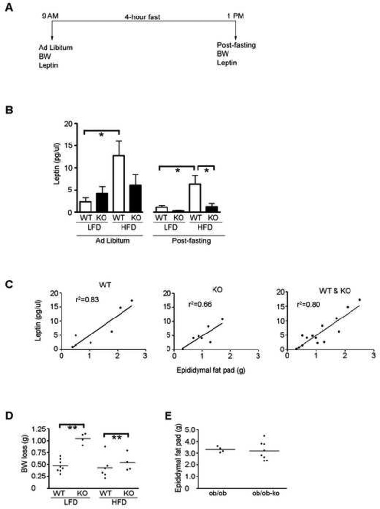
Leptin, which is synthesized in adipocytes, functions via different regions of the hypothalamus to, respectively, suppress appetite and enhance adipocyte lipolysis and hepatic fatty acid oxidation [21]. Under the non-fasting conditions, serum leptin levels in WT mice increased 5-fold on the HFD (p<0.05), but remained unchanged in the Cyp1b1-ko mice (Figures 4A and 4B), and correlated with adipose tissue mass (Figure 4C).
Figure 4. Enhanced decline of serum leptin and body weight in Cyp1b1-ko mice during fasting. Obesity of leptin-deficient Cyp1b1-ko mice.
A. Experimental design of serum leptin and body weight (BW) evaluation.
B. Ad libitum and fasting serum leptin levels in C57BL/6j (WT) and Cyp1b1-ko (KO) mice after 11 weeks on LFD or HFD. The bar graph represents the average leptin concentration ± SEM of WT (n=4) and KO (n=3) ad libitum fed and fasted (n=4 in each of the respective groups) mice.
C. Correlation between ad libitum serum leptin level and epididymal fat mass (n=7 in each of the respective WT and KO groups).
D. BW loss during acute fasting (9AM to 1PM). Each dot represents an individual animal. The horizontal line represents the average BW loss for WT (n=8 LFD, n=6 HFD) and KO (n=4 LFD and HFD) mice.
E. Leptin-deficient (ob/ob) mice are resistant to the adiposity-suppression effects of Cyp1b1 depletion introduced by cross-breeding. Each dot represents an individual animal. The horizontal line represents the average epididymal fat pad mass for ob/ob (n=4) and ob/ob-ko (n=8) mice.
*p<0.05, ** p<0.01.
Serum leptin levels decline substantially during fasting, largely through increased uptake across the blood brain barrier, into the hypothalamus [21]. This decline was observed in our WT mice during a 4 hour morning fast (Figure 4B), but was substantially enhanced in the Cyp1b1-ko mice, particularly when fed the LFD. This was accompanied by a loss of BW, which was more than doubled in the Cyp1b1-ko mice on the LFD (Figure 4D). This acceleration of BW loss in Cyp1b1-ko mice was similar to losses observed in WT mice after a 16 hour fast.
To test whether these large differences in leptin turnover contribute to Cyp1b1 effects on adiposity, we bred the Cyp1b1-ko allele into ob/ob mice, which are leptin deficient. The ob/ob mice develop extreme obesity due to enhanced food consumption and reduced metabolic rate and thermogenesis [21]. The enhanced adiposity of ob/ob mice was completely unaltered by Cyp1b1–deletion (Figure 4E), in contrast to a similar intervention produced by Scd1 deletion [40]
Cyp1b1 deletion prevents hepatic steatosis and attenuates liver mass increases produced by HFD
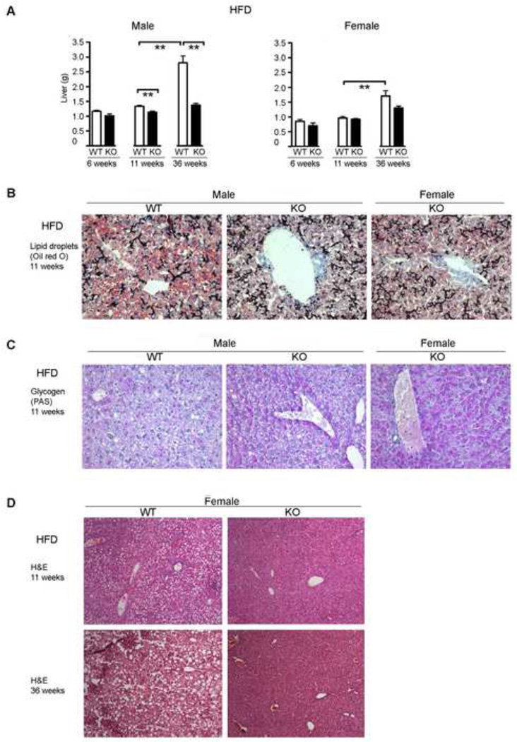
Prolonged exposure of C57BL/6j mice to HFD causes NASH [30]. The chronic accumulation of triglycerides in the liver causes oxidative stress and associated inflammation, which leads to considerable liver enlargement. The liver mass held constant in both genders of each of the respective strains between 6 and 11 weeks of the HFD challenge, but increased 2-fold in male and female WT mice between 11 and 36 weeks (Figure 5A). Due to the previously mentioned delay in female DIO responses, the effects on 36 week females were similar to those on 11 week males. These increases were all substantially attenuated by Cyp1b1 deletion. The weights of other organs (heart, spleen and thymus) were not affected either by HFD or by Cyp1b1 deletion (unpublished data).
Figure 5. Cyp1b1-ko mice exhibit decreased liver steatosis, but increased glycogen retention.
A. Liver mass at 6, 11 and 36 weeks of HFD exposure in C57BL/6j (WT) and Cyp1b1-ko (KO) mice. The bar graphs indicate the average liver mass ± SEM for male WT (n=19, 6-week; n=22, 11-week; n=11, 36-week) and KO (n=4, 6-week; n=19, 11 week; n=6, 36-week) and female WT (n=13, 6-week; n=12, 11-week; n=4, 36-week) and KO (n=4, 6-week; n=10, 11-week; n=3, 36-week) mice.
B. Oil Red O staining for hepatic sinusoidal lipid droplets in representative WT and KO mice fed the HFD for 11 weeks.
C. Pas staining for hepatic sinusoidal glycogen granules in representative male WT and KO mice fed the HFD for 11 weeks (for expanded region see Figure S4A).
D. H&E stained liver sections of representative female WT and KO mice after 11 and 36 weeks of HFD exposure. Increased lipid droplets in WT livers are shown by lipid vacuoles.
*p<0.05, ** p<0.01.
Hepatic triglycerides increased in male WT mice in response to the HFD, while the Cyp1b1-ko mice were unaffected (Table 1). These triglycerides are primarily located in lipid droplets, which are visualized directly by Oil red O staining (Figure 5B) or as large vacuoles (ghosts) with H&E staining, after the ethanol extraction of the lipids (Figure 5D).
Glycogen granules, generated from polymerization of glucose 6-phosphate by glycogen synthase and the glycogen branching enzyme [41], were increased in the sinusoids of Cyp1b1-ko mice (Figure 5C). However, unlike lipid droplets, glycogen granules were more highly concentrated in the peripheral hepatocytes in the WT mice, where they were unaffected by Cyp1b1-ko deletion (Figure S4A). This change suggests a redirection of fatty acid metabolism in the Cyp1b1-ko mice from triglyceride synthesis to mitochondrial oxidation and glycogenesis.
The hepatic lipid droplets increased in size in the WT female mice between 11 and 36 weeks of HFD exposure (Figure 5D), but were scarcely detectable at either time point in the Cyp1b1-ko mice. Small vacuoles were also evident in the sinusoidal region of WT mice on the LFD, but were appreciably decreased in Cyp1b1-ko mice (Figure S4B). This may reflect effects of Cyp1b1 deletion on sinusoidal morphology [42], since there are no differences in levels of liver triglycerides on the LFD. This extensive attenuation of hepatocyte lipid droplets is a more general phenotypic characteristic of Cyp1b1-ko mice than suppressed adipose volume.
Cyp1b1 deletion has a greater effect on liver gene expression than a switch to the HFD
To further probe the altered energy homeostasis that is demonstrated in the Cyp1b1-ko mice, we compared liver gene expression profiles from mice with extensive adipose suppression on HFD to WT and LFD counterparts. Agilent microarray profiles have been analyzed for three individual mice in each of the four treatment groups. Each gene is represented as one of three expression ratios: the mean for WT mice on the HFD is compared to the WT mean on the LFD (designated WT/HFD); the mean for Cyp1b1-ko mice is compared to the mean for WT mice on the same diet (designated KO/LFD and KO/HFD, respectively). The Cy5 binding provides a measure of relative basal gene expression in the WT mice. The LFD/HFD diet change and Cyp1b1 deletion mediated 681 gene responses that differed between the treatment groups by 2-fold or greater, with p<0.05 (Table S2). Remarkably, there was extensive overlap between their effects, with three times more genes responding to Cyp1b1 deletion (KO/LFD or KO/HFD) (560) than to this major diet change in WT mice (WT/HFD) (176). Accordingly, Cyp1b1 expression in the WT mice was not affected by the diet change (WT HFD/LFD=1.22, p=0.625).
The gene responses to Cyp1b1 deletion were distinguished by being largely independent of the diet change (122) or preferentially observed on the HFD (94) or LFD (326), respectively. Approximately a quarter of this final group of genes (75) comprises most of the genes for which the effect of Cyp1b1 deletion mimics the effect of the change to HFD in the WT animals. An additional 18 genes that responded to the HFD in the WT mice responded in the opposing direction (with equal magnitude) in the Cyp1b1-ko mice.
Gene expression changes in Cyp1b1-ko mice indicate a suppression of PPARα activity
In order to better understand how Cyp1b1 deletion suppresses DIO, we have focused on the gene responses to Cyp1b1 deletion that are, respectively, either largely diet-independent or occur primarily on the HFD (Table 2). A substantial proportion of the diet-independent effects of Cyp1b1 deletion represent suppressions of endogenous PPARα activity, which are opposed by the administration of the specific PPARα ligand, Wy-14,643 (Table 3). PPARα expression is, however, scarcely affected by Cyp1b1 deletion.
Table 2.
Liver microarray expression data for genes linked to obesity and diabetes.
| Gene | Gene Symbol |
KO LFDa Malec |
WT HFDa Malec |
KO HFDb Malec |
KO LFDa Femaled |
KO HFDb Femaled |
|---|---|---|---|---|---|---|
| Endocrine Regulated | ||||||
| Cytochrome P450 7b1 | Cyp7b1 | 11.4** | 3.4 | 4.6* | 2.7 | 2.9 |
| Adropin | Enho | 2.2 | 2.1 | 2.0 | 5.5 | 4.3 |
| Epidermal growth factor receptor | Egfr | 3.1** | 1.4 | 2.2** | 2.7 | 6.4 |
| Growth hormone receptor | Ghr | 1.3 | 1.4 | 1.5 | −1.1 | 1.0 |
| Hepatocyte nuclear factor 6 | Onecut1 | 2.8** | 1.0 | 4.5** | 1.2 | 2.5 |
| Hydroxy-delta-5-steroid dehydrogenase | Hsd3b4 | 13.0* | 6.1 | 2.2 | 5.6 | 3.1 |
| Major urinary protein 1 | Mup1 | 7.5* | 3.8 | 3.7 | −1.2 | 1.5 |
| Solute carrier organic anion transporter | Slco1a1 | 5.2* | 4.2 | 1.7 | 6.6 | 6.9 |
| Peroxisome proliferator-activated receptor γ | Pparγ | −4.5** | −1.2 | −4.6** | −1.5 | −2.7 |
| Energy Homeostasis | ||||||
| Acetyl-CoA carboxylase beta | Acacb | 1.4 | −3.2* | −1.9 | −1.3 | 1.5 |
| Fatty acid synthase | Fasn | 1.3 | −2.0 | −1.5 | −2.9 | 1.2 |
| Malic enzyme 1 | Me1 | 1.0 | −1.7 | −7.5* | −2.2 | −1.9 |
| Stearoyl-CoA desaturase 1 | Scd1 | 1.3 | −3.2* | −6.0** | −1.7 | −1.9 |
| Pyruvate dehydrogenase kinase 4 | Pdk4 | −1.9 | −3.1* | −3.7** | −2.4 | −1.9 |
| Elongation of very long chain fatty acids | Elovl5 | −1.5 | −1.6 | −2.1* | −1.3 | −1.7 |
| Fibroblast growth factor 21 | Fgf21 | −3.1 | −2.9* | −2.9* | −1.8 | −2.9 |
| Insulin-like growth factor binding protein 1 | Igfbp1 | −1.7 | −3.5 | −5.1* | 1.0 | −2.3 |
| Insulin-like growth factor 1 | Igf1 | −1.1 | 1.0 | 1.1 | −1.5 | 1.4 |
| STAT-induced STAT inhibitor 2 | Socs2 | 1.8 | −2.3 | 5.4* | 3.4 | 3.9 |
| Glycogen Synthesis | ||||||
| PP1 regulatory subunit 3b | Ppp1r3b | 2.2 | 1.8 | 1.0 | −3.7 | 1.0 |
| PP1 regulatory subunit 3c | Ppp1r3c | 3.8* | 1.6 | 2.4 | −2.7 | −1.3 |
| PP1 regulatory subunit 3g | Ppp1r3g | −4.6* | −2.4 | −5.9** | 9.7 | −4.3 |
| Glycogen-branching enzyme | Gbe1 | −1.2 | 1.2 | −2.3 | −2.2 | −1.6 |
| Liver glucose transporter, type 2 | Slc2a2 | 1.7 | 1.9 | 1.0 | −1.3 | −1.3 |
| Glucose transporter type 4 | Slc2a4 | 2.1 | −3.2 | −1.4 | −6.0 | −23.5 |
| PPARα activity | ||||||
| Peroxisome proliferator-activated receptor α | Pparα | 1.0 | 1.0 | −1.2 | 1.8 | −1.2 |
| Peroxisomal CoA diphosphatase 7 | Nudt 7 | 2.3** | 1.3 | 2.4** | −1.3 | 1.8 |
| Acyl-CoA thioesterase 1 | Acot1 | −4.4** | 1.5 | −4.7 | 3.0 | −1.6 |
| Cytochrome P450 4a10 | Cyp4a10 | −11.7 | 1.7 | −11.2 | 1.6 | −1.5 |
| Cytochrome P450 4a14 | Cyp4a14 | −43.4** | 1.4 | −66.3** | 1.4 | −1.8 |
| Fat stimulatory protein/FSP27 | Cidec | −4.7* | −1.9 | −4.8** | −2.0 | 1.1 |
| Fatty acid translocase | CD36 | −7.6** | −1.7 | −5.5** | −7.5 | −6.3 |
| G0/G1 Switch 2/Lipase activator | G0s2 | −2.5* | 1.3 | −2.8* | −2.3 | −1.8 |
| Kruppel-like factor 6 | Klf6 | −2.9** | −2.4 | −1.6 | −1.4 | 1.1 |
| Lipin phosphatidase 2 | Lpin 2 | −1.7* | 1.4 | −2.4 | 2.1 | −1.1 |
| Osteopontin | Spp1 | −2.6* | −2.0 | −1.2 | −1.5 | −1.9 |
| P8 transcription factor | Nupr1 | −5.1** | −5.5 | −1.3 | −4.7 | 1.3 |
| Very-low-density-lipoprotein receptor | Vldlr | −9.1** | −1.2 | −3.1* | −4.3 | −2.4 |
| Phosphatidyl choline transferase protein | Pctp | −1.9* | 1.7 | −2.3** | −1.2 | −1.3 |
| Peroxisomal biogenesis factor 11 alpha | Pex11a | −2.8** | 1.2 | −2.4 | 1.0 | −2.2 |
| Acyl-CoA thioesterase 13 | Them2 | −1.9 | 2.0 | −1.8 | −1.5 | −1.2 |
| Oxidant Stress | ||||||
| Glutathione S-transferase pi 1 | Gstp1 | 2.1** | −1.1 | 3.1** | 1.0 | −1.2 |
| Selenium binding protein 1 | Selenbp1 | 2.4** | 1.2* | 2.5** | −1.3 | 1.1 |
| Gamma-interferon-induced monokine | Cxcl9 | −3.0** | −1.4 | −4.0** | −2.2 | −2.3 |
| Gamma-interferon-induced protein | Cxcl10 | −3.8* | −1.9 | −3.6* | −3.2 | −2.3 |
Expression levels are calculated relative to WT, LFD
Expression levels are calculated relative to WT, HFD.
Male (n=3)
female (n=2) mice were fed the diets for 11 and 36 weeks, respectively, post-weaning. Data represents fold change.
p-value < 0.05
p-value < 0.01.
Table 3.
Many genes stimulated by PPARα are suppressed in Cyp1b1-ko mice.
| Gene | Gene Symbol |
WT LFDa Malec Wy-14,643e |
KO LFDa Malec |
KO LFDa Femalec |
KO HFDb Femaled |
|---|---|---|---|---|---|
| Acetyl-Coenzyme A acyltransferase 1B | Acaa1b | 3.6** | −2.1* | 1.0 | −2.0 |
| Acyl-CoA thioesterase 1 | Acot1 | 14.1** | −4.4** | 3.0 | −1.6 |
| Acyl-CoA thioesterase 2 | Acot2 | 3.3* | −15.6** | −1.1 | −1.9 |
| Aldo-keto reductase | Akr1c18 | 8.6** | −9.1** | −2.3 | 2.1 |
| Aldehyde dehydrogenase | Aldh3a2 | 2.1 | −3.0** | −1.1 | −1.4 |
| Cytochrome P450 4a10 | Cyp4a10 | 17.8* | −11.7 | 1.6 | −1.5 |
| Cytochrome P450 4a14 | Cyp4a14 | 75.6** | −43.4** | 1.4 | −1.8 |
| Cytochrome P450 4a31 | Cyp4a31 | 16.8** | −5.1** | 1.1 | −2.1 |
| Fatty acid translocase | Cd36 | 4.1** | −7.6** | −7.5 | −6.3 |
| Lectin, galactose binding, soluble | Lgals1 | 2.8* | −5.8** | −3.4 | −4.1 |
| Macrophage receptor, collagenous structure | Marco | 8.1** | −3.0 | −6.2 | −1.8 |
| Membrane spanning 4-domain A7 | Ms4a7 | 2.4* | −4.9** | −4.9 | −2.2 |
| Metallothionein 2 | Mt2 | 7.2* | −4.1 | 3.6 | 1.1 |
| Peroxisomal biogenesis factor 11 alpha | Pex11a | 2.7* | −2.8** | 1.0 | −2.2 |
| Retinol saturase | Retsat | 3.4* | −1.7 | −1.3 | −1.7 |
| T4 binding globulin | Serpina7 | 2.3** | −3.5** | −2.8 | −2.4 |
| Vanin 1 | Vnn1 | 6.2** | −10.9** | −1.7 | −2.9 |
| Very low density lipoprotein receptor | Vldlr | 3.1 | −9.1** | −4.3 | −2.4 |
| Cytochrome P450 2C54 | Cyp2c54 | −6.1** | 3.5 | 1.0 | 2.0 |
| Retinal-specific regulator of G-protein signaling | Rgs16 | −15.2** | 3.6 | −1.9 | −4.7 |
| Solute carrier organic anion transporter | Slco1a1 | −3.4* | 5.2* | 6.6 | 6.9 |
| Solute carrier sodium phosphate transporter | Slc17a3 | −3.0* | 2.7** | −1.1 | 1.5 |
| UDP glucuronosyltransferase 2 | Ugt2b1 | −2.8* | 2.9** | −1.2 | 1.1 |
| Fat stimulatory protein/ Fsp27 | Cidec | +++f | −4.7* | −2.0 | 1.1 |
| Fibroblast growth factor 21 | Fgf21 | +++f | −3.1 | −1.8 | −2.9 |
| G0/G1 switch 2/ lipase activator | G0s2 | +++f | −2.5* | −2.3 | −1.8 |
| Insulin-like growth factor binding protein 1 | Igfbp1 | +++f | −1.7 | 1.0 | −2.3 |
| Kruppel-like factor 6 | Klf6 | +++f | −2.9** | −1.4 | 1.1 |
| Peroxisomal CoA diphosphatase 7 | Nudt7 | -- -- | 2.3** | −1.3 | 1.8 |
| Pyruvate dehydrogenase kinase 4 | Pdk4 | +++f | −1.9 | −2.4 | −1.9 |
| Selenium binding protein 1 | Selenbp1 | -- -- | 2.4** | −1.3 | 1.1 |
Fold change relative to WT, LFD.
Fold change relative to WT, HFD.
Male (n=3) mice were fed the respective diets for 11 weeks, post-weaning.
Female (n=2) mice were fed the respective diets for 36 weeks, post-weaning.
Wy-14,643 treatment for 48 hours.
Genes respond at 6 hours [58] but not at 48 hours.
p-value < 0.05
p-value < 0.01.
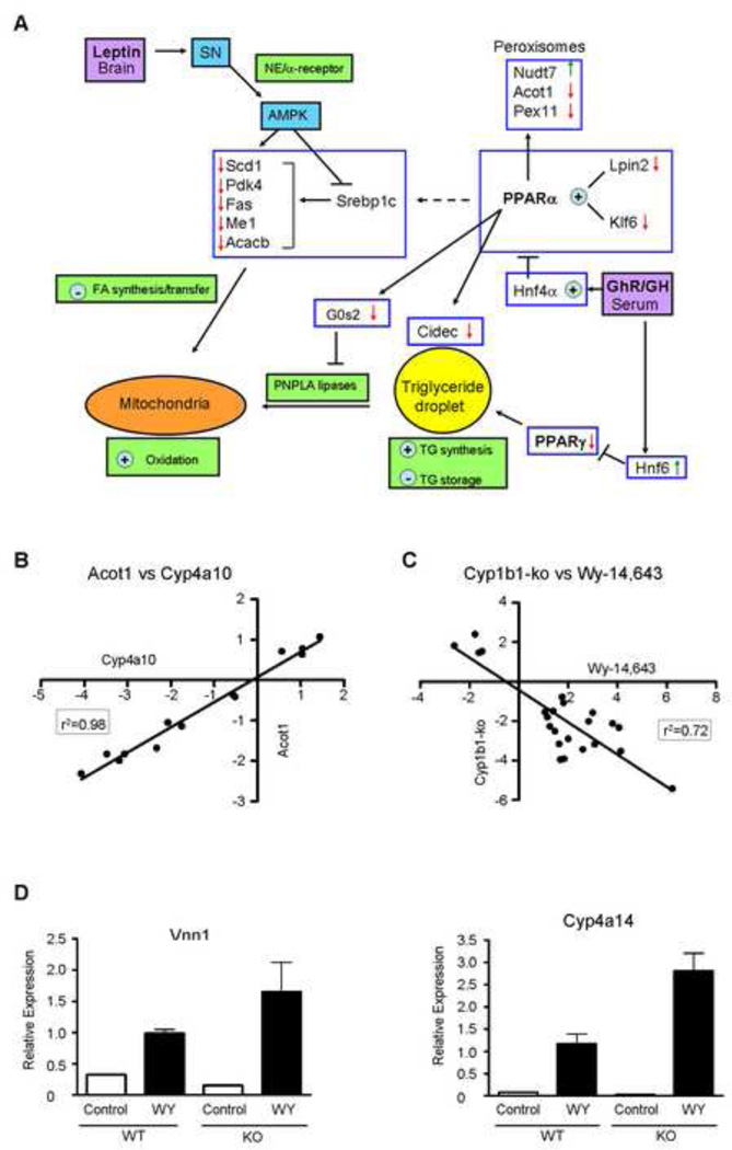
Many of the genes that show diet-independent responses to Cyp1b1 deletion are regulated by PPARα and affect steps in fatty acid homeostasis (Tables 2 and 3). Examples include CD36, which contributes to triglyceride homeostasis through fatty acid transport across the plasma membrane [43], cytochrome P450 4a14 (Cyp4a14), which hydroxylates long chain fatty acids [44] and several genes involved in peroxisome function, including the peroxisomal transporter 11 (Pex11a) and Acyl CoA transferase I (Acot 1) [45–46]. Several other gene changes that enhance triglyceride accumulation are stimulated by PPARα activation, but are reversed in Cyp1b1-ko mice. These include Vldlr (increases triglyceride uptake), Cidec/Fsp27 (stabilizes lipid droplets) and Klf6 (promotes adipocyte differentiation) [47–49]. Lpin2, a phosphatidic acid phosphatase that activates PPARα, is also decreased by Cyp1b1 deletion [50]. G0s2, an inhibitor of adipose triglyceride lipase (Pnpla2) is suppressed by PPARα activation [51] and Cyp1b1 deletion. The histological data (Figure 5) suggests that these effects on the control of fatty acid metabolism combine to suppress lipid droplets in Cyp1b1-ko livers as shown in Figure 6A.
Figure 6. Effects of constitutive PPARα suppression on liver gene expression in Cyp1b1-ko mice.
A. Functional association of Cyp1b1 deletion-responsive genes involved in fatty acid homeostasis, which are controlled either by leptin and AMPK or by PPARα activity.
B. Expression of PPARα-regulated genes, Acot1 versus Cyp4a10, for individual mice from the 4 treatment groups can be highly correlated (n=13 mice).
C. Effect of stimulation of PPARα activity by Wy-14,643 treatment (WT males, 48 hours, oral gavage) is inversely correlated with the effect of Cyp1b1 deletion on the constitutive expression of 23 genes shown in Table 3.
D. PPARα-responsive gene stimulation (Vnn1 and Cyp4a14) is retained in KO mice treated with Wy-14,463. WT and KO (n=2) mice were treated with Wy-14,463 for 48 h.
Selenium binding protein 1 (Selenbp1), which is suppressed by PPARα activation [52], is increased by Cyp1b1 deletion. Several selenium binding proteins function collectively to regulate liver oxidative stress, which is associated with PPARα activity. Other genes that show reversal of PPARα suppression in Cyp1b1-ko mice include Nudt7 [53] and various Cyp2c forms [54].
We examined two additional PPARα-sensitive genes in relation to lipid regulation: Pctp (StARD2) and acyl-CoA thioesterase 13 (Acot13/Them2) (Table 2) [55]. Pctp is not only a specific PC transfer protein, but also complexes with Acot13 to enhance mitochondrial thioesterase activity. This local activity prevents entry of long-chain fatty acids into the mitochondria for oxidation (via carnitine/palmitoyltransferase protein). In Cyp1b1-ko mouse liver, their dual suppression should enhance mitochondrial oxidation.
The enhanced glycogen storage observed with Cyp1b1 deletion is consistent with substantial increases in the stimulatory glycogen-targeting subunits of protein phosphatase 1 (PP1); Ppp1r3b and Ppp1r3c, and decline of the inhibitory subunit, Ppp1r3g [56].
There were considerable differences in gene expression between individual mice within each treatment group. However, expression was often highly correlated between genes in the same family for the individual mice in the four treatment groups (Acot1/Acot4, r2=0.87; Cyp4a10/Cyp4a14, r2=0.92), confirming expected co-regulation. These correlations further extended to PPARα-regulated genes in different families (Acot1/Cyp4a10, r2=0.98) (Figure 6B).
Our single 48 hour treatment with Wy-14,643 produced fewer responses than the standard two week multi-treatment protocol or a 6 hour acute stimulation [57–58]. However, 23 genes responded to Wy-14,643 in the opposing direction as the differential expression between Cyp1b1-ko and WT mice (Table 3). This includes 5 genes that were suppressed by Wy-14,643 treatment, but exhibited elevated expression in Cyp1b1-ko mice. Remarkably, the WT Wy-14,643 responses were highly correlated with suppression of constitutive expression in Cyp1b1-ko mice (r2=0.72) (Figure 6C). In addition, 8 genes that exhibited a large suppression response to Cyp1b1 deletion responded to chronic PPARα activation in the opposite direction [44], even though unresponsive to our 48 hour Wy-14,643 exposure. Accordingly, Cidec, G0s2, Pdk4 and Igfbp1 are stimulated by Wy-14,643 with these alternative protocols [57], wherein the single oral gavage of Wy-14,643 is more effective after 6 hours than after the 48 hours used here. Conversely, Cyp4a and CD36 gene stimulations are greater under our 48 hour treatment strategy.
The close inverse relationship between Wy-14,643 stimulation and constitutive suppression in Cyp1b1-ko mice strongly supports the hypothesis that these genes are stimulated by endogenous activation of PPARα, and that this activation is extensively suppressed in Cyp1b1-ko livers. To test whether these genes retained responsiveness in Cyp1b1-ko mice, we measured responses of three strongly expressed genes that are most highly suppressed by Wy-14,643 treatment in the WT mice by real-time qPCR. Vnn1 and Cyp4a14 (Figure 6D) and Cyp4a10 (data not shown) were each induced by at least 7-fold in Cyp1b1-ko mice.
Gene changes that redirect fatty acid homeostasis in Cyp1b1-ko mice on the HFD
Several key genes involved in fatty acid homeostasis were also suppressed extensively by the combination of HFD and Cyp1b1 deletion (Table 2). The largest response of this group involves the synergistic suppression of Scd1, which diverts fatty acids from oxidation to storage as triglycerides [16–17]. Malic enzyme 1 (Me1), acetyl-CoA carboxylase beta (Acacb), pyruvate dehydrogenase kinase 4 (Pdk4) and elongation of very long chain fatty acids protein 5 (Elovl5) are additional genes, which exhibit pivotal roles in stimulating fatty acid synthesis, that also decline selectively on the HFD [59–62]. The serum regulators of liver metabolism, insulin-like growth factor binding protein 1 (Igfbp1), which decreases IGF1 bioavailability, and fibroblast growth factor 21 (Fgf21), another important regulator of fatty acid metabolism [63–64], each demonstrated paralleled suppression with HFD and Cyp1b1 deletion. The expression levels of Igfbp1 and Fgf21 were highly correlated among the mice of the 4 treatment groups (r2=0.55) (data not shown).
While most of these genes can also be stimulated by activation of PPARα, none were stimulated by the present 48 hour administration of Wy-14,643. They are also unaffected by Cyp1b1 status on the LFD. This may be accounted for by differences in the timing of Wy-14,643 administration [65]. Each of these genes exhibits strong circadian suppression in the morning when the mice were sacrificed. This may diminish the constitutive activation by PPARα that is susceptible to Cyp1b1 deletion and Wy-14,643 activation.
Expression differences in 11 week Cyp1b1-ko male mice that are retained in 36 week females
The decreases in hepatocyte lipid droplets, adiposity and serum glucose in 36 week Cyp1b1-ko female mice, compared to WT counterparts, were similar to those seen in males at 11 weeks. Nevertheless, Cyp1b1 deletion mediated substantially fewer gene expression changes on the LFD in these females. On the other hand almost 60 percent retained their responses to Cyp1b1 deletion in both genders when fed the HFD (CD36, serpina7, Lgals, Ms4a7) (Tables 2 and 3). We have focused our analyses on genes that are conserved between the male and female animals as they are more likely to be linked to core Cyp1b1 regulation of energy homeostasis.
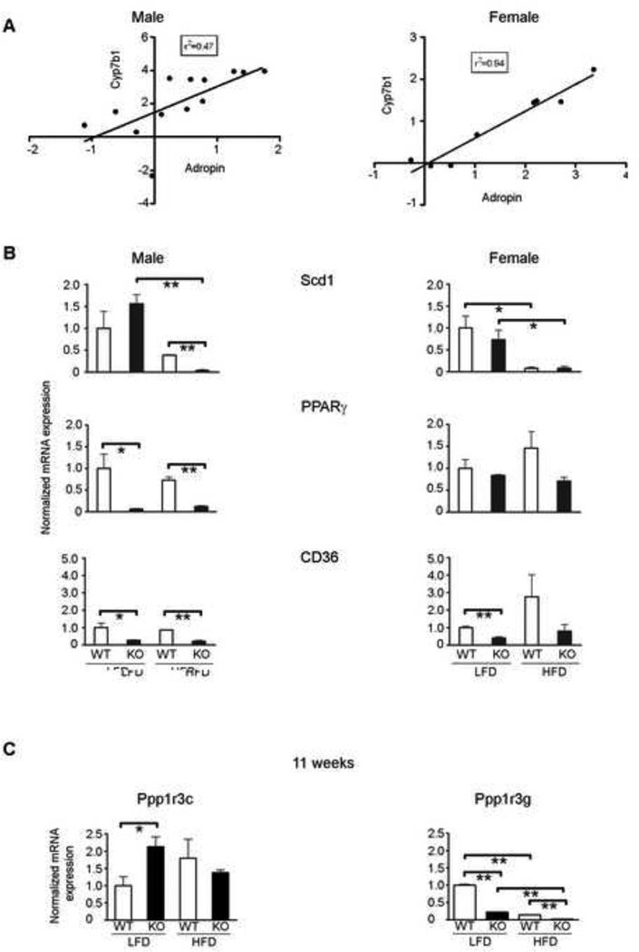
Several important male-selective genes, which demonstrated substantially increased expression in Cyp1b1-ko mice on the LFD, retained these stimulations in the female group, despite their lower expression. These genes include Cyp7b1, Hsd3b4, Slco1a1, Egfr, adropin and Socs2. Adropin, a peptide associated with insulin sensitivity and protection from liver steotosis, is secreted from hepatocytes [66]. Adropin expression correlates well with Cyp7b1 in individual mice for the male and female treatment cohorts suggesting similar regulation (Figure 7A).
Figure 7. Real-time PCR quantification and correlation of microarray gene expression of liver genes that control triglyceride and glycogen levels.
A. Correlation of adropin and Cyp7b1 gene expression in, respectively, male and female C57BL/6j (WT) and Cyp1b1-ko (KO) mice fed the LFD and HFD for 11 and 36 weeks, respectively (n=13 male and n=8 female mice).
B. Real-time PCR (qPCR) quantification of the relative mRNA expression of Scd1, PPARγ and CD36 in livers from three C57BL/6j (WT) or Cyp1b1-ko (KO) male and female mice after 11 or 36 weeks of LFD or HFD exposure, respectively. The 11 week mRNA samples were those examined in the microarray analyses. Microarray and qPCR responses are also compared in Tables S4 and S5.
C. qPCR quantification of the relative mRNA expression for three mice in each of the treatment groups of the glycogen synthase regulators, Ppp1r3c and Ppp1r3g.
*p<0.05, ** p<0.01.
Genes that retained decreased expression in both male and female Cyp1b1-ko mice include the chemokines CxCl9 and Cxcl10, as well as several key regulators of liver fatty acid homeostasis (notably Scd1, Pdk4, Elovl5, Fgf21, Igfbp1, PPARγ and Me1).
In addition, many cytochrome P450s exhibited previously characterized dimorphisms on the LFD, despite the age difference between the male and female groups (Male:7b1, 2c39, 2c44, 2c54, 2d22, 2d34, 4a12 and 4a31; Female: 2a4, 2a5, 2b9, 2b13, 2c29, 2c40, 2c70, 3a41, 2g1 and 17a1) [67].
Gene changes that are not conserved between 11 week males and 36 week females
Despite this important conservation, most gene changes observed in the 11 week Cyp1b1-ko male mice on the LFD are substantially attenuated in the 36 week females. Notable examples include two glycogen-associated phosphatases (Ppp1r3 forms b and c) and the glucose transporter, Slc2a4, which were each stimulated in the males, but suppressed in the females. These changes in Cyp1b1-ko mice not only indicate a loss of differences between the groups, but substantial changes in hepatic energy regulation, despite the conserved suppression of triglyceride synthesis in the Cyp1b1-ko mice. We cannot, however, presently resolve the extent to which Cyp1b1 deletion is affecting aging or sexual dimorphism.
qPCR analysis of liver gene expression
We applied real-time qPCR analyses to three cohorts of mice. In the first group, we examined gene responses for three key genes from the same male mice (11 week treatment group) that were used for microarray analysis. These were compared with the 36 week females. These Cyp1b1-ko mice exhibited appreciable suppression of adiposity (Figure 1D). Figure 7B shows the relative gene expression for Scd1, PPARγ and CD36 in the 11 and 36 week groups. At 11 weeks, these results largely replicate the microarray data. At 36 weeks, the HFD-mediated suppression of Scd1 was so enhanced that Cyp1b1 deletion has no further effect. CD36 expression is relatively unchanged between 11 and 36 weeks of age, whereas the suppression of PPARγ is relieved in the female Cyp1b1-ko mice. The real-time qPCR also confirms the microarray expression results of Ppp1r3c and Ppp1r3g, mediators of glycogen synthesis (Figure 7C).
We also applied qPCR analyses to examine the expression levels of a set of 12 genes in all of the available livers of the mice shown in Figure 1 (Table S3), as well as in mice that were provided a delayed 6 week exposure to HFD (weeks 6–12 of age) (Table S4), which by-passed the final period of liver maturation (weeks 3–6) [31]. These analyses showed that differences in Cyp1b1-ko mice that were selected for their robust suppression of BW were retained when averaged for the full groups shown for the diet responses in Figure 1.
qPCR analyses further demonstrated that HFD suppressed Scd1 in both adipose and muscle tissue, but that there was no effect of Cyp1b1 deletion on either tissue. Pparγ and CD36 expression were not affected by either HFD or Cyp1b1 deletion in these tissues (data not shown).
Relationship of LPC changes to PPARα activity
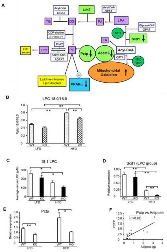
The analyses of serum LPC levels in these mice showed that Cyp1b1 deletion exerts a general suppression effect that was reversed by a human CYP1B1 transgene [38]. These LPC levels are of additional interest since the minor phosphatidyl choline, 16:0/18:1 diacyl glycerophosphatidyl choline (diacyl GPC), is a selective activator of PPARα. Thus, the 16:0 LPC and 18:1 LPC are potential indicators of suppression of diacyl GPC and of endogenous liver PPARα activity. The relationship between these genes with respect to synthesis of diacyl GPC, LPC and triglycerides versus fatty acid oxidation is shown in Figure 8A. Serum 18:0 LPC [38] or the ratio to the largely constant 16:0 LPC (18:0/16:0; preferred measure) provides an accessible marker of both adiposity and hepatic steatosis, and is suppressed on both LFD and HFD in Cyp1b1-ko mice (Figure 8B). The 18:1 LPC levels decline on a HFD and are further attenuated when combined with Cyp1b1 deletion (Figure 8C), paralleling changes in Scd1 expression in the same mice (Figure 8D).
Figure 8. Serum lysophosphatidylcholine (LPC) as a marker of obesity and PPARα activity. Potential role of PCTP in mediating the effects of Cyp1b1 deletion on triglyceride and LPC synthesis.
A. In the hepatocyte, the phosphatidyl choline (PC) transfer protein (PCTP) and its partnering protein, Acot13, mediates the transfer of PC between membranes and affects synthesis of PC, lysophosphatidyl choline (LPC) and triglyceride (TG) from acyl-CoA by decreasing mitochondrial oxidation. Direct interaction of PCTP with Acot13 on the outer mitochondrial membrane increases cleavage of acyl CoA, thus decreasing formation of acyl carnitines and transfer into the mitochondria and subsequent oxidation. Both PCTP and Acot13 are stimulated by PPARα [55] and fall within the group of genes (depicted in light green) that exhibit constitutive suppression in Cyp1b1-ko mice.
B. The ratio of 18:0/16:0 LPC provides an optimal marker for suppression of DIO and PPARα activity in Cyp1b1-ko (KO) mice, relative to their C57BL/6j (WT) counterparts. Serum LPC levels were measured in the mice depicted in Figure 1. The 18:0/16:0 LPC ratio parallels the trend observed in BW gain and adiposity. The bar graph represents the ratio of the average 18:0 concentration to the average 16:0 concentration ± SEM in the blood serum of WT (n=4 LFD, n=6 HFD) and KO (n=7 LFD, n=6 HFD) mice.
C. Serum 18:1 LPC levels decline in WT mice fed a HFD for 11 weeks, and are further attenuated with Cyp1b1 deletion. The bar graph represents the average 18:1 LPC serum concentration ±SEM in WT (n=4 LFD, n=5 HFD) and KO (n=7 LFD, n=6 HFD) mice.
D. Hepatic Scd1 expression parallels serum 18:1 LPC levels. The bar graph represents the average hepatic Scd1 mRNA expression level ±SEM in WT (n=11 LFD, n=13 HFD) and KO (n=12 LFD, n=9 HFD) mice.
E. Hepatic Pctp gene expression, measured by microarray analysis, parallels serum 18:0 LPC levels [38]. The bar graph represents the average hepatic Pctp mRNA expression level ±SEM in WT (n=3 LFD and HFD) and KO (n=4 LFD, n=3 HFD) mice.
F. Hepatic Pctp gene expression is highly correlative with adiposity. Data represents the compilation of WT and KO mice across all treatment groups (n=13 mice).
*p<0.05, ** p<0.01.
The PC exchange protein, Pctp, was one of very few genes whose liver expression was highly correlated with serum 18:0/16:0 LPC levels and adiposity (Figure 8E and 8F).
DISCUSSION
Despite minimal expression in mouse hepatocytes, Cyp1b1 deletion substantially impacts fatty acid homeostasis and the expression of associated liver genes. This is accompanied by extensive suppression of obesity that is induced by a high fat diet, as well as the subsequent liver enlargement and lipid infiltration that precedes NASH [30] (Figures 1, 2, 3 and 5). Although the DIO response is delayed, it is similarly suppressed in the Cyp1b1-ko female mice. The depletion of lipid droplets in Cyp1b1-ko livers was consistently observed, even when adiposity suppression was modest (Figure S4B). The hepatocytes of the Cyp1b1-ko mice are primarily affected within the sinusoidal region, where glycogen granules accumulate, replacing the triglycerides of lipid droplets (Figures 5 and Figure S4A). This replacement is consistent with the gene expression changes (Tables 2 and 3, Figure 7), particularly the near complete suppression of constitutive PPARα activity and Scd1 on the HFD. Scd1 re-directs fatty acid metabolism from mitochondrial oxidation to storage as triglycerides. Adenoviral expression of human SCD1 in these Cyp1b1-ko mice restored the WT level of obesity [38].
Remarkably, 17 major gene changes identified in the Cyp1b1-ko liver have been associated with the reversal of conditions associated with metabolic syndrome or Type 2 diabetes, when introduced as single genetic modifications (Table 4). Eighty percent of these changes are conserved in the older female mice, which retain the suppression of liver steatosis and adiposity with Cyp1b1 deletion. Cyp1b1 metabolites may, therefore, modulate processes that are central to the adverse effects of NASH and Type 2 diabetes.
Table 4.
Liver genes that are conserved between male and female mice fed the LFD, but are affected by Cyp1b1 deletion, and their impact on Type 2 diabetes.
| Gene | Effect of Cyp1b1-ko |
Gene Function | Impact of gene on Type 2 Diabetes |
Ref |
|---|---|---|---|---|
| Scd1 | − | FA desaturation | KO increases GT | 16 |
| Pdk4 | − | Pyruvate to Acetyl CoA | KO increases GT | 61 |
| Me1 | − | Malate/NADP to Pyruvate | KO increases GT | 59 |
| Map4k4 | − | TNF-activated kinase inhibits insulin | KO increases GT | 86 |
| Pparγ | − | Stimulates TG synthesis | Liver KO lowers SH | 87 |
| Spp1 | − | Sustains PPARγ/lipid droplets | KO lowers SH, glucose | 88 |
| Gas6 | − | Released from Kupffer cells/ increase FA oxidation | KO lowers SH, inflamation | 89 |
| Rgs16 | − | G protein activator | KO increases FA oxidation | 90 |
| Vldlr | − | Very low density lipoprotein receptor | KO lowers SH | 91 |
| Socs2 | + | Ubiquitin ligase, negative regulator of JAK/STAT | KO decreases insulin sensitivity | 92 |
| G0s2 | − | PNPLA2 inhibitor | Tg stimulates SH | 51 |
| Cidec | − | Binds lipid droplets, mediates PPARγ effects | Tg stimulates SH | 93 |
| Lpin2 | − | Lipid phosphatase/PPAR co-activator | SiRNA suppresses SH | 50 |
| Cyp7b1 | + | Bile acid synthesis | Tg stimulation lowers glucose | 94 |
| Slco1a1 | + | Bile acid transport | Tg stimulation lowers glucose | 94 |
| Cyp1b1-effects directly link to decreased T2D/SH | ||||
| Hamp2 | − | Hepcidin2/ Fe stimulant | Serum hepcidin linked to T2D | 95 |
| Hhex | + | Liver homeobox regulator | Mutant human marker for T2D | 96 |
Type 2 diabetes = T2D
Glucose tolerance = GT
Steatotic hepatitis = SH
Transgene = Tg
Sources of systemic Cyp1b1 intervention
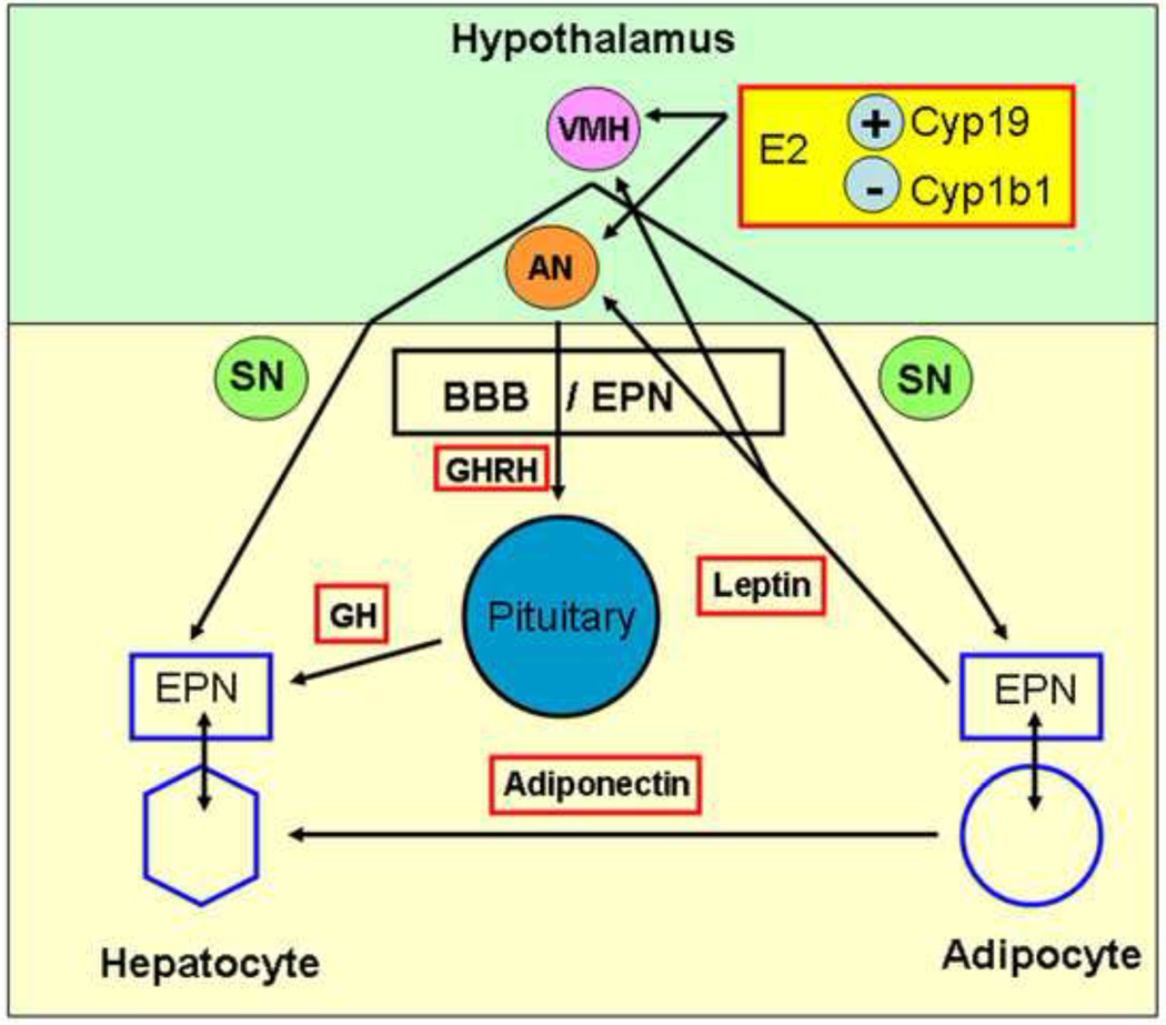
The absence of expression in mouse hepatocytes indicates that Cyp1b1 impacts hepatic gene expression through extra-hepatocyte signaling processes. This signaling can proceed locally via adjacent endothelia and stellate cells or systemically through endocrine and neuronal control mechanisms. These effects may occur directly or be realized through developmental intervention. Cyp1b1-ko mice retain normal caloric intake (Figure S1A), indicating improved energy utilization. This metabolic homeostasis is also regulated via ER-α, aromatase and VMH leptin receptors [33–34, 68]. Cyp1b1 is appreciably expressed in the hypothalamus [69] and diminished metabolism of estradiol in Cyp1b1-ko mice has been shown to affect blood pressure [12].
Growth hormone (GH), which suppresses obesity, is also linked to hypothalamic ER-α through control of GH neurons in the arcuate nucleus [70]. GH suppresses PPARα activation [71] and targets many liver genes affected by Cyp1b1 deletion. Male-selective expression derives from regulated, intermittent GH pulses, which contrast a continual, moderate secretion in female mice [72]. The minimal effect of Cyp1b1 deletion on the major functional target of GH in the liver, IGF-1 [73], suggests that Cyp1b1 is not simply elevating GH levels.
Cyp1b1 also influences the vasculature. Deficient expression in endothelia and associated pericytes disrupts capillary assembly in vitro and vasculogenesis in vivo through increased oxidative signaling [8–9, 18–19]. Liver stellate cells are specialized pericytes [74], positioned in the sinusoids between endothelia and hepatocytes, adjacent to sympathetic nerve terminals [75]. Endothelial-pericyte interactions are crucial to the blood-brain barrier, which controls leptin transfer to the hypothalamus [76–77]. The unexpected correlation of increased leptin clearance and enhanced hypothalamic activity in Cyp1b1-ko mice can be explained by such changes. These potential influences of Cyp1b1 deletion on the metabolic parameters are summarized in Figure 9.
Figure 9. Representation of proposed systemic effects of Cyp1b1 deletion that increase effects of leptin and growth hormone on the liver.
Cyp1b1 deletion effects on liver gene expression indicate increased leptin and growth hormone action, each of which are regulated in the hypothalamus. Hypothesis: Cyp1b1 deletion targets two important activities A) The endothelial-pericyte niche (EPN), which provides local regulation of adjacent cells, is disrupted in Cyp1b1-ko mice, B) Estradiol (E2) levels are increased, particularly in the hypothalamus, when local removal of E2 (produced by aromatase) via Cyp1b1 metabolism is decreased. Increased transfer of leptin across the blood brain barrier (BBB) increases activation of sympathetic neurons via the ventromedial hypothalamus (VMH) and release of GH releasing hormone (GHRH) from the arcuate nucleus (AN), which stimulates pulsatile release of GH from the pituitary [21, 101]. Similar EP niche activity functions in liver sinusoids (endothelia and stellate pericytes) [71] and adipose [32] to provide local controls over adjacent hepatocytes and adipocytes, respectively.
Interplay between Cyp1b1 and dietary fat in the control of gene expression
Cyp1b1 expression in WT mice is not sensitive to the dietary fat content. In fact, more genes in male mice (560) are sensitive to the deletion of Cyp1b1 than to the increase in dietary fat (176). These Cyp1b1-deletion-sensitive genes resolve into three main groups that are, respectively, independent of dietary fat or are diet-selective (LFD or HFD). Surprisingly, only about 10 percent of genes that respond to HFD in WT mice exhibit suppression in Cyp1b1-ko mice (18), thus paralleling the changes in obesity. Many genes (75), however, respond similarly to Cyp1b1 deletion and to HFD in the WT mice, suggesting that these genes exhibit an enhanced sensitivity to dietary fat in Cyp1b1-ko mice. In the 36 week female mice, there are many fewer responses to Cyp1b1 deletion on the LFD, but the number of these responses increases on the HFD. Nevertheless, an appreciable number of highly expressed genes conserve their responses to Cyp1b1 deletion on both diets, suggesting their linkage to the core Cyp1b1-sensitive control processes. Many of these changes also reflect an extensive attenuation of expression differences between these male and female mice. A more complete resolution of sex and age effects will be aided by ongoing analysis of age-matched 11 week females.
Genes regulated by endogenous PPARα activity are suppressed in Cyp1b1-ko mice
The inverse relationship of many gene responses to Wy-14,643 and to Cyp1b1 deletion (Table 3, Figure 6C) points to extensive suppression of their endogenous regulation by PPARα. Some genes (Cyp4a forms) are also substantially suppressed in PPARα-ko livers, indicative of endogenous PPARα stimulation [44]. Others (CD36) are more suppressed in Cyp1b1-ko mice, possibly due to the additional suppression of PPARγ (Tables 2 and 3). Much shorter exposures to Wy-14,643 targeted several additional PPARα-responsive genes that exhibit inverse responses to Cyp1b1 deletion (Tables 2 and 3) [58]. These metabolic genes often exhibit circadian fluctuation and rapid turnover that benefit from a shorter agonist exposure [65].
PC synthesis and endogenous PPARα activation
PPARα is stimulated by various fatty acid derivatives during fasting, but specifically by 1-palmitoyl-2-oleoyl-sn-glycero-3-phosphocholine [78]. This loss of endogenous PPARα regulation in fed Cyp1b1-ko mice could arise from lower activation by these endogenous ligands or partnering co-activator proteins [79]. PPARγ, which stimulates some of these genes, provides compensatory stimuli in PPARα-null mice [44], but is highly suppressed in Cyp1b1-ko livers. However, genes that were suppressed in Cyp1b1-ko mice were fully stimulated by Wy-14,643 administration (Figure 6C), demonstrating that exogenous PPARα activation can overcome this constitutive deficiency.
Both 18:0 LPC and 18:1 LPC serum levels decline in the Cyp1b1-ko mice described in Figure 1, whereas the introduction of human CYP1B1 restored both serum LPC content and low adiposity [38]. This diacyl GPC activation of PPARα is likely to depend on an exchangeable pool. The phosphatidyl choline transport protein, PCTP, not only has this exchange function, but also partners with Acot13 to suppress mitochondrial oxidation of fatty acids [55] (Figure 8A). Pctp gene expression and serum 18:0/16:0 LPC each correlate with these adiposity changes (Figures 8B and 8E). Serum 18:1 LPC decreases in Cyp1b1-ko mice in parallel with the loss of Scd1. This suppression plays an important role in this extra-hepatic control by Cyp1b1 deletion, since normal DIO is restored when Scd1 levels are increased by adenoviral transfection [38].
Gene changes that control metabolism in Cyp1b1-ko livers
The link between low PPARα-activity in Cyp1b1-ko mice and lipid droplet depletion is further established by several gene expression decreases that are conserved in male and female mice. These include CD36 (membrane transfer of fatty acids) [43], Vldlr (triglyceride uptake) [47], Cidec (lipid droplet stabilization) [48] and G0s2 (Pnpla triglyceride lipase inhibitor) [51] (Table 2). The decline of Igfbp1 in Cyp1b1-ko mice releases of sequestered IGF1, which then stimulates glucose uptake [63]. The conserved stimulation of adropin mRNA, when reproduced by a transgene, recapitulates many of the changes seen in Cyp1b1-ko mice [66] and also targets endothelial functions [80]. The strong correlation between the increased adropin and Cyp7b1 expression across treatment groups for both males and females (Figure 7A) may reflect their shared suppression by LXR activity, which could also be suppressed in Cyp1b1-ko mice.
Is leptin a mediator of Cyp1b1 deletion effects on hepatic fatty acid metabolism?
The lipogenic genes Scd1, Pdk4, Elovl5 and Me1 are suppressed in both male 11 week and female 36 week Cyp1b1-ko mice on the HFD. Scd1 suppression increases fatty acid oxidation, while decreasing storage as triglycerides in lipid droplets. This lipogenic gene expression is directed by Srebp-1c, which is inhibited by AMP-activated protein kinase (AMPK) [81–82]. AMPK is activated in hepatocytes, in vivo, by leptin activity, which is directed from the VMH via the sympathetic nervous system (Figure 9). Leptin also similarly stimulates lipolysis in the fat pad [21, 83–85]. An enhanced uptake of leptin into the VMH in Cyp1b1-ko mice is supported by acceleration of the normal fasting-induced decline of serum leptin [21] (Figure 4B). This uptake may be enhanced if the blood brain barrier is indeed altered by effects of Cyp1b1 deletion on pericyte-endothelial coupling. Leptin participation is also supported by the continued obesity of leptin-deficient Cyp1b1-ko mice (Figure 4E).
Future outlook
Cyp1b1 and undefined substrates play a central role in the systemic, external regulation of hepatic fatty acid metabolism. The extensive sexual dimorphism of the hepatic gene changes points towards estradiol metabolism as a contributor. Removal of Cyp1b1 activity produces an extensive suppression of endogenous PPARα signaling in males, under ad libitum feeding conditions, which becomes more selective in 36 week females. There is evidence for suppression of other lipid-controlled receptors, including CAR, PXR and LXR. ChIPseq analyses of specific epigenetic changes in responsive genes in Cyp1b1-ko hepatocytes provides the means to examine this possibility. These effects may also be developmentally determined, notably by effects of Cyp1b1 metabolism on local estradiol levels in the hypothalamus. Retention of in vivo changes in Cyp1b1-ko hepatocytes in culture or effects of non-parenchymal cells from Cyp1b1-ko mice on co-cultured WT hepatocytes may each provide insight into how Cyp1b1 is functioning. Adropin has emerged as a strong candidate for mediation of Cyp1b1-deletion effects [66]. The conserved effects of Cyp1b1-deletion on hepatic gene expression are remarkably linked to extensive anti-diabetic effects (Table 4). These beneficial effects could be realized with Cyp1b1 inhibitors in vivo. Vascular and mammary effects of Cyp1b1 have been linked to estradiol hydroxylation, and are reversed with the selective inhibitor tetramethoxystilbene [12]. Selective inhibition may, therefore, have therapeutic potential, including with dietary flavonoids.
Supplementary Material
Cyp1b1 deletion suppresses diet-induced obesity and liver steatosis
Cyp1b1 deletion replaces liver lipid droplet formation with glycogen retention
Extra-hepatic Cyp1b1 deletion extensively redirects liver gene expression
Cyp1b1 deletion suppresses constitutive liver PPARalpha activity
ACKNOWLEDGMENTS
The authors wish to thank Dr. Richard Halberg and Christopher Zahm for their resources and assistance in photographing the histological images presented in this manuscript. We also thank Drs. Li Fei and Frank Gonzalez for further discussion of Mass Spectrometry data generated from these mice and published recently [38].
This research was supported by NIH grants: R01 DK090249 (CRJ and NS), R01 DK072749 (CRJ), R01 EY018179 (NS and CRJ), R01 EY016695 (NS), R01 RCA EY021357 (NS), T32 HD041921, T32 ES007015, the Department of Defense grant PC050578 (CRJ) and the University of Wisconsin George M O’Brien Urology Research Center (CRJ).
Footnotes
Publisher's Disclaimer: This is a PDF file of an unedited manuscript that has been accepted for publication. As a service to our customers we are providing this early version of the manuscript. The manuscript will undergo copyediting, typesetting, and review of the resulting proof before it is published in its final citable form. Please note that during the production process errors may be discovered which could affect the content, and all legal disclaimers that apply to the journal pertain.
REFERENCES
- 1.Chambers D, Wilson L, Maden M, Lumsden A. RALDH-independent generation of retinoic acid during vertebrate embryogenesis by CYP1B1. Development. 2007;134:1369–1383. doi: 10.1242/dev.02815. [DOI] [PubMed] [Google Scholar]
- 2.Chambers D, Wilson LJ, Alfonsi F, Hunter E, Saxena U, Blanc E, Lumsden A. Rhombomere-specific analysis reveals the repertoire of genetic cues expressed across the developing hindbrain. Neural Dev. 2009;4:6. doi: 10.1186/1749-8104-4-6. [DOI] [PMC free article] [PubMed] [Google Scholar]
- 3.Lopez-Garrido MP, Medina-Trillo C, Morales-Fernandez L, Garcia-Feijoo J, Martinez-de-la-Cassa JM, Garcia-Anton M, Escribano J. Null CYP1B1 genotypes in primary congenital and nondominant juvenile glaucoma. Ophthalmology. 2013;120:716–723. doi: 10.1016/j.ophtha.2012.09.016. [DOI] [PubMed] [Google Scholar]
- 4.Jeannot E, Poussin K, Chiche L, Bacq Y, Sturm N, Scoazec JY, Buffet C, Van Nhieu JT, Bellanne-Chantelot C, de Toma C, Laurent-Puig P, Bioulac-Sage P, Zucman-Rossi J. Association of CYP1B1 germ line mutations with hepatocyte nuclear factor 1α-mutated hepatocellular adenoma. Cancer Res. 2007;67:2611–2616. doi: 10.1158/0008-5472.CAN-06-3947. [DOI] [PubMed] [Google Scholar]
- 5.Cho YC, Zheng W, Yamamoto M, Liu X, Hanlon PR, Jefcoate CR. Differentiation of pluripotent C3H10T1/2 cells rapidly elevates CYP1B1 through a novel process that overcomes a loss of Ah receptor. Arch Biochem Biophys. 2005;439:139–153. doi: 10.1016/j.abb.2005.04.025. [DOI] [PubMed] [Google Scholar]
- 6.D’Ambrosio DN, Walewski JL, Clugston RD, Berk PD, Rippe RA, Blaner WS. Distinct populations of hepatic stellate cells in the mouse liver have different capacities for retinoid and lipid storage. PLoS ONE. 2011;6:1–16. doi: 10.1371/journal.pone.0024993. [DOI] [PMC free article] [PubMed] [Google Scholar]
- 7.Piscaglia F, Knittel T, Kobold D, Barnikol-Watanabe S, Ki Rocco P, Ramadori G. Cellular localization of hepatic cytochrome 1B1 expression and its regulation by aromatic hydrocarbons and inflammatory cytokines. Biochem Pharmacol. 1999;58:157–165. doi: 10.1016/s0006-2952(99)00066-0. [DOI] [PubMed] [Google Scholar]
- 8.Tang Y, Scheef EA, Wang S, Sorenson CM, Marcus CB, Jefcoate CR, Sheibani N. CYP1B1 expression promotes the proangiogenic phenotype of endothelium through decreased intracellular oxidative stress and thrombospondin-2 expression. Blood. 2009;113:744–754. doi: 10.1182/blood-2008-03-145219. [DOI] [PMC free article] [PubMed] [Google Scholar]
- 9.Tang Y, Scheef EA, Gurel Z, Sorenson CM, Jefcoate CR, Sheibani N. CYP1B1 and endothelial nitric oxide synthase combine to sustain proangiogenic functions of endothelial cells under hyperoxic stress. Am J Physiol Cell Physiol. 2010;298:C665–C678. doi: 10.1152/ajpcell.00153.2009. [DOI] [PMC free article] [PubMed] [Google Scholar]
- 10.Shawahna R, Uchida Y, Decleves X, Ohtsuki S, Yousif S, Dauchy S, Jacob A, Chassoux F, Daumas-Duport C, Couraud PO, Terasaki T, Scherrmann JM. Transcriptomic and quantitative proteomic analysis of transporters and drug metabolizing enzymes in freshly isolated human brain microvessels. Mol Pharm. 2011;8:1332–1341. doi: 10.1021/mp200129p. [DOI] [PubMed] [Google Scholar]
- 11.Choudhary D, Jansson I, Schenkman JB, Sarfarazi M, Stoilov I. Comparative expression profiling of 40 mouse cytochrome P450 genes in embryonic and adult tissues. Arch Biochem Biophys. 2003;414:91–100. doi: 10.1016/s0003-9861(03)00174-7. [DOI] [PubMed] [Google Scholar]
- 12.Jennings BL, George LW, Pingili AK, Khan NS, Estes AM, Fang XR, Gonzalez FJ, Malik KU. Estrogen metabolism by cytochrome P450 1B1 modulates the hypertensive effect of angiotensin II in female mice. Hypertension. 2014;64:134–140. doi: 10.1161/HYPERTENSIONAHA.114.03275. [DOI] [PMC free article] [PubMed] [Google Scholar]
- 13.Goto T, Kim Y, Takahashi N, Kawada T. Natural compounds regulate energy metabolism by modulating the activity of lipid-sensing nuclear receptors. Mol Nutr Food Res. 2013;57:20–33. doi: 10.1002/mnfr.201200522. [DOI] [PubMed] [Google Scholar]
- 14.Wada T, Gao J, Xie W. PXR and CAR in energy metabolism. Trends Endocrinol Metabol. 2009;20:273–279. doi: 10.1016/j.tem.2009.03.003. [DOI] [PubMed] [Google Scholar]
- 15.Reddy JK, Rao MS. Lipid metabolism and liver inflammation. II. Fatty liver disease and fatty acid oxidation. Am J Physio Gastrointest Liver Physiol. 2006;290:G852–G858. doi: 10.1152/ajpgi.00521.2005. [DOI] [PubMed] [Google Scholar]
- 16.Ntambi JM, Miyazaki M, Stoehr JP, Lan H, Kendziorski CM, Yandell BS, Song Y, Cohen P, Friedman JM, Attie AD. Loss of stearoyl-CoA desaturase-1 function protects mice against adiposity. Proc Natl Acad Sci. 2002;99:11482–11486. doi: 10.1073/pnas.132384699. [DOI] [PMC free article] [PubMed] [Google Scholar]
- 17.Mauvoisin D, Mounier C. Hormonal and nutritional regulation of SCD1 gene expression. Biochimie. 2011;93:78–86. doi: 10.1016/j.biochi.2010.08.001. [DOI] [PubMed] [Google Scholar]
- 18.Palenski TL, Gurel Z, Sorenson CM, Hankenson KD, Sheibani N. Cyp1b1 expression promotes angiogenesis by suppressing NF-κB activity. Am J Physiol Cell Physiol. 2013;305:C1170–C1184. doi: 10.1152/ajpcell.00139.2013. [DOI] [PMC free article] [PubMed] [Google Scholar]
- 19.Palenski TL, Sorenson CM, Jefcoate CR, Sheibani N. Lack of Cyp1b1 promotes the proliferative and migratory phenotype of perivascular supporting cells. Lab Invest. 2013;93:646–662. doi: 10.1038/labinvest.2013.55. [DOI] [PMC free article] [PubMed] [Google Scholar]
- 20.Jefcoate CR, Liehr JG, Santen RJ, Sutter TR, Yager JD, Yue W, Satner SJ, Tekmal R, Demers L, Pauley R, Naftolin F, Mor G, Berstein L. Tissue specific synthesis and oxidative metabolism of estrogens. J Natl Can Inst Mono. 2000;27:95–112. doi: 10.1093/oxfordjournals.jncimonographs.a024248. [DOI] [PubMed] [Google Scholar]
- 21.Casaneuva FF, Dieguez C. Neuroendocrine regulation and actions of leptin. Frontiers Neuroendocrinol. 1999;20:317–363. doi: 10.1006/frne.1999.0187. [DOI] [PubMed] [Google Scholar]
- 22.Rettberg JR, Yao J, Brinton RD. Estrogen: a master regulator of bioenergetic systems in the brain and body. Frontiers Neuroendocrinol. 2014;35:8–30. doi: 10.1016/j.yfrne.2013.08.001. [DOI] [PMC free article] [PubMed] [Google Scholar]
- 23.Buters JT, Sakai S, Richter T, Pineau T, Alexander DL, Savas U, Doehmer J, Ward JM, Jefcoate CR, Gonzalez FJ. Cytochrome P450 CYP1B1 determines susceptibility to 7,12-dimethylbenz[a]anthracene-induced lymphomas. PNAS. 1999;96:1977–1982. doi: 10.1073/pnas.96.5.1977. [DOI] [PMC free article] [PubMed] [Google Scholar]
- 24.Cikos S, Bukovska A, Koppel J. Relative quantification of mRNA: comparison of methods currently used for real-time PCR data analysis. BMC Mol Biol. 2007;8:113–127. doi: 10.1186/1471-2199-8-113. [DOI] [PMC free article] [PubMed] [Google Scholar]
- 25.Hines CD, Yu H, Shimakawa A, McKenzie CA, Warner TF, Brittian JH, Reeder SB. Quantification of hepatic steatosis with 3-T MR imaging: validation in ob/ob mice. Radiology. 2010;254:119–128. doi: 10.1148/radiol.09090131. [DOI] [PMC free article] [PubMed] [Google Scholar]
- 26.Hines CD, Agni R, Roen C, Rowland I, Hernando D, Bultman E, Horng D, Yu H, Shimakawa A, Brittain JH, Reeder SB. J Magn Reson Imaging. 2012;35:844–851. doi: 10.1002/jmri.22890. [DOI] [PMC free article] [PubMed] [Google Scholar]
- 27.Hu HH, Hines CDG, Smith DL, Jr, Reeder SB. Variations in T2* and fat content of murine brown and white adipose tissues by chemical-shift MRI. Magnetic Resonance Imag. 2012;30:323–329. doi: 10.1016/j.mri.2011.12.004. [DOI] [PMC free article] [PubMed] [Google Scholar]
- 28.Vollrath AL, Smith AA, Craven M, Bradfield CA. EDGE3: A web-based solution for management and analysis of Agilent two color microarray experiments. BMC Bioinformatics. 2009;10:280. doi: 10.1186/1471-2105-10-280. [DOI] [PMC free article] [PubMed] [Google Scholar]
- 29.Galjda AM, Pellizzon MA, Ricci MR, Ulman EA. Diet-induced metabolic syndrome in rodent models. Animal Lab News. 2007 [Google Scholar]
- 30.Duval C, Thissen U, Keshtkar S, Accart B, Stienstra R, Boekschoten MV, Roskams T, Kersten S, Muller M. Adipose tissue dysfunction signals progression of hepatic steatosis towards nonalcoholic steatohepatitis in C57Bl/6 mice. Diabetes. 2010;59:3181–3191. doi: 10.2337/db10-0224. [DOI] [PMC free article] [PubMed] [Google Scholar]
- 31.Conforto TL, Waxman DJ. Sex-specific mouse liver gene expression: genome-wide analysis of developmental changes from pre-pubertal period to young adulthood. Biol Sex Differences. 2012;3:9. doi: 10.1186/2042-6410-3-9. [DOI] [PMC free article] [PubMed] [Google Scholar]
- 32.Lasar D, Julius A, Fromme T, Klingenspor M. Browning attenuates murine white adipose tissue expansion during postnatal development. Biochimica et Biophysica Acta. 2013;1831:960–968. doi: 10.1016/j.bbalip.2013.01.016. [DOI] [PubMed] [Google Scholar]
- 33.Heine PA, Taylor JA, Iwamoto GA, Lubahn DB, Cooke PS. Increased adipose tissue in male and female estrogen receptor-α knockout mice. PNAS. 2000;97:12729–12734. doi: 10.1073/pnas.97.23.12729. [DOI] [PMC free article] [PubMed] [Google Scholar]
- 34.Misso ML, Murata Y, Boon WC, Jones MEE, Britt KL, Simpson ER. Cellular and molecular characterization of the adipose phenotype in the aromatase-deficient mouse. Endocrinol. 2003;144:1474–1480. doi: 10.1210/en.2002-221123. [DOI] [PubMed] [Google Scholar]
- 35.Sun K, Kusminski CM, Scherer PE. Adipose tissue remodeling and obesity. J Clin Invest. 2013;121:2094–2101. doi: 10.1172/JCI45887. [DOI] [PMC free article] [PubMed] [Google Scholar]
- 36.Jeong S, Yoon M. Fenofibrate inhibits adipocyte hypertrophy and insulin resistance by activating adipose PPARα in high fat diet-induced obese mice. Exp Mol Med. 2009;41:397–405. doi: 10.3858/emm.2009.41.6.045. [DOI] [PMC free article] [PubMed] [Google Scholar]
- 37.de Meijer VE, Le HD, Meisel JA, Sharif MRA, Pan A, Nose V, Puder M. Dietary fat intake promotes the development of hepatic steatosis independently from excess caloric consumption in a murine model. Metabol Clin Experimental. 2010;59:1092–1105. doi: 10.1016/j.metabol.2009.11.006. [DOI] [PMC free article] [PubMed] [Google Scholar]
- 38.Li F, Jiang C, Larsen MC, Bushkofsky J, Krausz KW, Wang T, Jefcoate CR, Gonzalez FJ. Lipidomics reveals a link between Cyp1b1 and SCD1 in promoting obesity. J Proteome Res. 2014;13:2679–2687. doi: 10.1021/pr500145n. [DOI] [PMC free article] [PubMed] [Google Scholar]
- 39.Ferrara CT, Wang P, Neto EC, Stevens RD, Bain JB, Wenner BR, Ilkayeva OR, Keller MP, Blasiole DA, Kendziorski C, Yandell BS, Newgard CB, Attie AD. Genetic networks of liver metabolism revealed by integration of metabolic and transcriptional profiling. PLOS Genet. 2008;4:e1000034. doi: 10.1371/journal.pgen.1000034. [DOI] [PMC free article] [PubMed] [Google Scholar]
- 40.Cohen P, Miyazaki M, Siccu ND, Hagge-Greenberg A, Liedtke W, Soukas AA, Sharma R, Hudgins LC, Ntambi JM, Friedman JM. Role for stearoyl-CoA desaturase-1 in leptin-mediated weight loss. Science. 2002;297:240–243. doi: 10.1126/science.1071527. [DOI] [PubMed] [Google Scholar]
- 41.Dashty M. A quick look a biochemistry: carbohydrate metabolisim. Clin Biochem. 2013;46:1339–1352. doi: 10.1016/j.clinbiochem.2013.04.027. [DOI] [PubMed] [Google Scholar]
- 42.Farrell GC, Teoh NC, McCuskey RS. Hepatic microcirculation in fatty liver disease. The Anatomical Record. 2008;291:684–692. doi: 10.1002/ar.20715. [DOI] [PubMed] [Google Scholar]
- 43.Goudriaan JR, den Boer MAM, Rensen PCN, Febbraio M, Kuipers F, Romijn JA, Havekes LM, Voshol PJ. CD36 deficiency in mice impairs lipoprotein lipase-mediated triglyceride clearance. J Lipid Res. 2005;46:2175–2181. doi: 10.1194/jlr.M500112-JLR200. [DOI] [PubMed] [Google Scholar]
- 44.Patsouris D, Reddy JK, Muller M, Kersten S. Peroxisome proliferators-activated receptor α mediates the effects of high-fat diet on hepatic gene expression. Endocrinol. 2006;147:1508–1516. doi: 10.1210/en.2005-1132. [DOI] [PubMed] [Google Scholar]
- 45.Visser WF, van Roermund CWT, Ijlst L, Waterham HR, Wanders RJA. Metabolite transport across the peroxisomal membrane. Biochem J. 2007;401:365–375. doi: 10.1042/BJ20061352. [DOI] [PMC free article] [PubMed] [Google Scholar]
- 46.Dongol B, Shah Y, Kim I, Gonzalez FJ, Hunt MC. The acyl-CoA thioesterase I is regulated by PPARα and HNF4α via a distal response element in the promoter. J Lipid Res. 2007;48:1781–1791. doi: 10.1194/jlr.M700119-JLR200. [DOI] [PubMed] [Google Scholar]
- 47.Tacken PJ, Hofker MH, Havekes LM, van Dijk KW. Living up to a name: the role of the VLDL receptor in lipid metabolism. Curr Opin Lipidol. 2001;12:275–279. doi: 10.1097/00041433-200106000-00006. [DOI] [PubMed] [Google Scholar]
- 48.Yang H, Galea A, Sytnyk V, Crossley M. Controlling the size of lipid droplets: lipid and protein factors. Curr Opin Cell Biol. 2012;24:509–516. doi: 10.1016/j.ceb.2012.05.012. [DOI] [PubMed] [Google Scholar]
- 49.Bechmann LP, Vetter D, Ishida J, Hannivoort RA, Lang UE, Kocabayoglu P, Fiel MI, Munoz U, Patman GL, Ge F, Yakar S, Li X, Agius L, Lee YM, Zhang W, Hui KY, Televantou D, Schwartz GJ, LeRoith D, Berk PD, Nagai R, Suzuki T, Reeves HL, Friedman SL. Post-transcriptional activation of PPAR alpha by KLF6 in hepatic steatosis. J Hepatol. 2013;58:1000–1006. doi: 10.1016/j.jhep.2013.01.020. [DOI] [PMC free article] [PubMed] [Google Scholar]
- 50.Gropler MC, Harris TE, Hall AM, Wolins NE, Gross RW, Han X, Chen Z, Finck BN. Lipin 2 is a liver-enriched phosphatidate phosphohydrolase enzyme that is dynamically regulated by fasting and obesity in mice. J Biol Chem. 2009;284:6763–6772. doi: 10.1074/jbc.M807882200. [DOI] [PMC free article] [PubMed] [Google Scholar]
- 51.Zhang X, Xie X, Heckmann BL, Saarinen AM, Czyzyk TA, Liu J. Targeted disruption of G0/G1 switch gene 2 enhances adipose lipolysis, alters hepatic energy balance, and alleviates high-fat diet-induced liver steatosis. Diabetes. 2014;63:934–946. doi: 10.2337/db13-1422. [DOI] [PMC free article] [PubMed] [Google Scholar]
- 52.Giometti CS, Liang X, Tollaksen SL, Wall DB, Lubman DM, Subbarao V, Rao MS. Mouse liver selenium-binding protein decreased in abundance by peroxisome proliferators. Electrophoresis. 2000;21:2162–2169. doi: 10.1002/1522-2683(20000601)21:11<2162::AID-ELPS2162>3.0.CO;2-S. [DOI] [PubMed] [Google Scholar]
- 53.Reilly SJ, Tillander V, Ofman R, Alexson SE, Hunt MC. The nudix hydrolase 7 is an Acyl- CoA diphosphatase involved in regulating peroxisomal coenzyme A homeostasis. J Biochem. 2008;144:655–663. doi: 10.1093/jb/mvn114. [DOI] [PubMed] [Google Scholar]
- 54.Anderson SP, Dunn C, Laughter A, Yoon L, Swanson C, Stulnig TM, Steffensen KR, Chandraratna RA, Gustafsson JA, Corton JC. Overlapping transcriptional programs regulated by the nuclear receptors peroxisome proliferator-activated receptor alpha, retinoid X receptor, and liver X receptor in mouse liver. Mol Pharmacol. 2004;66:1440–1452. doi: 10.1124/mol.104.005496. [DOI] [PubMed] [Google Scholar]
- 55.Kang HW, Wei J, Cohen DE. PC-TP/StARD2: Of membranes and metabolism. Trends Endocrinol Metab. 2010;21:449–456. doi: 10.1016/j.tem.2010.02.001. [DOI] [PMC free article] [PubMed] [Google Scholar]
- 56.Luo X, Zhang Y, Ruan X, Jiang X, Zhu L, Wang X, Ding Q, Liu W, Pan Y, Wang Z, Chen Y. Fasting-induced protein phosphatase 1 regulatory subunit contributes to postprandial blood glucose homeostasis via regulation of hepatic glycogenesis. Diabetes. 2011;60:1435–1445. doi: 10.2337/db10-1663. [DOI] [PMC free article] [PubMed] [Google Scholar]
- 57.Cherkaoui-Malki M, Meyer K, Cao WQ, Latruffe N, Yeldandi AV, Sambasiva Rao M, Bradfield CA, Reddy JK. Identification of novel peroxisome proliferator-activated receptor α (PPARα) target genes in mouse liver using cDNA microarray analysis. Gene Expression. 2001;9:291–304. doi: 10.3727/000000001783992533. [DOI] [PMC free article] [PubMed] [Google Scholar]
- 58.Sandersonn LM, Degenhardt T, Koppen A, Kalkhoven E, Desvergne B, Muller M, Kersten S. Peroxisome proliferator-activated receptor β/δ (PPARβ/δ) but not PPARα serves as a plasma free fatty acid sensor in liver. Mol Cell Biol. 2009;29:6257–6267. doi: 10.1128/MCB.00370-09. [DOI] [PMC free article] [PubMed] [Google Scholar]
- 59.Al-Dwairi A, Pabona JMP, Simmen RCM, Simmen FA. Cytosolic malic enzyme 1 (ME1) mediates high fat diet-induced adiposity, endocrine profile, and gastrointestinal tract proliferation-associated biomarkers in male mice. PLoS ONE. 2012;7:e46716. doi: 10.1371/journal.pone.0046716. [DOI] [PMC free article] [PubMed] [Google Scholar]
- 60.Abu-Elheiga L, Wu H, Gu Z, Bressler R, Wakil SJ. Acetyl-CoA carboxylase 2-/- mutant mice are protected against fatty liver under high-fat, high-carbohydrate dietary and de novo lipogenic conditions. J Biol Chem. 2012;287:12578–12588. doi: 10.1074/jbc.M111.309559. [DOI] [PMC free article] [PubMed] [Google Scholar]
- 61.Tao R, Xiong X, Harris RA, White MF, Dong XC. Genetic inactivation of pyruvate dehydrogenase kinases improves hepatic insulin resistance induced diabetes. PLoS ONE. 2013;88:e71997. doi: 10.1371/journal.pone.0071997. [DOI] [PMC free article] [PubMed] [Google Scholar]
- 62.Tripathy S, Lytle KA, Stevens RD, Bain JR, Newgard CB, Greenberg AS, Huang LS, Jump DB. Fatty acid elongase-5 (Elovl5) regulates hepatic triglyceride catabolism in obese C57BL/6J mice. J Lipid Res. 2014;55:1448–1464. doi: 10.1194/jlr.M050062. [DOI] [PMC free article] [PubMed] [Google Scholar]
- 63.Wheatcroft SB, Kearney MT, Shah AM, Grieve DJ, Williams IL, Miell JP, Crossey PA. Vascular endothelial function and blood pressure homeostasis in mice overexpressing IGF binding protein-1. Diabetes. 2003;52:2075–2082. doi: 10.2337/diabetes.52.8.2075. [DOI] [PubMed] [Google Scholar]
- 64.Badman MK, Pissios P, Kennedy AR, Koukos G, Flier JS. Hepatic fibroblast growth factor 21 is regulated by PPARα and is a key mediator of hepatic lipid metabolism in ketotic states. Cell Metabol. 2007;5:426–437. doi: 10.1016/j.cmet.2007.05.002. [DOI] [PubMed] [Google Scholar]
- 65.Chen L, Yang G. PPARs integrate the mammalian clock and energy metabolism. PPAR Res 2014, article ID 653017, 6 pages. 2014 doi: 10.1155/2014/653017. [DOI] [PMC free article] [PubMed] [Google Scholar]
- 66.Kumar KG, Trevaskis JL, Lam DD, Sutton GM, Koza RA, Chouljenko VN, Kousoulas KG, Rogers PM, Kesterson RA, Thearle M, Ferrante AW, Jr, Mynatt RL, Burris TP, Dong JZ, Halem HA, Culler MD, Heisler LK, Stephens JM, Butler AA. Identification of Adropin as a Secreted Factor Linking Dietary Macronutrient Intake with Energy Homeostasis and Lipid Metabolism. Cell Metab. 2008;8:468–481. doi: 10.1016/j.cmet.2008.10.011. [DOI] [PMC free article] [PubMed] [Google Scholar]
- 67.Cui JY, Renaud HJ, Klaassen CD. Ontogeny of novel cytochrome P450 gene isoforms during postnatal liver maturation in mice. Drug Metab Dispos. 2012;40:1226–1237. doi: 10.1124/dmd.111.042697. [DOI] [PMC free article] [PubMed] [Google Scholar]
- 68.McMinn JE, Liu SM, Liu H, Dragatsis I, Dietrich P, Ludwig T, Boozer CN, Chua SC., Jr Neuronal deletion of Lepr elicite diabesity in mice without affecting cold tolerance or fertility. Am J Physiol Endocrinol Metab. 2005;289:E403–E411. doi: 10.1152/ajpendo.00535.2004. [DOI] [PubMed] [Google Scholar]
- 69.Desaulniers D, Xiao GH, Leingartner K, Chu I, Musicki B, Tsang BK. Comparisons of brain, uterus, and liver mRNA expression for cytochromes P450s, DNA methyltransferases-1, and catechol-o-methyltransferase in prepubertal female sprague dawley rats exposed to a mixture of aryl hydrocarbon receptor agonists. Tox Sci. 2005;86:175–184. doi: 10.1093/toxsci/kfi178. [DOI] [PubMed] [Google Scholar]
- 70.Addison ML, Rissman EF. Sexual dimorphism of growth hormone in the hypothalamus: Regulation by estradiol. Endocrinol. 2012;153:1898–1907. doi: 10.1210/en.2011-1982. [DOI] [PMC free article] [PubMed] [Google Scholar]
- 71.Stauber AJ, Brown-Berg H, Liu J, Waalkes MP, Laughter A, Staben RA, Coley JC, Swanson C, Voss KA, Kopchick JJ, Corton JC. Constitutive expression of peroxisome proliferator-activated receptor α-regulated genes in dwarf mice. Mol Pharmacol. 2005;67:681–694. doi: 10.1124/mol.104.007278. [DOI] [PubMed] [Google Scholar]
- 72.Wauthier V, Sugathan A, Meyer RD, Dombkowski AA, Waxman DJ. Intrinsic sex differences in the early growth hormone responsiveness of sex-specific genes in mouse liver. Mol Endocrinol. 2010;24:667–678. doi: 10.1210/me.2009-0454. [DOI] [PMC free article] [PubMed] [Google Scholar]
- 73.Chia DJ. Minireview: Mechanisms of growth hormone-mediated gene regulation. Mol Endocrinol. 2014;28:1012–1025. doi: 10.1210/me.2014-1099. [DOI] [PMC free article] [PubMed] [Google Scholar]
- 74.Yin C, Evason KJ, Asahina K, Stainier DYR. Hepatic stellate cells in liver development, regeneration, and cancer. J Clin Investigation. 2013;123:1902–1910. doi: 10.1172/JCI66369. [DOI] [PMC free article] [PubMed] [Google Scholar]
- 75.Kordes C, Haussinger D. Hepatic stem cell niches. J Clin Invest. 2013;123:1874–1880. doi: 10.1172/JCI66027. [DOI] [PMC free article] [PubMed] [Google Scholar]
- 76.Hsuchou H, Jayaram B, Kastin AJ, Wang Y, Ouyang S, Pan W. Endothelial cell leptin receptor mutant mice have hyperleptinemia and reduced tissue uptake. J Cell Physiol. 2013;228:1610–1616. doi: 10.1002/jcp.24325. [DOI] [PMC free article] [PubMed] [Google Scholar]
- 77.Armulik A, Genove G, Mae M, Nisancioglu MH, Wallgard E, Niaudet C, He L, Norlin J, Lindblom P, Strittmatter K, Johansson BR, Betsholtz C. Pericytes regulate the blood-brain barrier. Nature. 2010;468:557–562. doi: 10.1038/nature09522. [DOI] [PubMed] [Google Scholar]
- 78.Chakravarthy MV, Lodhi IJ, Yin L, Malapaka RV, Xu HE, Turk J, Semenkovich CF. Identification of a physiologically relevant endogenous ligand for PPARα in liver. Cell. 2009;138:476–488. doi: 10.1016/j.cell.2009.05.036. [DOI] [PMC free article] [PubMed] [Google Scholar]
- 79.Viswakarma N, Jia Y, Bai L, Vluggens A, Borensztajn J, Xu J, Reddy JK. Coactivators in PPAR-regulated gene expression. PPAR Res vol. 2010, Article ID 250126, 21 pages. 2010 doi: 10.1155/2010/250126. [DOI] [PMC free article] [PubMed] [Google Scholar]
- 80.Lovren F, Pan Y, Quan A, Singh KK, Shukla PC, Gupta M, Al-Omran M, Teoh H, Verma S. Adropin Is a Novel Regulator of Endothelial Function. Circulation. 2010;122:S185–S192. doi: 10.1161/CIRCULATIONAHA.109.931782. [DOI] [PubMed] [Google Scholar]
- 81.Kim E, Lee JH, Ntambi JM, Hyun CK. Inhibition of stearoyl-CoA desaturase1 activates AMPK and exhibits beneficial lipid metabolic effects in vitro. Eur J Pharmacol. 2011;672:38–44. doi: 10.1016/j.ejphar.2011.09.172. [DOI] [PubMed] [Google Scholar]
- 82.Houten SM, Chegary M, Te Brinke H, Wijnen WJ, Glatz JFC, Luiken JJFP, Wijburg FA, Wanders RJA. Pyruvate dehydrogenase kinase 4 expression is synergistically induced by AMP-activated protein kinase and fatty acids. Cell Mol Life Sci. 2009;66:1283–1294. doi: 10.1007/s00018-009-9066-x. [DOI] [PMC free article] [PubMed] [Google Scholar]
- 83.Li X, Wu X, Camacho R, Schwartz GJ, LeRoith D. Intracerebroventricular leptin infusion improves glucose homeostasis in lean type 2 diabetic MKR mice via hepatic vagal and non-vagal mechanisms. PLoS ONE. 2011;6:e17058. doi: 10.1371/journal.pone.0017058. [DOI] [PMC free article] [PubMed] [Google Scholar]
- 84.Miyamoto L, Ebihara K, Kusakabe T, Aotani D, Yamamoto-Katoaka S, Sakai T, Aizawa-Abe M, Yamamoto Y, Fujikura J, Hayashi T, Hosoda K, Nakao K. Leptin activates hepatic 5’-AMP-activated protein kinase through sympathetic nervous system and α1-adrenergic receptor. J Biol Chem. 2012;287:40441–40447. doi: 10.1074/jbc.M112.384545. [DOI] [PMC free article] [PubMed] [Google Scholar]
- 85.Childs GV, Akhter N, Haney A, Syed M, Odle A, Cozart M, Brodrick Z, Gaddy D, Suva LJ, Akel N, Crane C, Benes H, Charlesworth A, Luque R, Chua S, Kineman RD. The somatotrope as a metabolic sensor: deletion of leptin receptors causes obesity. Endocrinol. 2011;152:69–81. doi: 10.1210/en.2010-0498. [DOI] [PMC free article] [PubMed] [Google Scholar]
- 86.Bouzakri K, Ribaux P, Halban PA. Silencing mitogen-activated protein 4 kinase 4 (MAP4K4) protects beta cells from tumor necrosis factor-alpha-induced decrease in IRS-2 and inhibition of glucose-stimulated insulin secretion. J Biol Chem. 2009;284:27892–27898. doi: 10.1074/jbc.M109.048058. [DOI] [PMC free article] [PubMed] [Google Scholar]
- 87.Gavrilova O, Haluzik M, Matsusue K, Cutson JJ, Johnson L, Dietz KR, Nicol CJ, Vinson C, Gonzalez FJ, Reitman ML. Liver peroxisomal proliferators-activated receptor g contributes to hepatic steatosis, triglyceride clearance and regulation of body fat mass. J Biol Chem. 2003;278:34268–34276. doi: 10.1074/jbc.M300043200. [DOI] [PubMed] [Google Scholar]
- 88.Kiefer FW, Neschen S, Pfau B, Legerer B, Neuhofer A, Kahle M, Hrabe de Angelis M, Schlederer M, Mair M, Kenner L, Plutzky J, Zeyda M, Stulnig TM. Osteopontin deficiency protects against obesity-induced hepatic steatosis and attenuates glucose production in mice. Diabetologia. 2011;54:2132–2142. doi: 10.1007/s00125-011-2170-0. [DOI] [PMC free article] [PubMed] [Google Scholar]
- 89.Fourcot A, Couchie D, Chobert MN, Zafrani ES, Mavier P, Laperche Y, Brouillet A. Gas6 deficiency prevents liver inflammation, steatohepatitis, and fibrosis in mice. Am J Physiol Gastrointest Liver Physiol. 2011;300:G1043–G1053. doi: 10.1152/ajpgi.00311.2010. [DOI] [PubMed] [Google Scholar]
- 90.Pashkov V, Huang J, Parameswara VK, Kedzierski W, Kurrasch DM, Tall GG, Esser V, Gerard RD, Uyeda K, Towle HC, Wilkie TM. Regulator of G protein signaling (RGS16) inhibits hepatic fatty acid oxidation in a carbohydrate response element-binding protein (ChREBP)-dependent manner. J Biol Chem. 2011;286:15116–15125. doi: 10.1074/jbc.M110.216234. [DOI] [PMC free article] [PubMed] [Google Scholar]
- 91.Jo H, Choe SS, Shin KC, Jang H, Lee JH, Seong JK, Back SH, Kim JB. Endoplasmic reticulum stress induces hepatic steatosis via increased expression of the hepatic very low-density lipoprotein receptor. Hepatology. 2013;57:1366–1377. doi: 10.1002/hep.26126. [DOI] [PubMed] [Google Scholar]
- 92.Zadjali F, Santana-Farre R, Vesterlund M, Carow B, Mirecki-Garrido M, Hernandez-Hernandez I, Flodstrom-Tullberg M, Parini P, Rottenberg M, Norstedt G, Fernandez-Perez L, Flores-Morales A. SOCS2 deletion protects against hepatic steatosis but worsens insulin resistance in high-fat-fed mice. FASEB J. 2012;26:3282–3291. doi: 10.1096/fj.12-205583. [DOI] [PubMed] [Google Scholar]
- 93.Vila-Brau A, De Sousa-Coelho AL, Goncalves JF, Haro D, Marrero PF. Fsp27/CIDEC is a CREB target gene induced during early fasting in liver and regulated by FA oxidation rate. J Lipid Res. 2013;54:592–601. doi: 10.1194/jlr.M028472. [DOI] [PMC free article] [PubMed] [Google Scholar]
- 94.Fu S, Fan J, Blanco J, Gimenez-Cassina A, Danial NN, Watkins SM, Hotamisligil GS. Polysome profiling in liver identifies dynamic regulation of endoplasmic reticulum translatome by obesity and fasting. PLoS Genet. 2012;8:e1002902. doi: 10.1371/journal.pgen.1002902. [DOI] [PMC free article] [PubMed] [Google Scholar]
- 95.Jiang F, Sun ZZ, Tang YT, Xu C, Jiao XY. Hepcidin expression and iron parameters change in Type 2 diabetic patients. Diabetes Res Clin Practice. 2011;93:43–48. doi: 10.1016/j.diabres.2011.03.028. [DOI] [PubMed] [Google Scholar]
- 96.Chauhan G, Spurgeon CJ, Tabassum R, Bhaskar S, Kulkarni SR, Mahajan A, Chavali S, Kumar MVK, Prakash S, Dwivedi OP, Ghosh S, Yajnik CS, Tandon N, Bharadwaj D, Chandak GR. Impact of common variants of PPARG, KCNJ11, TCF7L2, SLC30A8, HHEX, CDKN2A, IGF2BP2, and CDKAL1 on the risk of type 2 diabetes in 5,164 Indians. Diabetes. 2010;59:2068–2074. doi: 10.2337/db09-1386. [DOI] [PMC free article] [PubMed] [Google Scholar]
Associated Data
This section collects any data citations, data availability statements, or supplementary materials included in this article.


