Abstract
Background.
Incidental 18F-FDG uptake in the thyroid on PET-CT examinations represents a diagnostic challenge. The maximal standardized uptake value (SUVmax) is one possible parameter that can help in distinguishing between benign and malignant thyroid PET lesions.
Patients and methods.
We retrospectively evaluated 18F-FDG PET-CT examinations of 5,911 patients performed at two different medical centres from 2010 to 2011. If pathologically increased activity was accidentally detected in the thyroid, the SUVmax of the thyroid lesion was calculated. Patients with incidental 18F-FDG uptake in the thyroid were instructed to visit a thyroidologist, who performed further investigation including fine needle aspiration cytology (FNAC) if needed. Lesions deemed suspicious after FNAC were referred for surgery.
Results.
Incidental 18F-FDG uptake in the thyroid was found in 3.89% — in 230 out of 5,911 patients investigated on PET-CT. Malignant thyroid lesions (represented with focal thyroid uptake) were detected in 10 of 66 patients (in 15.2%). In the first medical centre the SUVmax of 36 benign lesions was 5.6 ± 2.8 compared to 15.8 ± 9.2 of 5 malignant lesions (p < 0.001). In the second centre the SUVmax of 20 benign lesions was 3.7 ± 2.2 compared to 5.1 ± 2.3 of 5 malignant lesions (p = 0.217). All 29 further investigated diffuse thyroid lesions were benign.
Conclusions.
Incidental 18F-FDG uptake in the thyroid was found in 3.89% of patients who had a PET-CT examination. Only focal thyroid uptake represented a malignant lesion in our study — in 15.2% of all focal thyroid lesions. SUVmax should only serve as one of several parameters that alert the clinician on the possibility of thyroid malignancy.
Keywords: thyroid, 18F-FDG, PET-CT, PET incidentaloma, thyroid cancer
Introduction
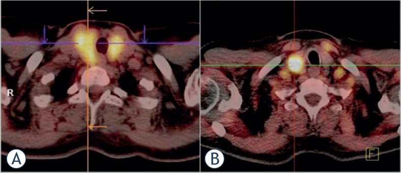
Incidental uptake of 18F-fluorodeoxyglucose (18F-FDG) in the thyroid is sometimes found during positron emission tomography - computed tomography (PET-CT)1–3, which is mostly used in cancer staging and diagnostics.4–6 Throughout the literature the reported incidence of incidental thyroid uptake of 18F-FDG on PET-CT varies between 0.2% and 8.9%.2 Thyroid lesions on PET-CT can be either diffuse or focal (Figure 1). Diffuse 18F-FDG uptake is usually associated with autoimmune thyroiditis or Graves’ disease7–9, whereas focal 18F-FDG uptake can be either due to a benign or malignant process in the thyroid.10–19
FIGURE 1.
Fusion (PET-CT) scans of the thyroid. A diffuse 18F-FDG accumulation in the thyroid is presented on scan (A) (this scan was done at the Institute of Oncology Ljubljana). A focal 18F-FDG accumulation in the thyroid is presented on scan (B) (this scan was done at the University Medical Centre Ljubljana).
A semi-quantitative parameter that could help in differentiating thyroid lesions on PET-CT is the standardized uptake value (SUV), often expressed as the maximal SUV (SUVmax) or mean SUV (SUVmean).20 However, the discriminating power of this parameter is still unclear, as some studies have reported a statistically significant difference between SUV values of benign and malignant thyroid lesions13,16,21,22, whilst others have shown no statistically significant difference.17,23–28 Moreover, the SUV of benign and malignant thyroid lesions varied greatly between these studies. We also know that the calculated SUV is highly dependent on the scanner type, reconstruction algorithms and software packages used, which prevents the comparisons of studies conducted at different centres using different equipment.29–31 This represented a challenge for our study.
The aims of this study were to (i) determine the incidence of thyroid lesions incidentally found on 18F-FDG PET-CT, (ii) identify what diffuse and focal thyroid lesions represent, and (iii) what is the optimal SUVmax that can discriminate between benign and malignant focal thyroid lesions incidentally found on PET-CT. This study was conducted at two PET-CT centres (having different PET-CT scanners) in Slovenia: the Department of nuclear medicine at the University Medical Centre Ljubljana (UMC) and the Institute of Oncology Ljubljana (IO).
Patients and methods
Subjects and study design
We retrospectively evaluated the medical records of 5,911 patients (2,840 patients from UMC and 3,071 patients from IO) who underwent an 18F-FDG PET-CT investigation between January 2010 and December 2011. Only patients (males and non-pregnant females) aged 18 years or more were included in this study. The 18F-FDG PET-CT investigation of patients included in the study was performed for different purposes, mainly because of oncologic indications. The study was approved by the Ethics Committee at the Ministry of Health, Republic of Slovenia (No.: 53/04/12).
Methods employed
Patients from both centres fasted for at least 6 hours, ideally having a blood glucose level less than 7 mmol/l, before receiving 370 MBq of 18F-FDG. The acquisition on the PET-CT scanner started 60 minutes after the radiotracer administration. The PET-CT scanners used were different: at UMC a Siemens Biograph mCT and at IO a Philips Gemini 16 GXL. In all patients, the localisation and attenuation correction CT was first done, followed by the PET scan itself. The CT acquisition parameters in both centres were fairly similar. Also, the PET acquisition parameters did not differ a lot; at UMC a bed position of 2 min with 45% overlap and at IO a bed position of 2 min with 50% overlap was used. The acquired PET-CT data was processed using similar iterative reconstruction algorithms.
Nuclear medicine doctors at both centres used visual and semi-quantitative data analysis (SUVmax) for creating a final report. They had access to relevant patient history and previous examination reports. Patients with thyroid lesion incidentally found on 18F-FDG PET-CT were referred to a thyroidologist.
Thyroid investigation normally included the patient’s history, clinical examination, relevant laboratory workup, ultrasound examination and 99mTc scintigraphy of the thyroid. For a final diagnosis of suspicious thyroid lesions, patients were further investigated using fine needle aspiration cytology (FNAC). A histological report was obtained for lesions that were surgically removed. All data (PET-CT reports, reports of thyroid examinations, cytological and histological reports) were obtained only from patients treated and followed-up at UMC and IO.
Statistical analyses
Statistical analysis was performed using IBM SPSS Statistics 22.0 and Microsoft Excel for Mac 14.1. The SUVmax of benign and malignant thyroid lesions were compared using Student’s t-test. Results were deemed statistically significant for p < 0.05. A receiver operating characteristic (ROC) analysis was performed to determine a SUVmax cut-off point that differentiates between suspicious and unsuspicious focal thyroid lesions.
Results
Characteristics of patients
The mean age of 2,840 patients who had a PET-CT investigation at UMC was 61.2 ± 12.9 years; the mean age of 3,071 patients at IO was 64.4 ± 12.1 years. Fifty per cent of UMC patients were males and 50% females. The percentage of males and females in the IO group was 52.5% and 47.5% respectively. Patients at UMC underwent an 18F-FDG PET-CT investigation mainly for cancer-related diagnostics or inflammatory/infection problems. On the other side, patients at IO underwent an 18F-FDG PET-CT investigation almost exclusively because of cancer-related diagnostics.
Incidentally detected thyroid lesions
Incidental 18F-FDG uptake in the thyroid was found in 230 out of 5,911 investigated patients (in 3.89%). Focal thyroid uptake represented 64.3% and diffuse thyroid uptake 35.7% of detected thyroid lesions. 56.1% of all focal lesions and 81.7% of all diffuse lesions were detected in female patients. More detailed information about patients with incidentally found thyroid lesions on 18F-FDG PET-CT is presented in Table 1.
TABLE 1.
Patients and characteristics of incidental 18F-FDG uptake in the thyroid detected by PET-CT
| Patients with incidental thyroid uptake | Incidental thyroid uptake | ||||
|---|---|---|---|---|---|
|
| |||||
| Number (m/f) | Incidence (%) | Age (year) (average ± SD) | Type | SUVmax (average ± SD) | |
| UMC | 61 (24/37) | 2.15 | 63.6 ± 12.1 | Focal | 6.6 ± 4.4 |
| 21 (4/17) | 0.74 | 57.5 ± 14.4 | Diffuse | 7.9 ± 4.0 | |
| (all) | 82 (28/54) | 2.89 | 62 ± 12.9 | 6.9 ± 4.3 | |
| IO | 87 (41/46) | 2.83 | 64.2 ± 12.3 | Focal | 4.2 ± 2.1 |
| 61 (11/50) | 1.99 | 64.9 ± 11.2 | Diffuse | 4.3 ± 2.7 | |
| (all) | 148 (52/96) | 4.82 | 64.5 ± 11.8 | 4.2 ± 2.3 | |
UMC = University Medical Centre Ljubljana; IO = Institute of Oncology Ljubljana; SUVmax = maximal standardised uptake value
Data of further treatment were found for 58 out of 82 patients (in 70.7%) with increased 18F-FDG uptake in the thyroid investigated at UMC and for 46 out of 148 patients (in 31.1%) investigated at IO. Diffuse thyroid lesions in 14/58 patients (24.1%) from UMC (SUVmax range from 3.5 to 10.3) and in 15/46 (32.6%) patients from IO (SUVmax range from 1.9 to 9.2) were all benign. Hashimoto’s thyroiditis was diagnosed in 92.9% and 73.3% respectively.
At UMC, 44 patients with focal 18F-FDG uptake in the thyroid (SUVmax range from 2.3 to 31.9) were further investigated. Thyroid nodules were found in 30 patients (in 68.2%). Autoimmune thyroid disease was diagnosed in 29.5% – in 12 patients with Hashimoto’s thyroiditis and in one patient with Graves’ disease. One patient was diagnosed to have benign diffuse goitre. FNAC was performed in 28 of 44 patients (63.6%). Results of FNAC are presented in Table 2.
TABLE 2.
Results of fine needle aspiration cytology for focal thyroid lesions, classified according to the Bethesda classification
| Centre | FNAC (No.) | ND or UnS (No. (%)) | BEN (No. (%)) | AUS or FLUS (No. (%)) | FN (No. (%)) | SM (No. (%)) | M (No. (%)) |
|---|---|---|---|---|---|---|---|
| UMC | 28 | 2 (7.1) | 17 (60.8) | 2 (7.1) | 5 (17.9) | 0 | 2 (7.1) |
| IO | 24 | 5 (20.8) | 7 (29.2) | 1 (4.2) | 3 (12.5) | 3 (12.5) | 5 (20.8) |
| All | 52 | 7 (13.4) | 24 (46.2) | 3 (5.8) | 8 (15.4) | 3 (5.8) | 7 (13.4) |
FNAC = fine needle aspiration cytology, ND or UnS = non-diagnostic or unsatisfactory; BEN = benign; AUS or FLUS = atypia of undetermined significance or follicular lesion of undetermined significance; FN = follicular neoplasms and oncocytic tumours; SM = suspicious for malignancy; M = malignant
Out of 31 focal thyroid lesions diagnosed on PET-CT in patients from IO (SUVmax range from 1.5 to 8.7) thyroid nodules were found in 28 cases (in 90.3%). In two patients the focal lesion was caused by Hashimoto’s thyroiditis and in one by Graves’ disease. FNAC diagnostics were performed in 24 of 31 patients (77.4%) (Table 2).
The optimal SUVmax cut-off point for differentiating between suspicious and unsuspicious focal thyroid lesions incidentally detected on PET-CT, calculated using ROC analysis, was 5.4 for patients investigated at UMC (sensitivity 76.9%, specificity 61.3%, AUC = 0.785); the optimal differentiating SUVmax for patients investigated at IO was 4.0 (sensitivity 66.7%, specificity 73.7%, AUC = 0.754).
Surgically removed focal thyroid lesions
Malignant thyroid disease was found in 10 out of 18 patients (55.6%) who underwent surgery. Malignant thyroid disease was more common in males (8 cases) than in females (2 cases). Nine patients with focal thyroid lesions who were referred for surgery were lost to follow-up. Therefore in 10 out of 66 patients (15.2%) with focal thyroid lesion incidentally detected on 18F-FDG PET-CT malignant thyroid disease was confirmed. Detailed characteristics of all surgically removed thyroid lesions are presented in Table 3.
TABLE 3.
Characteristics of surgically removed thyroid lesions
| Centre | Referral diagnosis | Sex (m/f) | Age (year) | SUVmax | Size (mm) | Cytology | Histology |
|---|---|---|---|---|---|---|---|
| UMC | Gastric carcinoma | f | 71 | 5.5 | 10 | Oncocytic cells | Hürthle adenoma |
| Suspicious lesion in the right lungs | m | 68 | 4.8 | 12 | Unsatisfactory | Nodular goitre | |
| Tumour of the cardia | f | 48 | 8.9 | 9 | Oncocytic cells | Hürthle adenoma | |
| Erythema nodosum and pharyngitis | f | 40 | 7.5 | 22 | Unsatisfactory | Hürthle adenoma | |
| Pelvic inflammatory disease | f | 61 | 6.4 | 10 | Oncocytic cells | Nodular goitre | |
| Lung carcinoma | m | 70 | 15.2 | 30 | Oncocytic cells | Follicular carcinoma | |
| Origo ignota malignant disease | m | 48 | 11 | 21 | Atypia of undetermined significance | Medullary carcinoma | |
| Histiocytosis | m | 41 | 11 | 10 | Papillary carcinoma | Papillary carcinoma | |
| GIT malignancy | f | 64 | 31.9 | 52 | Atypia of undetermined significance | Papillary carcinoma | |
| Metastatic lesion on the left side of the neck | m | 74 | 10 | 30 | Planocelluar metastasis | Planocellular subglottic carcinoma — metastasis | |
| IO | Hodgkin’s lymphoma | f | 64 | 3.2 | 15 | Suspicious for malignancy (follicular or Hürthle) | Hyperplastic follicular benign nodule |
| Malignant melanoma | m | 71 | 2 | 23 | Suspicious for malignancy (follicular or papillary) | Multinodular colloid goitre | |
| Tumour of the GE junction | f | 62 | 8.7 | 35 | Oncocytic cells | Hürthle adenoma | |
| Tumour mass in the thigh | m | 22 | 7.8 | 9 | Papillary carcinoma | Follicular carcinoma | |
| Rectal carcinoma | m | 71 | 2.7 | 40 | Suspicious for follicular malignancy | Follicular carcinoma | |
| Malignant melanoma | f | 55 | 6 | 10 | Papillary carcinoma | Thyroid malignancy with elements of follicular, papillary and Hürthle carcinoma | |
| Rectal carcinoma | m | 59 | 6.4 | 10 | Papillary carcinoma | Papillary carcinoma | |
| Rectal carcinoma | m | 58 | 2.8 | 15 | Oncocytic cells | Papillary carcinoma |
UMC = University Medical Centre Ljubljana; IO = Institute of Oncology Ljubljana; SUVmax = maximal standardised uptake value; GE = gastro-oesophageal; GIT = gastro-intestinal tract
SUVmax of malignant and benign focal thyroid lesions
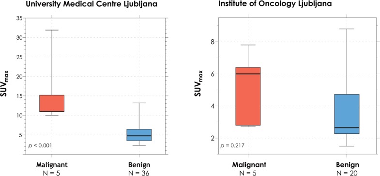
SUVmax of malignant focal lesions (histologically confirmed) was compared to SUVmax of benign focal lesions (the benign nature of a lesion was established either after a thorough thyroid examination with ultrasound, FNAC or surgical treatment) (Figure 2). A statistically significant (p < 0.001) difference was observed between 36 benign (SUVmax from 2.3 to 13.2) and 5 malignant (SUVmax from 10 to 31.9) focal thyroid lesions incidentally detected on PET-CT in patients from UMC. No statistically significant difference (p = 0.217) was observed between 20 benign (SUVmax from 1.5 to 8.8) and 5 malignant (SUVmax from 2.7 to 7.8) focal thyroid lesions in patients from IO.
FIGURE 2.
SUVmax of malignant and benign focal thyroid lesions (median, IQR and MIN/MAX values).
Discussion
Incidental 18F-FDG uptake in the thyroid was observed in 3.89% of 5,911 patients investigated; in 2.89% of patients investigated at UMC and in 4.82% of patients investigated at IO. This is in accordance with the present literature, where the incidence of such lesions varied from 0.2 to 8.9%, with most studies reporting a incidence between 2 and 3%.2,3,11–13,16,17,19,21–28,32–36 In a review article by Bertagna et al.2, the authors postulated that this variability in incidence could be attributed to population characteristics and background risk of thyroid disease related to specific geographic areas.
Slovenia, although not an endemic goitre region, still has a significant incidence of thyroid nodules in the general population.37 This could in part explain the slightly higher incidence of thyroid lesions incidentally found on PET-CT compared to some studies, where authors found a smaller incidence of thyroid lesions.11,12,17
According to the American Thyroid Association Guidelines Taskforce38 further investigation of incidentally found thyroid nodules is recommended. Adhering to these guidelines, all patients from our practices with an incidentally detected thyroid lesion on PET-CT were referred to a thyroidologist. Due to different reasons, not all patients had a consultation, mainly because of the management of their primary illness. In our study, 71% of patients from UMC and only 31% of patients from IO received additional thyroid diagnostics. Our explanation for this difference is that PET-CT examinations in patients at IO were done almost exclusively for staging of known primary malignant diseases – many of these patients had more severe primary malignancies that required more prompt treatment than potential thyroid neoplasms. In comparison at UMC, approximately one third of PET-CT examinations were done for non-oncologic indications in which cases additional thyroid diagnostics were more likely than in oncologic patients with more severe primary disease. Other studies also reported a similar percentage of patients with incidentally discovered thyroid PET lesions who were further investigated, with follow-up rates in the ranks of 50%.11–13,16–18,23–25,28
Experts agree that diffuse thyroid uptake of 18F-FDG on PET-CT is associated with Hashimoto’s thyroiditis.9 This was also confirmed by our results, where most diffuse lesions were caused by Hashimoto’s thyroiditis and no malignancy was found in patients with diffuse thyroid PET lesions.
According to the literature, the rate of focal lesions ranges from 14% to 73% of all thyroid PET lesions8,16,24,32 with a risk of malignancy in further investigated lesions of about 33%.2,38 In our study, focal thyroid lesions were present in 64.3% of all cases with incidental thyroid uptake. These lesions represented a thyroid nodule in 68.2% (UMC patients) and in 90.3% (IO patients). We histologically confirmed thyroid malignancy in 5 of 10 surgically treated patients from UMC and in 5 of 8 patients from IO. Altogether, malignant disease was observed in 10 of 66 patients (in 15.2%) with a focal 18F-FDG uptake in the thyroid. In comparison to other reports, the incidence of thyroid malignancy in our study was somewhat lower.2,12,13,16,17,21–28,34 This is, in our opinion, mainly due to higher goitre prevalence in our population.37
Autoimmune thyroid disease was present in 29.5% of focal thyroid lesions from UMC patients. This finding is quite different from data published in the literature.18,23 Our explanation for this discrepancy is in the different diagnostic process that was used in different institutions. At UMC, a thorough thyroid examination with relevant laboratory workup and an ultrasound examination of the thyroid, irrespective of the use of FNAC, was in most patients enough to make a final diagnosis of thyroid disease. The decision regarding FNAC examination was undertaken by the consulting thyroidologist on a patient by patient basis. Most studies, like the one conducted by Chu et al.12, were more in line with the IO group, where only 3 of 31 focal PET lesions proved to be of autoimmune origin.
According to the literature, Graves’ disease is demonstrated most commonly by diffusely increased 18F-FDG uptake in the thyroid.39,40 However, in our study, we found two cases of Graves’ disease with focal 18F-FDG uptake.
One of the main goals of our study was to determine whether it would be possible to differentiate between benign and malignant thyroid lesions using SUVmax. The literature is quite divided on this topic, with studies claiming to being able to differentiate between benign and malignant lesions13,21,22,41 and others whose conclusions were the exact opposite.17,23–28 This was also the case in our study, where the UMC group presented a statistically significant difference between benign and malignant lesions, whereas no such difference was found in the IO group. Even though the mean SUVmax of malignant lesions were on average higher than benign lesions, the overlap between both sets of lesions was considerable. For example, a Hürthle adenoma had a relatively high SUVmax of 8.9 while on the other side; a papillary thyroid carcinoma had a SUVmax of only 2.8.
It should also be noted, that calculated SUVmax is highly dependent on the type of PET-CT scanner, reconstruction algorithms and software packages used20,29–31, as was the case in our study, which included two centres with different equipment. The newer Siemens Biograph® mCT used at UMC had a better detector system and time of flight technology compared to the older Philips Gemini 16 GXL. These might be some of the factors resulting in different SUVmax readings at both centres. Therefore, the SUVmax of a thyroid lesion should only serve as one of several parameters that alert the clinician on the possibility of thyroid malignancy. The correct protocol in this situation is, as recommended by the American Thyroid Association Guidelines, to promptly investigate all focal thyroid PET lesions with additional diagnostics.38
Conclusions
Incidental 18F-FDG uptake in the thyroid on PET-CT was found in 3.89%. Only focal thyroid uptake represented a malignant lesion in our study – in 15.2% of all focal thyroid lesions. SUVmax should only serve as one of several parameters that alert the clinician on the possibility of thyroid malignancy and as such must be used with caution in the interpretation of PET-CT studies.
Footnotes
Disclosure: No potential conflicts of interest were disclosed.
References
- 1.Jin J, McHenry CR. Thyroid incidentaloma. Best Pract Res Clin Endocrinol Meta. 2012;26:83–96. doi: 10.1016/j.beem.2011.06.004. [DOI] [PubMed] [Google Scholar]
- 2.Bertagna F, Treglia G, Piccardo A, Giubbini R. Diagnostic and clinical significance of F-18-FDG-PET/CT thyroid incidentalomas. J Clin Endocrinol Metab. 2012;97:3866–75. doi: 10.1210/jc.2012-2390. [DOI] [PubMed] [Google Scholar]
- 3.Treglia G, Muoio B, Giovanella L, Salvatori M. The role of positron emission tomography and positron emission tomography/computed tomography in thyroid tumours: an overview. Eur Arch Otorhinolaryngol. 2012;270:1783–7. doi: 10.1007/s00405-012-2205-2. [DOI] [PubMed] [Google Scholar]
- 4.Cistaro A, Quartuccio N, Mojtahedi A, Fania P, Filosso PL, Campenni A, et al. Prediction of 2 years-survival in patients with stage I and II non-small cell lung cancer utilizing (18)F-FDG PET/CT SUV quantification. Radiol Oncol. 2013;47:219–23. doi: 10.2478/raon-2013-0023. [DOI] [PMC free article] [PubMed] [Google Scholar]
- 5.Maza S, Buchert R, Brenner W, Munz DL, Thiel E, Korfel A, et al. Brain and whole-body FDG-PET in diagnosis, treatment monitoring and long-term follow-up of primary CNS lymphoma. Radiol Oncol. 2013;47:103–10. doi: 10.2478/raon-2013-0016. [DOI] [PMC free article] [PubMed] [Google Scholar]
- 6.Moon E-H, Lim ST, Han Y-H, Jeong YJ, Kang Y-H, Jeong H-J, et al. The usefulness of F-18 FDG PET/CT-mammography for preoperative staging of breast cancer: comparison with conventional PET/CT and MR-mammography. Radiold Oncol. 2013;47:390–7. doi: 10.2478/raon-2013-0031. [DOI] [PMC free article] [PubMed] [Google Scholar]
- 7.Karantanis D, Bogsrud TV, Wiseman GA, Mullan BP, Subramaniam RM, Nathan MA, et al. Clinical Significance of diffusely increased 18F-FDG uptake in the thyroid gland. J Nucl Med. 2007;48:896–901. doi: 10.2967/jnumed.106.039024. [DOI] [PubMed] [Google Scholar]
- 8.Kurata S, Ishibashi M, Hiromatsu Y, Kaida H, Miyake I, Uchida M, et al. Diffuse and diffuse-plus-focal uptake in the thyroid gland identified by using FDG-PET: prevalence of thyroid cancer and Hashimoto’s thyroiditis. Ann Nucl Med. 2007;21:325–30. doi: 10.1007/s12149-007-0030-2. [DOI] [PubMed] [Google Scholar]
- 9.Yasuda S, Shohtsu A, Ide M, Takagi S, Takahashi W, Suzuki Y, et al. Chronic thyroiditis: diffuse uptake of FDG at PET. Radiology. 1998;207:775–8. doi: 10.1148/radiology.207.3.9609903. [DOI] [PubMed] [Google Scholar]
- 10.Lowe VJ, Mullan BP, Hay ID, McIver B, Kasperbauer JL. 18F-FDG PET of patients with Hürthle cell carcinoma. J Nucl Med. 2003;44:1402–6. [PubMed] [Google Scholar]
- 11.Chen Y-K, Ding H-J, Chen K-T, Chen Y-L, Liao AC, Shen Y-Y, et al. Prevalence and risk of cancer of focal thyroid incidentaloma identified by 18F-fluorodeoxyglucose positron emission tomography for cancer screening in healthy subjects. Anticancer Res. 2005;25:1421–6. [PubMed] [Google Scholar]
- 12.Chu QD, Connor MS, Lilien DL, Johnson LW, Turnage RH, Li BDL. Positron emission tomography (PET) positive thyroid incidentaloma: the risk of malignancy observed in a tertiary referral center. Am Surg. 2006;72:272–5. [PubMed] [Google Scholar]
- 13.Choi JY, Lee KS, Kim H-J, Shim YM, Kwon OJ, Park K, et al. Focal thyroid lesions incidentally identified by integrated 18F-FDG PET/CT: clinical significance and improved characterization. J Nucl Med. 2006;47:609–15. [PubMed] [Google Scholar]
- 14.Katz SC, Shaha A. PET-associated incidental neoplasms of the thyroid. J Am Coll Surg. 2008;207:259–64. doi: 10.1016/j.jamcollsurg.2008.02.013. [DOI] [PubMed] [Google Scholar]
- 15.Barnabei A, Ferretti E, Baldelli R, Procaccini A, Spriano G, Appetecchia M. Hurthle cell tumours of the thyroid. Personal experience and review of the literature. Acta Otorhinolaryngol Ital. 2009;29:305–11. [PMC free article] [PubMed] [Google Scholar]
- 16.Pagano L, Samà MT, Morani F, Prodam F, Rudoni M, Boldorini R, et al. Thyroid incidentaloma identified by 18F-fluorodeoxyglucose positron emission tomography with CT (FDG-PET/CT): clinical and pathological relevance. Clin Endocrinol. 2011;75:528–34. doi: 10.1111/j.1365-2265.2011.04107.x. [DOI] [PubMed] [Google Scholar]
- 17.Bonabi S, Schmidt F, Broglie MA, Haile SR, Stoeckli SJ. Thyroid incidentalomas in FDG-PET/CT: prevalence and clinical impact. Eur Arch Otorhinolaryngol. 2012;269:2555–60. doi: 10.1007/s00405-012-1941-7. [DOI] [PubMed] [Google Scholar]
- 18.Ohba K, Sasaki S, Oki Y, Nishizawa S, Matsushita A, Yoshino A, et al. Factors associated with fluorine-18-fluorodeoxyglucose uptake in benign thyroid nodules. Endocr J. 2012;60:985–90. doi: 10.1507/endocrj.ej13-0155. [DOI] [PubMed] [Google Scholar]
- 19.Bertagna F, Treglia G, Piccardo A, Giovannini E, Bosio G, Biasiotto G, et al. F18-FDG-PET/CT thyroid incidentalomas: a wide retrospective analysis in three Italian centres on the significance of focal uptake and SUV value. Endocrine. 2013;43:678–85. doi: 10.1007/s12020-012-9837-2. [DOI] [PubMed] [Google Scholar]
- 20.Huang SC. Anatomy of SUV. Standardized uptake value. Nucl Med Biol. 2000;27:643–6. doi: 10.1016/s0969-8051(00)00155-4. [DOI] [PubMed] [Google Scholar]
- 21.Kang BJ, O JH, Baik JH, Jung SL, Park YH, Chung SK. Incidental thyroid uptake on F-18 FDG PET/CT: correlation with ultrasonography and pathology. Ann Nucl Med. 2009;23:729–37. doi: 10.1007/s12149-009-0299-4. [DOI] [PubMed] [Google Scholar]
- 22.Kim BH, Na MA, Kim IJ, Kim S-J, Kim Y-K. Risk stratification and prediction of cancer of focal thyroid fluorodeoxyglucose uptake during cancer evaluation. Ann Nucl Med. 2010;24:721–8. doi: 10.1007/s12149-010-0414-6. [DOI] [PubMed] [Google Scholar]
- 23.Kim TY, Kim WB, Ryu JS, Gong G, Hong SJ, Shong YK. 18F-fluorodeoxyglucose uptake in thyroid from positron emission tomogram (PET) for evaluation in cancer patients: high prevalence of malignancy in thyroid PET incidentaloma. Laryngoscope. 2005;115:1074–8. doi: 10.1097/01.MLG.0000163098.01398.79. [DOI] [PubMed] [Google Scholar]
- 24.Are C, Hsu JF, Ghossein RA, Schöder H, Shah JP, Shaha AR. Histological aggressiveness of fluorodeoxyglucose positron-emission tomogram (FDGPET)-detected incidental thyroid carcinomas. Ann Surg Oncol. 2007;14:3210–5. doi: 10.1245/s10434-007-9531-4. [DOI] [PubMed] [Google Scholar]
- 25.Bogsrud TV, Karantanis D, Nathan MA, Mullan BP, Wiseman GA, Collins DA, et al. The value of quantifying 18F-FDG uptake in thyroid nodules found incidentally on whole-body PET–CT. Nucl Med Comun. 2007;28:373–81. doi: 10.1097/MNM.0b013e3280964eae. [DOI] [PubMed] [Google Scholar]
- 26.Kwak JY, Kim E-K, Yun M, Cho A, Kim MJ, Son EJ, et al. Thyroid incidentalomas identified by 18F-FDG PET: sonographic correlation. Am J Roentgenol. 2008;91:598–603. doi: 10.2214/AJR.07.3443. [DOI] [PubMed] [Google Scholar]
- 27.Chen W, Parsons M, Torigian DA, Zhuang H, Alavi A. Evaluation of thyroid FDG uptake incidentally identified on FDG-PET/CT imaging. Nucl Med Commun. 2009;30:240–4. doi: 10.1097/MNM.0b013e328324b431. [DOI] [PubMed] [Google Scholar]
- 28.Pampaloni MH, Win AZ. Prevalence and Characteristics of Incidentalomas Discovered by Whole Body FDG PETCT. Int J Mol Imaging. 2012;18:1–6. doi: 10.1155/2012/476763. [DOI] [PMC free article] [PubMed] [Google Scholar]
- 29.Nguyen NC, Kaushik A, Wolverson MK, Osman MM. Is there a common SUV threshold in oncological FDG PET/CT, at least for some common indications? A retrospective study. Acta Oncol. 2011;50:670–7. doi: 10.3109/0284186X.2010.550933. [DOI] [PubMed] [Google Scholar]
- 30.de Langen AJ, Vincent A, Velasquez LM, van Tinteren H, Boellaard R, Shankar LK, et al. Repeatability of 18F-FDG uptake measurements in tumors: a metaanalysis. J Nucl Med. 2012;53:701–8. doi: 10.2967/jnumed.111.095299. [DOI] [PubMed] [Google Scholar]
- 31.Makris NE, Huisman MC, Kinahan PE, Lammertsma AA, Boellaard R. Evaluation of strategies towards harmonization of FDG PET/CT studies in multicentre trials: comparison of scanner validation phantoms and data analysis procedures. Eur J Nucl Med Mol Imaging. 2013;40:1507–15. doi: 10.1007/s00259-013-2465-0. [DOI] [PMC free article] [PubMed] [Google Scholar]
- 32.King D, Stack B, Spring P, Walker R, Bodenner D. Incidence of thyroid carcinoma in fluorodeoxyglucose positron emission tomography-positive thyroid incidentalomas. Otolaryngol Head Neck Surg. 2007;137:400–4. doi: 10.1016/j.otohns.2007.02.037. [DOI] [PubMed] [Google Scholar]
- 33.Bertagna F, Giubbini R. F18-FDG-PET/CT thyroid incidentalomas and their benign or malignant nature: a critical and debated issue. Ann Nucl Med. 2010;25:151–2. doi: 10.1007/s12149-010-0443-1. [DOI] [PubMed] [Google Scholar]
- 34.Ohba K, Nishizawa S, Matsushita A, Inubushi M, Nagayama K, Iwaki H, et al. High incidence of thyroid cancer in focal thyroid incidentaloma detected by 18F-fluorodeoxyglucose [corrected] positron emission tomography in relatively young healthy subjects: results of 3-year follow-up. Endocr J. 2010;57:395–401. doi: 10.1507/endocrj.k10e-008. [DOI] [PubMed] [Google Scholar]
- 35.Nishimori H. Incidental thyroid “PETomas”: clinical significance and novel description of the self-resolving variant of focal FDG-PET thyroid uptake. Can J Surg. 2011;54:83–8. doi: 10.1503/cjs.023209. [DOI] [PMC free article] [PubMed] [Google Scholar]
- 36.Treglia G, Annunziata S, Muoio B, Salvatori M, Ceriani L, Giovanella L. The role of fluorine-18-fluorodeoxyglucose positron emission tomography in aggressive histological subtypes of thyroid cancer: An overview. Int J Endocrinol. 2013;2013:1–6. doi: 10.1155/2013/856189. [DOI] [PMC free article] [PubMed] [Google Scholar]
- 37.Zaletel K, Gaberscek S, Pirnat E. Ten-year follow-up of thyroid epidemiology in Slovenia after increase in salt iodization. Croat Med J. 2011;52:615–21. doi: 10.3325/cmj.2011.52.615. [DOI] [PMC free article] [PubMed] [Google Scholar]
- 38.Cooper DS, Haugen BR, Hauger BR, Kloos RT, Lee SL, Mandel SJ, et al. Revised American Thyroid Association management guidelines for patients with thyroid nodules and differentiated thyroid cancer. Thyroid. 2009;19:1167–214. doi: 10.1089/thy.2009.0110. [DOI] [PubMed] [Google Scholar]
- 39.Boerner AR, Voth E, Theissen P, Wienhard K, Wagner R, Schicha H. Glucose metabolism of the thyroid in Graves’ disease measured by F-18-fluorodeoxyglucose positron emission tomography. Thyroid. 1998;8:765–72. doi: 10.1089/thy.1998.8.765. [DOI] [PubMed] [Google Scholar]
- 40.Liu Y. Clinical significance of thyroid uptake on F18-fluorodeoxyglucose positron emission tomography. Ann Nucl Med. 2009;23:17–23. doi: 10.1007/s12149-008-0198-0. [DOI] [PubMed] [Google Scholar]
- 41.Ho T-Y, Liou M-J, Lin K-J, Yen T-C. Prevalence and significance of thyroid uptake detected by 18F-FDG PET. Endocrine. 2011;40:297–302. doi: 10.1007/s12020-011-9470-5. [DOI] [PubMed] [Google Scholar]


