

Key message
Congenital heart disease affects almost 1% of live births. The majority of these cases are syndromic. A large no. of them have associated ocular pathology like retinal vascular anomalies, optic nerve hypoplasia, ptosis, squint. Cataracts, especially bilateral, are seen almost exclusively in syndromic patients. Here, we report a case of bilateral non-syndromic cataract with tetralogy of fallot, which had been undiagnosed till the age of three years. The child had been examined at various centres, however, an ocular examination had not been carried out. All children with congenital heart disease, even non-syndromic, warrant a detailed ocular examination to rule out common treatable ocular pathologies.
Introduction
Paediatric cataract is one of the major causes of childhood blindness, constituting almost 10–12% of cases. A number of these cases are associated with syndromes, especially bilateral cases. Common syndromes associated are Downs, patau, trisomy 18 etc. Congenital heart disease is one of the commonest childhood birth defects which affects almost 1% of live child births.1,2 A number of these cases, especially syndromic, have a number of ocular pathologies like ptosis, squint, cataract, Optic disc hypoplasia, retinal haemorrhages and vascular tortuosity. However, there are very few studies which document ocular pathologies and congenital heart disease. A.M. Mansour et al.3 in 2004 studied 240 cases of congenital heart disease and ocular pathologies. Only six of their cases had congenital cataract, of which only one was non-syndromic. There are no other case reports available which document a case of bilateral cataract in a child of cyanotic congenital heart disease, which is non-syndromic.
Case Report
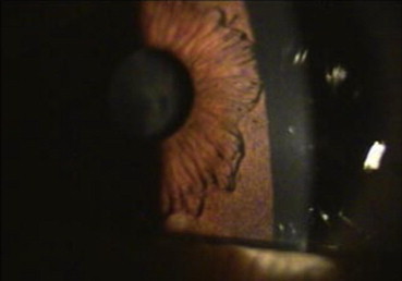
A 3 year old child patient was brought to our outpatient department by the parents with history of intermittent deviation of eyes while watching TV and bringing small objects close to her face to see them. She was also watching TV from a very close distance. There was no other associated ocular history. The mother was 27 year old and the child was a full term caesarean delivery with no maternal problems during ante-natal period. There was no history of anoxia, seizures at birth. She had normal developmental milestones. Child was a known case of tetralogy of fallot (TOF), a cyanotic congenital heart disease, which was detected during the first year of life. She had been taken up for surgery for correction of the TOF but was declared inoperable. There was no mention of any ocular problems in her earlier documents, which could have been present at birth or could have developed later. On examination, she had a visual acuity of 20/400 in Rt eye and 20/500 in Lt eye. Ocular examination revealed bilateral central lamellar cataracts (Figs. 1–3). Rest anterior segment examination was normal. All ocular movements were normal. There was no manifest squint which was seen during examination; however, there was intermittent exophoria of 15 degrees on cover test. Fundus examination after dilatation revealed a hazy media with normal Optic Disc, no retinal abnormalities were noted. Ultrasound B scan revealed a normal posterior segment. There was no nystagmus. Pupillary dilatation was adequate. The child underwent investigations to rule out common causes of congenital cataracts, however, all relevant investigations, like urine for reducing substances, TORCH titre etc were normal.
Fig. 1.

Central Lamellar Cataract – Rt eye.
Fig. 2.
Developmental Cataract – Lt Eye.
Fig. 3.

Central Developmental Cataract.
IOL power calculation was done and an axial length of 22.51 was recorded in Rt eye and 22.56 in Lt eye. The IOL power in both eyes was +23.5 D. SRK II formula was used, 90% correction was given as the child was 3 yrs old. Child was taken up for surgery under GA on 14 Jan 2010 and phacoaspiration – Rt eye was done with implantation of a rigid PMMA IOL (+21.5 D) as the capsulorhexis was lost during surgery. Postoperatively, the child was managed with Topical Antibiotic and steroid eye drops, cycloplegics and NSAIDS. During the immediate Post Op period, child had two cyanotic episodes which were successfully managed by the paediatrician and anaesthetist. Child was discharged on 21 Jan 2010.
On the 15th Post Op day, child developed redness and pain in Rt eye. Examination showed ciliary congestion and cells and flare in ant chamber (Figs. 4 and 5). The child was managed with frequent steroid eye drops and atropine eye drops and improved after five days. Visual acuity after six weeks was 20/80 – unaided in the Rt eye.
Fig. 4.

Post Op Uveitis – Rt eye.
Fig. 5.

Post Op Uveitis – Rt eye.
Phacoaspiration and Hydrophobic acrylic foldable IOL implantation (+21.5 D) in Lt eye was done under GA on 15 march 2010. Post Op recovery was uneventful and after six weeks, child had an unaided visual acuity of 20/50 in the Lt Eye. All medications were stopped after eight weeks. Refraction revealed a refractive error of – 1.5 D sph/−0.50 D Cyl 75° in Rt eye and – 1.75 D Sph/−0.50 D Cyl 108° in Lt eye. The child is currently under three monthly follow-up for repeat refraction and treatment for amblyopia. There is no manifest squint or phoria after surgery. Fundus examination revealed a normal fundus in both eyes.
Discussion
Congenital heart disease affects almost 1–2% of live births. A large no of factors have been implicated in their aetiology, including both environmental and genetic. Most studies carried out earlier have concentrated on single associations, ocular and cardiac with various syndromes.4–8 Mansour et al, in their series of 240 patients described the various syndromes, common ocular findings and their genetic anomalies. The commonest defects noted in their study were ASD, VSD, Tetralogy of Fallot, Pulmonic stenosis and Transposition of great vessels. Ocular findings were varied and ranged from Retinal vascular anomalies, optic nerve hypoplasia, ptosis, squint, retinal haemorrhages, cataract and nystagmus. Cataracts were present in six out of 240 patients (2.5%), of which five were syndromic and only one was non-syndromic (0.41%). However, there is no mention of unilaterality or bilaterality in the series. Wirth,9 in his series of 29 congenital cataract cases, found Down's syndrome in 62% of cases.
With the improvement in operative techniques and management of congenital heart disease, the long term survival of these patients has improved. A large number of these cases are syndromic, the ocular pathology is varied and present in almost 50–60% of these children. A high index of suspicion, along with regular screening, is required to diagnose and manage the various ocular pathologies. As seen in our case report, even a non-syndromic CHD can be associated with an ocular pathology. Absence of screening or routine examination can result in such cases being missed, as in our case. Timely intervention, especially in surgically correctible ocular defects, is of utmost importance in the long term ocular and visual outcomes in these patients. There have been no reported cases of bilateral congenital cataracts in cases of congenital heart disease or Tetralogy of fallot specifically. Developmental or congenital cataracts should always be considered a possibility in these cases.
Conflicts of interest
All authors have none to declare.
References
- 1.Lewin M.B. The genetic basis of congenital heart disease. Ped Ann. 2000;19:469–480. doi: 10.3928/0090-4481-20000801-06. [DOI] [PubMed] [Google Scholar]
- 2.Goldmuntz E. The epidemiology and genetics of congenital heart disease. Clin Perinatol. 2001;28:1–10. doi: 10.1016/s0095-5108(05)70067-1. [DOI] [PubMed] [Google Scholar]
- 3.Mansour A.M., Bitar F.F., Traboulsi E.I. Ocular pathology in congenital heart disease. Eye. 2005;19:29–34. doi: 10.1038/sj.eye.6701408. [DOI] [PubMed] [Google Scholar]
- 4.Gardiner P.A., Joseph M. Eye defects in children with congenital heart lesions: a preliminary study. Dev Med Child Neurol. 1968;10:4. doi: 10.1111/j.1469-8749.1968.tb02836.x. [DOI] [PubMed] [Google Scholar]
- 5.Mansour A.M., Goldberg R.B., Wang F.M., Shprintzen R.J. Ocular findings in the velo-cardio-facial syndrome. J Ped Ophthalmol Strab. 1987;24:263–266. doi: 10.3928/0191-3913-19870901-16. [DOI] [PubMed] [Google Scholar]
- 6.Cullum L., Liebman J. The association of congenital heart disease with Down's syndrome (mongolism) Am J Cardiol. 1969;24:354. doi: 10.1016/0002-9149(69)90428-7. [DOI] [PubMed] [Google Scholar]
- 7.Beauchamp G.R. Blepharophimosis and cardiopathy. J Pediatr Ophthalmol Strabismus. 1980;17:227–228. doi: 10.3928/0191-3913-19800701-07. [DOI] [PubMed] [Google Scholar]
- 8.Vander Veen D.K., Pasquale L.R., Fulton A.B. Central retinal vein occlusion in a young child with cyanotic heart disease. Arch Ophthalmol. 1997;115:1077. doi: 10.1001/archopht.1997.01100160247020. [DOI] [PubMed] [Google Scholar]
- 9.Wirth M.G. Aetiology of congenital and paediatric cataract in an Australian population. Br J Ophthalmol. 2002;86:782–786. doi: 10.1136/bjo.86.7.782. [DOI] [PMC free article] [PubMed] [Google Scholar]