Abstract
To evaluate the chemopreventive potential of quercetin in an experimental skin carcinogenesis mouse model. Skin tumor was induced by topical application of 7, 12-dimethyl Benz (a) anthracene (DMBA) and Croton oil in Swiss albino mouse. Quercetin was orally administered at a concentration of 200 mg/kg and 400 mg/kg body weight daily for 16 weeks in mouse to evaluate chemopreventive potential. Skin cancer was assessed by histopathological analysis. We found that quercetin reduced the tumor size and the cumulative number of papillomas. The mean latent period was significantly increased as compared to carcinogen treated controls. Quercetin significantly decreased the serum levels of glutamate oxalate transaminase, glutamate pyruvate transaminase, alkaline phosphatase and bilirubin. It significantly increased the levels of glutathione, superoxide dismutase and catalase. The elevated level of lipid peroxides in the control group was significantly inhibited by quercetin. Futhermore, DNA damage was significantly decreased in quercetin treated mice as compared to DMBA and croton oil treated mice. The results suggest that quercetin exerts chemopreventive effect on DMBA and croton oil induced skin cancer in mice by increasing antioxidant activities.
Keywords: quercetin, papilloma, skin carcinogenesis, chemoprevention
Introduction
Ocimum sanctum Linn, commonly known as Tulsi, belongs to the Lamiaceae family. It has a variety of constituents e.g. saponins, flavonoids, triterpenoids, tannins, eugenol, vicenin-2, dimethyl benzene, myrecene, ethyl benzene, limocene, linoleic acid, aesculin, and others[1]. Hence, it is an essential plant used in the preparation of several ayurvedic pharmacological products. A wide range of beneficial effects have been reported such as anti-cancer, analgesia and anti-inflammation. Leaves of Ocimum sanctum were investigated for its effect on male reproductive function (sperm count and reproductive hormones) in male albino rabbits. It has a good wound healing property[2].
In vitro and in vivo experiments have demonstrated that flavanoids produce a wide range of biological effects in several mammalian cell systems. Quercetin is a polyphenolic flavanoid extensively present in plants and plant food sources, such as Ocimum sanctum. This flavanoid has a broad range of biological activities, including anti-inflammatory, antioxidant, neuroprotective and antiviral activity. Quercetin has antioxidant properties and therefore may play an important role in preventing cancer. Some researchers reported that quercetin acts as an oxygen radical scavenger and an inhibitor of xanthine oxidase and lipid peroxidation in vitro. Independently and with ascorbic acid, quercetin reduced the occurrence of skin oxidative damage and inhibited neuron damage caused by glutathione depletion. Earlier studies has investigated quercetin in animal models and cancer cell lines, including cancer of the breast, the colon, and the ovary, and reported that it has antiproliferative effects[3]. The ability of quercetin to inhibit inflammatory leukotriene production may be a key to its beneficial impact on anti-inflammatory activity. The anti-inflammatory activity of quercetin is due to its antioxidant and inhibitory effects on inflammation-producing enzymes, such as cyclooxygenase and lipoxygenase and subsequent inhibition of inflammatory mediators, including leukotrienes and prostaglandins. Inhibition of histamine release by mast cells and basophils also contributes to the anti-inflammatory activity of quercetin[4]. Quercetin is a strong inhibitor of human lens aldose reductase[5].
The current study was aimed to understand the chemopreventive potential of quercetin isolated from Ocimum sanctum using a mouse model of skin cancer induced by 7, 12-dimethyl benz (a) anthracene (DMBA) and croton oil.
Materials and methods
Plant material and preparation of the methanolic extract
Leaves of Ocimum sanctum were collected from Sanjeevni, Bhopal, M.P., India. Plant material was taxonomically identified by Dr. Zia-UL-Hassan, Department of Botany, Saifia College of Science, Bhopal. Voucher specimen (351/Bot/Saifia/12) was preserved in the above herbarium for future reference. The leaves were dried under shade and pulverized using electric grinder. Firstly, dried sample was extracted with different solvents, such as petroleum ether, benzene, acetone, ethanol, methanol and hot water by using soxhlet apparatus. Then, the methanolic extract of leaves was fractionated by column chromatography. Slurry of silica gel with 60–120 mesh size prepared in petroleum ether was added to the column. Methanolic extract (15.0 gm) was loaded on the column and then eluted with various solvents, including petroleum ether, benzene, chloroform, chloroform:acetone (1:1), acetone, acetone:ethanol (1:1), ethanol, methanol and distilled water in the order of their increasing polarity. The methanolic fraction was preserved in refrigerator to obtain a yellow powder (2.2 gm)[6].
Characterization of quercetin
Isolated compound was analyzed using infrared, gas chromatography mass spectrometry and nuclear magnetic resonance spectroscopy. IR spectral analysis showed a broad peak for O-H stretch hydrogen bonded from 3412–2712 cm–1. The 1664, 1649, 1560 and 1450 cm–1 showed C = C stretch of benzene and 1242 cm–1 for C-O stretch. The 941, 864, 823, 794 and 702 cm–1 showed peaks for substituted benzene. 1H NMR spectrum of the isolated compound exhibited proton signals at δ7.23 (1H, H-6), δ7.28 (1H, H-8), δ7.02 (1H, H-2′), δ7.33 (1H, H-5′), δ7.54 (1H, H-6′); δ3.56 (1, OH-5), δ3.51 (1, OH-7), δ2.19 (1, OH-3), δ2.03(1, OH-3′) and δ1.56 (1, OH-4′). The negative-ion electrospray ionization (ESI) mass spectrum of the isolated compound showed a quasi molecular ion [M-H]– at m/e 303, indicating a relative molecular weight of 302. The data of spectroscopic analysis revealed the presence of a high content of the flavonoid 3,3′, 4′, 5,7-pentahydroxyflavone (quercetin). This material was further purified by recrystalization with methanol to obtain quercetin (99% purity).
Chemicals
DMBA and croton oil were purchased from Sigma Aldrich (St Louis, MO, USA). Other chemical reagents were commercially available and of analytical grade.
Animals
Swiss albino mice of either sex were randomly selected from the animal house of the Pinnacle Biomedical Research Institute (PBRI), Bhopal. Animals were housed in polypropylene cages with sterile husk and provided standard pellet (Golden Feeds, New Delhi, India) and water with ad libitum access. The animals were maintained with a 12 hour light/dark cycle at 22±2 °C at controlled condition. The study protocol was approved by the local institutional review board at the authors' affiliated insituttion. Animal welfare and the experimental procedures were carried out strictly in accordance with the Guide for Care and Use of Laboratory Animals of the Institutional Animal Ethics Committee (IAEC) of PBRI, Bhopal (Reg No. 1283/c/09/CPCSEA).
Determination of effect of quercetin on DMBA/croton oil induced skin carcinogenesis
Four groups (6 animals per group) of Swiss albino mice were used for the study. Mice were dorsally shaved with hair clipper. Group I mice were given MiliQ water (10 mL/kg body weight), a normal diet and tap water with ad libitum access daily. After 16 weeks, mice were autopsied and the skin of the dorsal area was taken for histopathological studies and blood for biochemical analysis. Group II mice were treated with a single dose of DMBA (100 μg/100 μL of acetone) over the shaven area of the skin of the mice; thereafter, 1% croton oil was applied to skin 3 times a week up to 16 weeks. After the single dose of DMBA, group III mice were treated with quercetin (200 mg/kg) orally each day untill completion of the experiment and 1% croton oil was applied on the skin 3 times a week 1 hour after quercetin was administered. Group IV mice were treated with the same procedure as that of group III, but the dose of quercetin was 400 mg/kg[7]. After 14 days of DMBA application, mice were observed each week for incidence and size of skin tumors, and the body weight and average latent period were recorded for 16 weeks[8].
Biochemical measurements
Mice were sacrificed by cervical dislocation on the last day of the experiment. The skin was removed and washed in ice-cold saline (0.9% NaCl). Then, it was weighed and blotted dry. A 10% tissue homogenate was prepared from the skin in 0.15 mol/L Tris-KCL (pH 7.4) and was centrifuged at 12000 rpm for 15 minutes for clarification. The levels of lipid peroxidase (LPO), GSH (Moron MA), SOD (Marklund) and catalases (Clainborn) were determined as described[9]-[12].
Furthermore, blood was collected from all mice via the retro-orbital plexus. At room temperature, the blood samples were clotted for 45 minutes. Serum was separated by centrifugation at 2500 rpm at 30°C for 15 minutes and utilized for the determination of various biochemical parameters including serum glutamate oxalate transaminase (GOT), glutamate pyruvate transaminase (GPT), alkaline phosphatase (ALP) and bilirubin by using the Span Diagnostic Kit.
Histopathology
Paraffin blocks were routinely prepared for processing formalin fixed tissue samples. Tissue sections (3–4 μm) were performed for hematoxylin-eosin (H&E) staining and epidermal thickness was determined by microscopic examination of H&E-stained tissue samples.
Alkaline single cell gel electrophoresis (SCGE) was performed as a 3-layer procedure[13] with slight modification[14]. The lymphocytes were separated from blood using histopaque density gradient centrifugation and were diluted 20-fold for the Comet assay. Viability of the lymphocytes was evaluated by trypan blue exclusion[15]. Cells with viability > 84% were further processed for Comet assay. In brief, 15 μL cell suspension (approx. 20,000 cells) was mixed with 85 μL 0.5% low melting-point agarose and layered on one end of a frosted glass slide, coated with a layer of 200 μL of 1% normal agarose. It was covered with a third layer of 100 μL low melting-pointagarose. After solidification of the gel, the slides were immersed in lysing solution (2.5 MNaCl, 100 mmol/LNa2 EDTA, 10 mmol/LTris, pH 10 with 10% DMSO and 1% Triton X-100 added fresh) overnight at 4°C. The slides were then placed in a horizontal gel electrophoresis unit, immersed in fresh cold alkaline electrophoresis buffer (300 mmol/L NaOH, 1 mmol/L Na2 EDTA and 0.2% DMSO, pH > 13.5), and left in solution for 20 minutes at 4°C for DNA unwinding and conversion of alkali-labile sites to single strand breaks. Electrophoresis was carried out using the same solution at 4°C for 20 minutes, using 15 V (0.8 V/cm) and 300 mA. The slides were neutralized gently with 0.4 mol/L Tris buffer at pH 7.5 and stained with 75 μL ethidium bromide (20 μg/mL). For positive control, lymphocytes cells were treated with 100 μM H2O2 for 10 minutes at 4°C. Two slides per mouse were prepared and 25 cells per slide (250 cells per group) were scored randomly and analyzed using an image analysis system (Komet-5.5, Kinetic Imaging) attached to a fluorescent microscope (Leica) equipped with appropriate filters. The parameter selected for quantification of DNA damage was percent tail DNA as determined by the software.
Statistical analysis
Values were recorded as mean ± SD. The data obtained from different groups was analyzed by ANOVA. P < 0.05 was considered statistically significant for all experiments.
Results
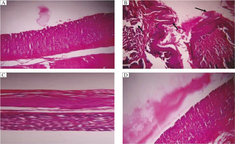
The normal control mice showed normal structure in the dermal layer (Fig. 1A). In DMBA and croton oil treated mice (Fig. 1B), the layers started differentiating in the form of papilloma with signs of abnormal architecture of the epidermal layer due to irregular proliferation of stratum spinosum cells, with abnormal thickening of the stratum corneum and stratum spinosum. Displastic changes in the squamous layer, damage in the stroma, hyperkeratosis, acanthosis and cysts with horns were observed in the DMBA and croton oil treated control group. In quercetin treated animals, histological observation revealed that signs of tumor were present, but hyperkeratosis and acanthosis were present, which, however, were less as compared to control treatment (Fig. 1C and D).
Fig. 1. H&E stained cross-sections of mouse skin.
A: Group I (water), B: Group II (water + DMBA + croton oil), C: Group III (quercetin 200 mg/kg+ DMBA + croton oil), D: Group IV (quercetin 400 mg/kg+ DMBA + croton oil). Arrow (→) shows damage in tissue.
The outcomes of the present study are shown in Table 1, 2 and 3. A gradual decrease in body weight was found in all mice of the different groups. Mice of group III and IV, given a continuous treatment of different doses of quercetin orally as mentioned above along with the repeated applications of croton oil, showed a significant reduction in the cumulative number of papillomas and tumor size (Table 1) as compared to the control group. The latency period was found to be 10.10 ± 5.17 weeks in the carcinogen treated control group, whereas it was significantly longer in the quercetin treated groups.
Table 1. Chemopreventive effect of quercetin on DMBA croton oil induced skin carcinogenesis in mice.
| Treatment | Body weight (g) | No. of papilloma | Tumor size (mm) | Average latent perioda | |
|---|---|---|---|---|---|
| Initial | Final | ||||
| Group I (normal) | 38.12±4.23 | 37.56±5.43 | |||
| Group II (control) | 26.26±2.16 | 22.75±11.24 | 10.16±5.03 | 2.06±0.37 | 10.10±5.17 |
| Group III (treated) | 29.57±1.18 | 26.15±12.88 | 1.50±1.04 | 0.93±0.24 | 13.10±0.76 |
| Group IV (treated) | 29.64±1.50 | 27.04±13.25 | 1.16±0.75 | 0.87±0.22 | 13.59±0.68 |
The lag between the application of the promoting agent and the appearance of 50% of tumors were determined. Average latent period = Σfx/n, f is the number of tumors appearing each week; x is the number of weeks and n is the total number of tumors.
Table 2. Effect of quercetin on oxidative enzyme levels.
| Treatment | GSH μmol/mg protein | SOD μmol/mg protein | Catalase U/mg protein | LPO nmol/mg protein |
|---|---|---|---|---|
| Group I (normal) | 24.28±1.60* | 74.42±2.06* | 36.60±1.72* | 1.82±0.86* |
| Group II (control) | 8.02±0.96 | 59.59±2.15 | 5.97±1.07 | 4.82±1.71 |
| Group III (treated) | 23.69±3.23* | 70.34±9.93* | 12.79±5.76* | 3.00±0.21* |
| Group IV (treated) | 29.09±3.86* | 76.25±13.80* | 18.72±3.99* | 2.32±0.27* |
P < 0.05. GSH: glutathione; LPO, lipid peroxidase; SOD, superoxide dismutase.
Table 3. Effect of quercetin on serum enzyme levels.
| Treatment | GOT IU/L | GPT IU/L | ALP IU/L | Bilurubbin mg/dL |
|---|---|---|---|---|
| Group I | 58.33±1.47* | 34.45±1.75* | 30.29±0.73* | 1.12±0.03* |
| Group II | 162.73±12.33 | 149.06±17.00 | 231.52±13.93 | 5.56±2.53 |
| Group III | 126.71±26.05* | 103.59±7.43* | 101.48±55.31* | 2.94±0.15* |
| Group IV | 114.68±11.53* | 82.62±18.54* | 68.85±7.05* | 2.50±0.43* |
P < 0.05.
A significant increase in GSH, SOD and catalase was found in the skin of quercetin administered mice (Group III and IV) than the control mice (Group II) (Table 2). On the contrary, lipid peroxide level was decreased significantly in quercetin administered mice as compared to the control mice. A significant decrease in serum glutamate oxalate transaminase and GPT, ALP and bilirubin level was found in quercetin administered mice (Group III and IV) than the control mice (Group I) (Table 3).
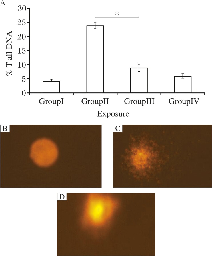
DNA damage was measured as % tail DNA in the control and treated groups. Higher DNA damage was observed in lymphocytes of DMBA and croton oil treated mice than quercetin treated Group III and IV mice (Fig. 2).
Fig. 2. DNA damage in lymphocytes after exposure to quercetin.
A: % tail DNA. B: Group I. C: Group II. D: Group III. Each value represents the mean ±S.E. of 3 experiments. *P < 0.05.
Discussion
Human epidemiological data indicate that regular use of certain medicinal plants suppresses carcinogenesis in various organs[16]. Therefore, it is becoming increasingly important to screen natural products which might suppress or reverse the process of carcinogenesis[17]. The results of the current study and other studies indicate several beneficial effects of quercetin. Skin carcinogenesis is based on the fact that initiation with sequential application of a subthreshold dose of a carcinogen such as DMBA, followed by repetitive treatments with a noncarcinogenic promoter like croton oil will lead to the development of skin tumors. Among the initiation and promotion steps, animal studies show that the promotion stage takes longer period to occur and it is reversible initially[18]. Therefore, cancer prevention by inhibition of tumor promotion is expected to be a resourceful approach. In the present study, quercetin administration could significantly inhibit DMBA induced papilloma formation by the incidence of tumor and the mean number of papillomas.
Lipid peroxidation is a free radical chain reaction and is known to cause 2 main steps of carcinogenesis i.e. initiation and propagation. It is a highly destructive process. During the carcinogenic process, lipid peroxidation is increased and more complex and reactive compounds such as malondialdehyde (MDA) and 4-hydroxynonenal were obtained. These products of lipid peroxidation were observed to be mutagenic and carcinogenic. Therefore, agents that can reduce the production of free radicals in vivo may be considered to have the potential for chemoprevention[19]. In the present study, administration of quercetin significantly reduced the level of LPO in mice exposed to DMBA and croton oil, and subsequently decreased the incidence of skin tumor.
GSH plays a crucial role in protecting cells against reactive oxygen species and free radical produced even in normal metabolism. In the control group, the activity of GSH was decreased while quercetin treatment increased the GSH level, which clearly suggests their antioxidant property. Antioxidants were reported to possess a chemopreventive property[20]. Antioxidants are generally regarded as the first line of defense against free radical stress and are useful in eliminating the risk of oxidative damage induced during carcinogenesis. SOD and catalase are acting as mutually supportive antioxidative enzymes, which provide protective defense against reactive oxygen species (ROS)[21]. There was a significant enhancement in the levels of GSH, SOD, and catalases level in the quercetin treated group compared to the control group.
ROS plays an important role in the process of apoptosis. ROS induces permeabilization of the outer mitochondrial membrane, which releases soluble proteins from the intermembrane space into the cytosol, where they promote caspase activation. ROS typically includes superoxide radical (O2−), H2O2, and the hydroxyl radical (OH•), which cause damage to cellular components, such as DNA, and ultimately lead to apoptotic cell death.
DMBA treatments generate LPO and ROS in the affected area of the skin and ultimately lead to carcinogenesis. This oxidative stress was easily observed in the control group as level of LPO was higher and the levels of catalase, SOD and GSH were lower. The beneficial action of quercetin is probably due to its ability to stimulate antioxidant enzymes in the cells. This increase in enzyme activity effectively reduced the generation of ROS and LPO in the skin and thus might reduce the incidences of skin papillomas on the treated areas.
In conclusion, quercetin isolated from Ocimum sanctum could significantly enhance antioxidant enzyme levels, including GSH, SOD, and catalase, and inhibited lipid peroxides, hence showing its role in detoxification pathway. Both histology and enzyme activities suggest that environmental effects that lead to skin carcinogenesis can be inhibited by oral combination of quercetin in the daily diet to achieve some protection against skin cancer. The results from the present study indicate that quercetin can inhibit papilloma growth.
Acknowledgments
The authors are grateful to Dr. Gajendra Dixit, Dean R & C, MANIT, Bhopal, India (Grant No. Dean R&C/2010/159) and Manvendra Karchuli, Research Officer, PBRI, Bhopal, India to support this research and that provided facilities to achieve the desired goals of the study.
References
- 1.Joseph B, Nair VM. Ethanopharmacological and phytochemical aspects of Ocimum sanctum Linn- the elixir of life. BJPR. 2013;3:273–292. [Google Scholar]
- 2.Singh V, Amdekar S, Verma O. Ocimum sanctum (tulsi): bio-pharmacological activities. Webmed Central Pharmacology. 2010;1:1–7. [Google Scholar]
- 3.Buer CS, Imin N, Djordjevic MA. Flavonoids: new roles for old molecules. JIPB. 2010;52:98–111. doi: 10.1111/j.1744-7909.2010.00905.x. [DOI] [PubMed] [Google Scholar]
- 4.Joshi UJ, Gadge AS, D'Mello P, et al. Anti-inflammatory, antioxidant and anticancer activity of quercetin and its analogues. IJRPBS. 2011;2:1756–1766. [Google Scholar]
- 5.Chaudry PS, Cabera J, Juliani HR, et al. Inhibition of human lens aldose reductase by flavonoids, sulindac, and indomethacin. Biochem Pharmacol. 1983;32:1995–1998. doi: 10.1016/0006-2952(83)90417-3. [DOI] [PubMed] [Google Scholar]
- 6.Chand MM, Patni V. Isolation and Identification of flavonoid “Quercetin” from Citrullus colocynthis (Linn.) Schrad. Asian J. Exp. Sci. 2008;22:137–142. [Google Scholar]
- 7.Shoskes DA, Zeitlin SI, Shahed A, et al. Quercetin in men with category III chronic prostatitis: a preliminary prospective, double blind, placebo-controlled trial. Urology. 1999;54:960–963. doi: 10.1016/s0090-4295(99)00358-1. [DOI] [PubMed] [Google Scholar]
- 8.Chaudhary G, Parmar J, Verma P, et al. Inhibition of DMBA/Croton oil induced tumorigenesis in Swiss albino mice by Aloe vera treatment. IJBMR. 2011;2:671–678. [Google Scholar]
- 9.Das I, Saha T. Effect of garlic on lipid peroxidation and antioxidation enzymes in DMBA-induced skin carcinoma. Nutrition. 2009;25:459–471. doi: 10.1016/j.nut.2008.10.014. [DOI] [PubMed] [Google Scholar]
- 10.Pandey S, Agrawal RC. Effect of Bauhinia Variegate bark extract on DMBA-induced mouse skin arcinogenesis: a preliminary study. GJP. 2009;3:158–162. [Google Scholar]
- 11.Blessy D, Suresh K, Manoharan S, et al. Evaluation of chemopreventive potential of Zingiber Officinale Roscoe ethanolic root extract on 7, 12-dimethyl Benz[a]anthracene induced oral carcinogenesis. Res. J. Agric. & Biol. Sci. 2009;5:775–781. [Google Scholar]
- 12.Das I, Das S, Saha T. Saffron suppresses oxidative stress in DMBA- induced skin carcinoma: a histopathological study. Acta histochem. 2010;112:317–327. doi: 10.1016/j.acthis.2009.02.003. [DOI] [PubMed] [Google Scholar]
- 13.Singh NP, McCoy MT, Tice RR, et al. A simple technique for quantization of low levels of DNA damage in individual cells. Exp Cell Res. 1988;175:184–191. doi: 10.1016/0014-4827(88)90265-0. [DOI] [PubMed] [Google Scholar]
- 14.Ali D, Ray RS, Hans RK. UVA-induced cyototoxicity and DNA damaging potential of Benz (e) acephenanthrylene in human skin cell line. Toxicol Lett. 2010;199:193–200. doi: 10.1016/j.toxlet.2010.08.023. [DOI] [PubMed] [Google Scholar]
- 15.Anderson D, Yu TW, Phillips BJ, et al. The effect of various antioxidants and other modifying agents on oxygen-radical generated DNA damage inhuman lymphocytes in the comet assay. Mutat Res. 1994;307:261–271. doi: 10.1016/0027-5107(94)90300-x. [DOI] [PubMed] [Google Scholar]
- 16.Mouli KC, Vijaya T, Rao SD. Phytoresources as potential therapeutic agents for cancer treatment and prevention. JGPT. 2009;1:4–18. [Google Scholar]
- 17.Roslida AH, Fezah O, Yeong LT. Suppression of DMBA/Croton Oil-induced mouse skin tumor promotion by Ardisia Crispa root hexane extract. APJCP. 2011;12:665–669. [PubMed] [Google Scholar]
- 18.Klaunig JE, Kamendulis LM, Hocevar BA. Oxidative stress and oxidative damage in carcinogenesis. J Toxicol Pathol. 2010;38:96–109. doi: 10.1177/0192623309356453. [DOI] [PubMed] [Google Scholar]
- 19.Ekin S, Ozdemir H, Demir H, et al. Plantago major protective effects on antioxidant status after administration of 7,12-Dimethylbenz(a)anthracene in rats. APJCP. 2011;12:531–535. [PubMed] [Google Scholar]
- 20.Blessy D, Suresh K, Manoharan S, et al. Evaluation of chemopreventive potential of Zingiber Officinale Roscoe ethanolic root extract on 7, 12-dimethyl Benz[a]anthracene induced oral carcinogenesis. Res. J. Agric. & Biol. Sci. 2009;5:775–781. [Google Scholar]
- 21.Parmar J, Sharma P, Verma P, et al. Anti-tumor and anti-oxidative activity of Rosmarinus officinalis in 7, 12 dimethyl Benz(a) anthracene induced skin carcinogenesis in mice. Am. J. Biomed. Sci. 2011;3:199–209. [Google Scholar]


