A 65-year-old man with end-stage renal disease secondary to nephroangiosclerosis on hemodialysis was admitted to undergo a cadaveric kidney transplant. He had a previous history of moderate-to-severe aortic stenosis. Twenty-four hours after surgery, the patient developed an acute pulmonary oedema followed by cardiac arrest. Advanced cardiopulmonary resuscitation and aggressive fluid removal through extracorporeal ultrafiltration were performed, resulting in a successful recovery and no apparent neurological consequences. A transesophageal echocardiography revealed the progression of the aortic stenosis to a critical stage. Balloon aortic valvuloplasty was performed as a bridge therapy to a transcatheter aortic valve replacement.
Over the next 5 days, the patient complained of dyspepsia, nausea, malaise and occasional heartburn. He remained afebrile, and the physical examination only revealed a pansystolic murmur in chest auscultation. Immunosuppression therapy included steroids, mycophenolate mofetil and tacrolimus.
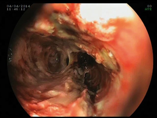
On the sixth post-operative day, the patient had a limited haematemesis (coffee-ground like material) and an upper endoscopy was performed, revealing diffuse, circumferential, black-appearing esophageal mucosa, suggestive of acute esophageal necrosis (AEN) (Figure 1). A computed tomography ruled out perforations or abscess around the esophageal tract.
Fig. 1.
Diffuse ischaemic lesions of the esophageal mucosa. The black, necrotic appearance of the base of the ulcers gives the name to this clinical condition (‘black esophagus’).
A biopsy was not done because it was considered unsafe due to the frailty and devitalized appearance of the esophageal mucosa; however, the microscopic examination of a smear obtained from an esophageal lavage did not reveal hyphae or eosinophils, and fungal cultures resulted negative.
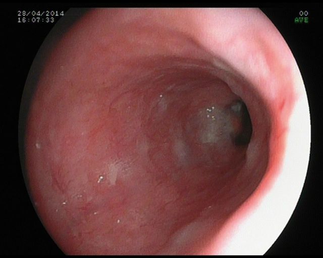
After 4 weeks of fasting, parenteral nutritional, antibiotics (no antifungal), proton-pump inhibitor and an oral suspension of sucralfate, the patient recovered, and a new upper endoscopy confirmed the complete healing of the esophageal lesions (Figure 2).
Fig. 2.
Four weeks later, a new endoscopic exploration revealed a complete healing of the esophageal mucosa, with isolated fibrin deposits.
AEN is a rare clinical condition, which may arise from the combination of several factors, such as the ischaemic damage in hypoperfusion states, direct injury to esophageal mucosa and defects in repair mechanisms and the healing process due to malnourishment and debilitating conditions [1]. AEN has also been reported as manifestation of primary cytomegalovirus infection (CMV) [2], although in the case here described the polymerase chain reaction (PCR) for CMV was negative. The clinical presentations range from asymptomatic to gastrointestinal bleeding. The prognosis of AEN depends upon the severity and extension of the necrotic lesions. Esophageal perforation or infections are infrequent but are accompanied by serious complications associated with high mortality [3].
Conflict of interest statement
None declared.
References
- 1.Gurvits GE. Black esophagus: acute esophageal necrosis syndrome. World J Gastroenterol. 2010;16:3219–3225. doi: 10.3748/wjg.v16.i26.3219. [DOI] [PMC free article] [PubMed] [Google Scholar]
- 2.Trappe R, Pohl H, Forberger A, et al. Acute esophageal necrosis (black esophagus) in the renal transplant recipient: manifestation of primary cytomegalovirus infection. Transpl Infect Dis. 2007;1:42–45. doi: 10.1111/j.1399-3062.2006.00158.x. [DOI] [PubMed] [Google Scholar]
- 3.Gurvits GE. Management of acute esophageal necrosis. J Thorac Cardiovasc Surg. 2011;4:955. doi: 10.1016/j.jtcvs.2011.03.039. [DOI] [PubMed] [Google Scholar]


