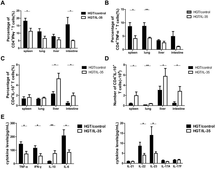
Figure 4. IL-35 reduced the presence of Th1 cells and enhanced IL-10 production.
Lymphocytes isolated from spleen, liver, lung, and small intestine were activated for 4h with PMA, ionomycin and Brefeldin A, and (A) the percentage of IFN-γ-secreting CD4+T cells from spleen, liver, lung, and small intestine were shown. (B) The percent of CD4+TNF-α+ T cells in the spleen, lung, liver and small intestine of recipients were shown. The percent (C) and absolute cell numbers (D) of CD4+T cells secreting IL-10 in the spleen, lung, liver and small intestine are shown. (E) Serum TNF-α, IFN-γ, IL-10, IL-6, IL-21, IL-22, IL-23, IL-17A and IL-17F levels were measured by CBA. Data are presented as the Mean± SEM. All the experiments were performed with eight mice per group. The data shown are the representative of three experiments. *p<0.05, **p<0.01.

