

Highlights
-
•
Digital squamous cell carcinoma commonly mimics chronic granulomatous or fungal infections.
-
•
Chronic non-healing lesions of the digits should be viewed with suspicion.
-
•
It usually has an indolent course but can be locally destructive.
-
•
Excisional biopsy, providing a clear margin, where possible is usually sufficient in the absence of metastatic spread.
Keywords: Squamous cell carcinoma, Chronic infection, Finger
Abstract
Introduction
Squamous cell carcinoma (SCC) of the finger, especially those arising from the nail bed matrix and lateral skin folds are common, especially after the fifth decade of life. A variety of aetiological factors and associations have been described. The appearance can be so ambiguous and appear benign that it can lead to a delay in presentation and diagnosis.
Presentation of the case
We report a case of a 66 year old retired Engineer who presented to our Emergency Department with a 2-year history of a painless swelling of his left ring finger. Examination revealed a diffuse circumferential swelling of the left ring finger involving the middle and distal phalanx. The tip was insensate. The differential diagnoses included pyogenic granuloma, soft tissue sarcoma and chronic granulomatous infection. An excisional biopsy confirmed the diagnosis of a well differentiated squamous cell carcinoma.
Discussion
This case highlights how digital squamous cell carcinoma can appear benign, mimic a wide variety of conditions leading to a delay in diagnosis and treatment. It usually runs an indolent course but can be locally destructive. Excisional biopsy, providing a clear margin where possible is usually sufficient in the absence of metastatic spread.
Conclusion
Chronic, non-healing lesions of the digits should be viewed with suspicion. Digital squamous cell carcinoma commonly mimics a variety of benign conditions and efforts should be made to rule out other possible diagnoses and to institute early treatment.
1. Introduction
Squamous cell carcinoma (SCC) of the finger, especially those arising from the nail bed matrix and lateral skin folds are common, especially after the fifth decade of life.
A variety of aetiological factors and associations have been described.
Clinical presentation can mimic a host of conditions ranging from infections to neoplasms of bone and soft tissue. The appearance can be so ambiguous and appear benign that it can lead to a delay in presentation and diagnosis.
We present a case of a retired Engineer who presented with an unusual lesion in his left ring finger who was referred to us following a delayed presentation to his General Practitioner.
2. Presentation of the case
We present a case of a 66 year old retired Engineer who presented to our Emergency Department with a 2-year history of a painless swelling of his left ring finger.
According to the patient, it all started as a small lesion around the finger tip. He thought nothing of it and expected it to resolve spontaneously. He did not seek medical attention at this time.
He further stated that the lesion got progressively larger over the subsequent 12 months and the finger started becoming deformed and slightly painful. He still did not seek medical help at this stage believing it will resolve. He self-managed the lesion by applying dressings and taking pain killers. He only presented to the ED when the swelling increased in size associated with a foul smelling discharge.
He smoked approximately 15 cigarettes per day and only drank alcohol occasionally.
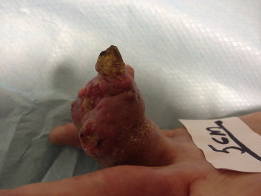
Examination revealed a diffuse circumferential swelling of the left ring finger involving the middle and distal phalanx with a lobulated firm to soft surface. There were multiple sinuses some of which had healed and some discharging foul smelling serous fluid. The tip was insensate.
The metacarpo-phalangeal joint function was normal whereas the proximal interphalangeal joint was held in fixed flexion at approximately 85° with no movement whatsoever. The distal interphalangeal joint was engulfed in the mass. A provisional diagnosis of a fungating neoplasm to rule out a chronic granulomatous infection was made Figs. 1 and 2.
Fig. 1.

Fig. 2.
He had an X-ray of the left hand and left ring finger which revealed a soft tissue swelling with complete destruction of the distal phalanx and partial destruction of the middle phalanx Figs. 3 and 4.
Fig. 3.

Fig. 4.

He was admitted for further investigations. He was commenced on intravenous antibiotics, and he had blood samples sent for routine tests and inflammatory markers. These all returned normal. Wound swabs grew mixed skin flora. No fungus was isolated.
An MRI scan of the finger showed an almost total destruction of the distal phalanx with attenuation of the tendons by the mass Fig. 5.
Fig. 5.

Whole body nuclear magnetic isotope scan showed an increased uptake around the middle phalanx of the left ring finger. The rest of the isotope scan did not reveal any pathological uptake.
A guillotine amputation was undertaken across the base of the middle phalanx of the ring finger and the whole sample sent for histology. Histological examination revealed a well differentiated squamous cell carcinoma with a tumour-free margin. The stump was left to heal by secondary intention with a view to flap closure in due course.
3. Discussion
Squamous cell carcinoma (SCC) of the finger, especially those arising from the nail bed matrix and lateral skin folds are more common after the fifth decade of life.
Clinical presentation can mimic a host of lesions ranging from infections to neoplasms of bone and soft tissue. The presentation can be so ambiguous that it can lead to a delay in presentation and diagnosis.
The course of the disease can be indolent and diagnosis may be not be made for up to 14 years [1]. Our patient only sought medical attention 2 years after the initial appearance of the lesion.
Various aetiological factors have been described including trauma, chronic infection (including viral infection) and radiation. Eliezri reported that they found the Human Papilloma Virus (HPV) 16 DNA which is restricted to neoplasms of the genital tract and finger using either the filter or in-situ hybridization technique [2].
Ashinoff et al. reported the use of polymerase chain reaction (PCR) to isolate the HPV type 16 in some of these tumours. In-situ hybridisation technique failed to identify HPV 16 in any of their patients [3].
In another study, HPV DNA was present in 80% of cases of subungual squamous cell carcinoma using blot dot analysis of frozen tissue [4].
According to Guitart et al., it is highly likely that up to 84% of subungual squamous cell carcimona occur in the finger; the rest in the toes. The majority of the subungual lesions occur in the thumb (44%) and those in the toes occur predominantly in the great toe (64%) [5].
The correct diagnosis is made following histological analysis after a biopsy of the lesion. In our patient, this involved an excisional biopsy.
It is imperative that efforts are made to rule out other possible diagnoses such as chronic granulomatous lesions, extra pulmonary tuberculosis and fungal infections, warts, chronic paronychia, whitlow and osteomyelitis.
Plain X-rays will determine if there is bony involvement. MRI scan and isotope bone scan will help to determine the extent of the disease. The diagnosis is usually more obvious in late presentations.
The conventional treatment option has been amputation; ensuring a safe resection margin. In early cases, Mohs micrographic surgery has been reported to achieve favourable outcomes in up to 96% of cases [6]. Intra-arterial infusion of methotrexate has been reported to achieve a successful outcome in squamous cell carcinoma of the great toe [7].
In a small series recently reported by Rosen et al., radiation therapy was found to be successful and could be considered as an alternative to amputation [8].
Following treatment, HPV-associated digital squamous cell carcinoma is more likely to recur after surgical treatment. The rate of recurrence exceeds that for cutaneous squamous cell carcinoma in general [9].
4. Conclusion
Chronic, non-healing lesions of the digits should be viewed with suspicion.
Digital squamous cell carcinoma commonly mimics a variety of benign conditions and efforts should be made to rule out other possible diagnoses and to institute early treatment. It can be locally destructive. Excisional biopsy with a clear margin is usually sufficient in the absence of metastatic spread.
Conflict of interest
We; Mr V Ameh and Dr A Afridi hereby declare that we have no conflict of interest to declare in the preparation of this manuscript.
Funding
There has been no source of funding for this article.
Consent
We, the Authors, hereby confirm that Written informed consent was obtained from the patient for publication of this case report and accompanying images. A copy of the written consent is available for review by the Editor-in-Chief of this journal on request.
Author contribution
The author contribution to the preparation of this manuscript are as follows; Mr Victor Ameh: case identification, obtaining consent from patient, literature search, preparation, editing and proof reading of script.
Dr Abdul Afridi: literature search, preparation and proof reading of script.
Acknowledgement
We will like to sincerely thank Mr. Raja Swaminathan for allowing us to use some of his clinical material in the preparation of this manuscript.
Contributor Information
Victor Ameh, Email: amehyaks@gmail.com.
Abdul Afridi, Email: apreedee@hotmail.com.
References
- 1.Carrol R.E. Squamous cell carcinoma of the nail. J. Hand Surg. 1976;1:92–97. doi: 10.1016/s0363-5023(76)80002-0. [DOI] [PubMed] [Google Scholar]
- 2.Eliezri Y.D. Occurrence of the HPV type 16 DNA in cutaneous and basal cell neoplasm. J. Am. Acad. Dermatol. 1990;23(5):836–842. doi: 10.1016/0190-9622(90)70299-w. [DOI] [PubMed] [Google Scholar]
- 3.Ashinoff R., Li J.J., Jacobson M., Friedmann-Klein A.E., Geronimus R.G. Detection of HPV DNV in squamous cell carcinoma of the nail bed and finger determined by polymerase chain reaction. Arch. Dermatol. 1991;127(12) [PubMed] [Google Scholar]
- 4.Attiyeh F.F., Shah I., Booker R.J., Knapper W.H. Subungual squamous cell carcinoma. J. Am. Med. Assoc. 1979;241:262–263. [PubMed] [Google Scholar]
- 5.Giutart J., Bergfeld W.F., Tuthill R.J., Tubbs R.R., Zienowicz R., Fleegler E.J. Subungual carcinoma of the nail bed: a clinico-pathological review of 12 cases. Br. J. Dermatol. 1990;123:215–222. doi: 10.1111/j.1365-2133.1990.tb01849.x. [DOI] [PubMed] [Google Scholar]
- 6.Zaiac M.N., Weiss E. Mohs micrographic surgery of the nail unit and squamous cell carcinoma. Dermatol. Surg. 2001;27:246–251. [PubMed] [Google Scholar]
- 7.Sheen Y.S., Sheen M.C., Sheu H.M. Squamous cell carcinoma of the big toe successfully treated by intra-arterial infusion of methotrexate. Dermatol. Surg. 2003;29:982–983. doi: 10.1046/j.1524-4725.2003.29267.x. [DOI] [PubMed] [Google Scholar]
- 8.Rosen L.R., Powell K., Katz S.R., Wu H.T., Durci M. Subungual squamous cell carcinoma: radiation therapy as an alternative to amputation and review of the literature. Am. J. Clin. Dermatol. 2010;11(4):285–288. doi: 10.2165/11311080-000000000-00000. [DOI] [PubMed] [Google Scholar]
- 9.Alam M., Caldwell J.B., Eliezri J. HPV-associated digital squamous cell carcinoma. Literature review and report of 21 cases. Am. J. Dermatol. 2003;48(March (3)):385–393. doi: 10.1067/mjd.2003.184. [DOI] [PubMed] [Google Scholar]