
Fig. 4.
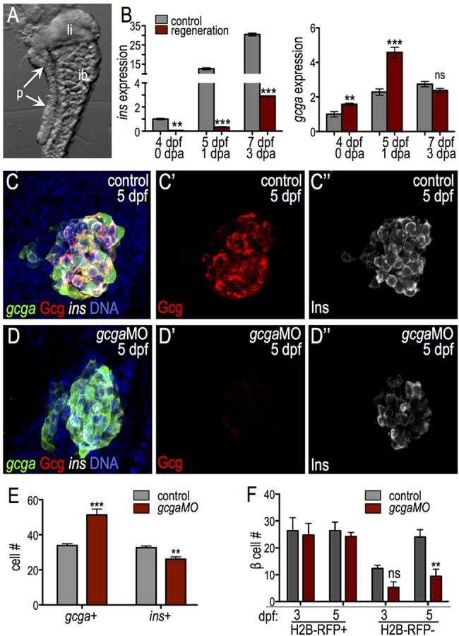
glucagon is required for islet development. (A) Endodermal organs isolated from 4-dpf larvae. p, pancreas; li, liver; ib, intestinal bulb. (B) qPCR of insulin (ins) and glucagon (gcga) in control and ablated larvae. ins expression was diminished and gcga expression transiently increased during regeneration (n=3 independent experiments). (C-E) gcgaMO blocks glucagon protein expression and increases the α to β cell ratio. (C-D″) Confocal projections of 5-dpf control MO-injected (C) and gcgaMO-injected (D) Tg(gcga:GFP); Tg(ins:dsRed) larvae stained for glucagon (red). (E) Quantification of gcga:GFP+ α cells and ins+ β cells in control MO-injected (n=15) and gcgaMO-injected (n=10) islets. (F) Quantification of H2B-RFP+ and H2B-RFP− β cells at 3 and 5 dpf in control MO-injected (n≥3) and gcgaMO-injected (n=5) islets. Ventral bud-derived H2B-RFP− β cells are specifically diminished in gcga morphants. *P≤0.05, **P≤0.01, ***P≤0.001; ns, not significant (two-way ANOVA followed by Bonferroni post-test).