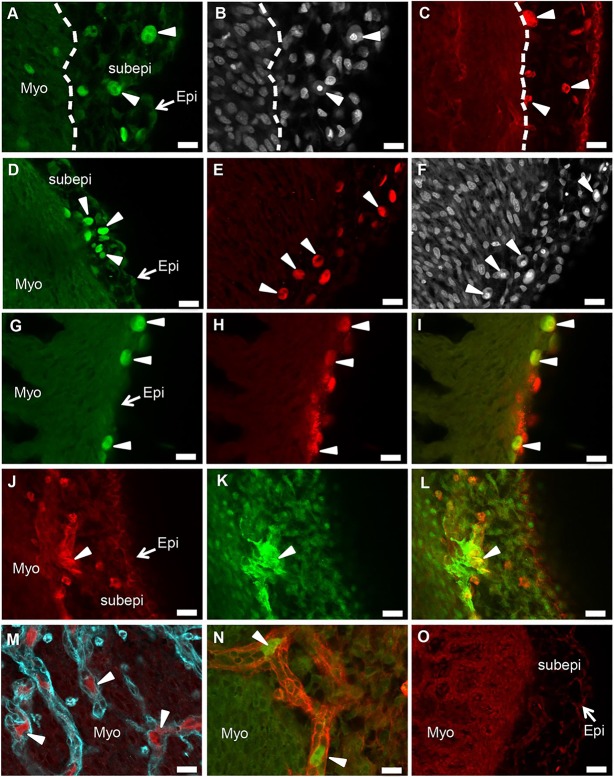
Fig. 4.
HESC-derived epicardial cells survive and differentiate in vivo. (A-C) Fluorescent (GFP+ and mStrawb+) human epicardial cells detected in the subepicardial region at the base of the chicken heart (arrowheads). (B) Human cells in A are identifiable by bright and distinct Hoechst 33342 staining. (D-F) Fluorescent human epicardial cells detected in the subepicardial region in the middle part of the heart (arrowheads). (F) Human cells in E are identifiable by bright and distinct Hoechst 33342 staining. (G,H) Epicardial cells (GFP+) that localised at the apex of the heart (G) expressed WT1 (H; indicated by arrowheads). (I) Co-expression of WT1+ and GFP+ human cells. (J,K) A few engrafted mStrawb+ epicardial cells (J) expressed ACTA2 (K), suggesting differentiation to SMCs in vivo. (L) Cells co-expressing mStrawb and ACTA2 (indicated by arrowheads). (M,N) GFP+ and mStrawb+ epicardial cells detected within lectin-stained (in cyan and red) coronary vessels. (O) Subepicardial region in a chicken embryo heart injected with mStrawb+ human neural crest cells. Scale bars: 100 μm. Myo, myocardium; Epi, epicardium; Subepi, subepicardium.

