Fig. 6.
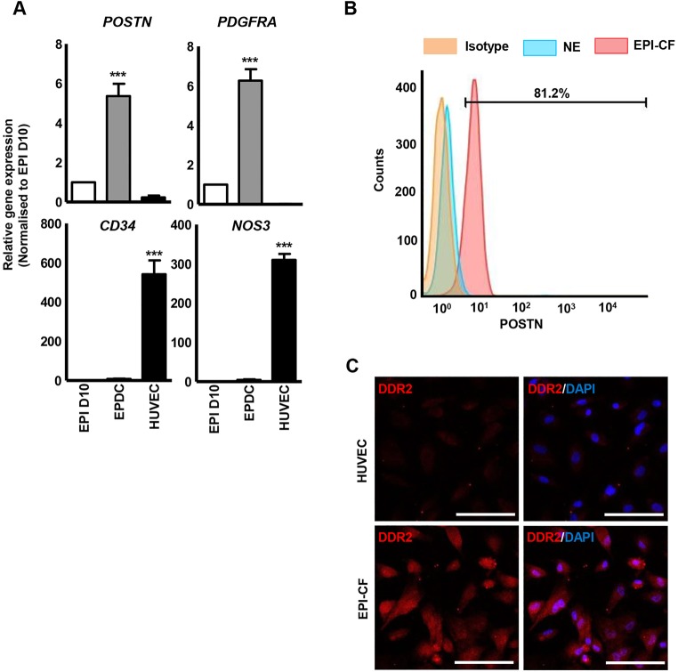
Epicardium-derived cardiac fibroblast differentiation. (A) Analysis of fibroblast (PDGFRA and POSTN) and endothelial (NOS3 and PECAM1) markers in epicardium-derived cells (EPDCs) obtained with VEGF and FGF treatment. ***P<0.001. (B) Percentage of POSTN+ cells in epicardium-derived cardiac fibroblasts (EPI-CFs) determined by flow cytometry. Rabbit IgG isotype and HUVECs were used as negative controls. (C) The majority of EPI-CFs immunostained positive for DDR2, whereas HUVECs displayed negligible expression. Scale bars: 100 μm.

