Abstract
Angong Niuhuang pill, a Chinese materia medica preparation, can improve neurological functions after acute ischemic stroke. Because of its inconvenient application and toxic components (Cinnabaris and Realgar), we used transdermal enhancers to deliver Angong Niuhuang pill by modern technology, which expanded the safe dose range and clinical indications. In this study, Angong Niuhuang stickers administered at different point application doses (1.35, 2.7, and 5.4 g/kg) were administered to the Dazhui (DU14), Qihai (RN6) and Mingmen (DU4) of rats with chronic cerebral ischemia, for 4 weeks. The Morris water maze was used to determine the learning and memory ability of rats. Hematoxylin-eosin staining and Nissl staining were used to observe neuronal damage of the cortex and hippocampal CA1 region in rats with chronic cerebral ischemia. The middle- and high-dose point application of Angong Niuhuang stickers attenuated neuronal damage in the cortex and hippocampal CA1 region, and improved the memory of rats with chronic cerebral ischemia with an efficacy similar to interventions by electroacupuncture at Dazhui (DU14), Qihai (RN6) and Mingmen (DU4). Our experimental findings indicate that point application with Angong Niuhuang stickers can improve cognitive function after chronic cerebral ischemia in rats and is neuroprotective with an equivalent efficacy to acupuncture.
Keywords: nerve regeneration, cerebral ischemia, point application, Angong Niuhuang sticker, brain injury, neurological functions, acupuncture, traditional Chinese medicine, NSFC grants, neural regeneration
Introduction
At present, point application as a route of administration is a complex therapeutic method combining points and meridians with drugs (Zhang and Shi, 2013). Point application to treat ischemia is a newly developed Cantonese style of acupuncture therapy by Jin. Jin's Three-Needle Therapy was clinically verified and showed advantages for the repeated treatment of stroke (Yuan, 2006; Zhang et al., 2012a). Can acupoint application therapy protect against ischemic brain damage? A recent clinical trial demonstrated that point application therapy significantly improved neurologic impairment and normal daily living and thinking in stroke patients, as well as reducing the recurrence rate of stroke (Zhang, 2008).
The Angong Niuhuang pill is a traditional Chinese medicine drug for the treatment of “febrile disease”, which originated from the “Wenbing Tiaobian” written by the physician Jutong Wu in the Qing Dynasty, and has been widely used for ischemic stroke in the clinic in China. The main components of Angong Niuhuang pills include Calculus Bovis, Radix Curcumae, rhinoceros horn, Moschus, Rhizoma Coptidis, Radix Scutellariae, Fructus Gardeniae, Cinnabaris, Margarita, Broneolum Syntheticum and realgar. This prescription has detoxification, resuscitation and anticonvulsant properties. A previous study showed that Angong Niuhuang pill improved neurological functional defects of patients with acute ischemic apoplexy (Cui, 2012). However, its formulation is inconvenient for administration to patients, as the effective components do not dissolve easily. Furthermore, some drug ingredients are toxic and the pill dosage is difficult to control, which is of concern for elderly patients who require lower doses (Yan et al., 1999). Thus, use of an Angong Niuhuang pill is used only for critical patients in the acute stage of heavy phlegm heat, and it is generally discouraged for long-term use. We extracted and purified components from Angong Niuhuang pill and removed cinnabar and realgar, toxic ingredients (Dai et al., 2009). Then we applied transdermal enhancers to Angong Niuhuang stickers using a cataplasm transdermal drug delivery system and Laurocapram, Mentholum, Broneolum Syntheticum and Moschus as transdermal enhancers. Angong Niuhuang sticker is a new preparation for transdermal absorption of traditional Chinese medicine and consists of curcuma, berberine hydrochloride, baicalin, geniposide, borneol, and musk. These components can induce resuscitation, activate blood circulation to dissipate blood stasis and cool blood and promote the circulation of Qi to relieve pain. Evidence from a pharmacological study in rats indicated the above herbs refreshed and restored consciousness, protected against anti-free radical damage, and was antipyretic, analgesic, antimicrobial, and anti-inflammatory (Zhao et al., 2002). Angong Niuhuang stickers might be more suitable for clinical treatment of cerebral ischemia as they allow an expanded safe dose range.
Dazhui (DU14), Qihai (RN6) and Mingmen (DU4) are important points in the Ren and Du meridians that coordinate the Yin and Yang, nourish the brains, and promote the circulation of Qi and blood, as well as recovery of function. Recent studies confirmed these points improved immunity and promoted recovery of neurological functions in mice (Hua and Ye, 2011; Kim et al., 2013). We previously showed that point application on Dazhui, Qihai, Mingmen significantly improved neurologic impairment scores in patients of ischemic stroke, significantly raised the activity of superoxide dismutase in the plasma of patients, and reduced the malonaldehyde content (Zhang et al., 2008). Furthermore, no negative reactions or side effects were observed after treatment. Previous studies demonstrated that Angong Niuhuang sticker significantly prolonged the breathing time and survival time after ligation of the bilateral common carotid artery in an acute cerebral ischemia mouse model. It also reduced neurobehavioral scores of stroke mice, prevented death of mice with pulmonary thrombosis and prolonged hypoxia tolerance time in mice, with a similar effect to the Angong Niuhuang pill (Zhang et al., 2008, 2012b).
This study investigated the protective effect of acupoint application with Angong Niuhuang sticker and elucidated its protective mechanism against damage of chronic cerebral ischemia, by measuring neurobehavioral scores and neural damage in brain tissues from rats with chronic cerebral ischemia.
Materials and Methods
Animals
Fifty male 8-month-old specific-pathogen-free Wistar rats, weighing 250 ± 20 g, were provided by the Experimental Animal Center of the Southern Medical University, China (certification No. SCXK (Yue) 2011-0015). The experiments were conducted after routine feeding for 7 days at 25°C and under a 12-hour light cycle. The study conformed to the Guide for the Care and Use of Laboratory Animals published by the US National Institutes of Health (NIH publication No. 85-23, revised 1996), and the experimental procedures were consistent with the ethical requirements established by Experimental Animal Ethics and Welfare Committee of Southern Medical University in China.
Wistar rats were fed for 1 week, then were trained in a Morris water maze (Feng et al., 2011) for 4 days. Rats that could not swim were excluded and the remaining rats were randomly divided into normal (n = 8) and operation groups (n = 42). Two rats in the operation group died. Surviving rats were randomized into five groups (n = 8 in each group) as follows: model group, acupuncture group, low-, medium- and high-dose point application groups.
Establishment of chronic cerebral ischemia model
The chronic cerebral ischemia model was prepared using the method by Sarti et al. (2002). In brief, rats were intraperitoneally anesthetized with 10% chloral hydrate (0.35 mL/kg) after fasting for 12 hours and water deprivation for 4 hours, then cervical unilateral incision and blunt dissection of the common carotid artery was preformed and the proximal part and distal end of the artery were ligated with No. 0 lines. Sulfa powder was used for disinfection of the operation area and the wound was stitched layer by layer. After 1 week, a common carotid artery ligation on the contralateral side was made using the above methods. After surgery, rats were housed singly in cages and were fed.
Treatment of point application
We previously improved the preparation of Angong Niuhuang stickers by substituting cremor with a cataplasm transdermal drug delivery system to better satisfy clinical requirements (Zhang et al., 2008). Curcuma, coptis, scutellaria and gardenia (provided and identified by the Dispensary of TCM in Nanfang Hospital, China) were heated and extracted under reflux by ethanol. The ethanol was recycled through decompression and the extracts were dried after filtration. Gelatin, sodium carboxymethylcellulose, polyvinylpyrrolidone and sorbitol were immersed in a certain volume of water (method is patent pending), and were then heated in a water bath at 60°C. Then we evenly mixed sodium polyacrylate, kaolin, propylene and glycol together, and added these components into the swelling matrix solution. Finally, a polyethylene glycol solution was added, and the matrix solution was mixed for several minutes. We mixed the matrix with azone, menthol, borneol, musk (Dispensary of TCM in Nanfang Hospital) and the drug extractions. These components were stirred in the matrix to obtain a gelatinous drug-containing matrix. The drug-containing matrices were smeared evenly on non-woven fabrics and dried in a well-ventilated place. After being covered with a protective membrane, they were divided into stickers (1 cm × 1 cm).
At 1 hour after induction of disease, rats received either low-dose (1.35 g/kg), medium-dose (2.7 g/kg) or high-dose (5.4 g/kg) Angong Niuhuang stickers (Sarti et al., 2002). Dazhui (DU14, at the central line of the back, the spinous process between the seventh cervical vertebra and the first thoracic vertebra), Mingmen (DU4, at the central line of the back, the spinous process between the second and third lumbar vertebra), and Qihai (RN6, at RN8, located at the intersection of upper 2/3 and below 1/3 on line of xiphoid and pubic symphysis, and RN6 is about 12 mm below RN8) points of rats, as referenced in the experimental animal points in Experimental Acupuncture Science (Li, 2010), were depilated with 8% sodium sulfide in a 1 cm × 1 cm area. For each of the three doses, each point was smeared evenly with the gelatinous drug-containing matrix by gentle massage for 5 minutes. The point then was covered with rubberized fabric, uncovered after 6 hours (Kong, 2008) and cleaned with clear water once a day. After continuous therapy for 4 weeks, rats were euthanized. Rats in the acupuncture group received straight acupuncture of 3–5 mm by needles 25 mm long with a 0.18 mm diameter carrying electricity (frequency = 10 Hz of continuous wave) and an intensity where the needle handle quivered for 15 minutes each time (acupuncture treatment times were the same for all groups, once per day, for 4 consecutive weeks) (Li, 2010) (Qingdao Xinsheng G6805–type I electro-acupuncture apparatus from Qingdao Xinsheng Co., Ltd.; Hua Tuo acupuncture needle from Suzhou Medical Appliance Factory). The points for the acupuncture group were the same as the point application group. In the model group and control group, rats only received needle acupuncture but no treatment.
Morris water maze test
The memory function of rats was measured using the Morris water maze test (Feng et al., 2011; Feng and Han, 2013). The Morris water maze (self-made) is a circular stainless steel pool with a diameter of 130 cm and height of 50 cm. The pool is divided into four quadrants by wall markings at four entry points. A platform with a diameter of 11 cm and height of 29 cm was placed in the median of any one of the quadrants. All rats received navigation training in the Morris water maze for four days, twice in the morning and afternoon, before being divided into groups. The rats were put into the water from any of the four entry points facing the pool wall, then the time to find and climb onto the platform (maximum time = 3 minutes) was recorded as the escape latency. If rats did not find the platform within 3 minutes, they were pulled to the platform by hand, left for 10 seconds, then put it back into the cage and excluded from the analysis. A latent period of less than 180 seconds was taken as the standard of learning to swim maze. Rats that were confirmed to have no significant differences in their intelligence (by assessment of escape latency) were randomized into groups and prepared for surgery. The navigation test was performed again 4 weeks after the operation for 4 days, twice a day. Escape latency was measured and a mean score of the time taken for rats to find and climb the platform within 3 minutes (two tests per day for 4 days) was calculated. A long escape latency indicated a worse learning and memory ability of rats.
Pathological analysis by hematoxylin-eosin staining and Nissl staining
All rats were decapitated immediately after the Morris water maze test. Eight rats per group were selected and the brains were removed and dehydrated, then cut into coronal slices showing the hippocampus and frontal cortex (Longa et al., 1989). Coronal slices then were embedded in paraffin and deparaffinized following routine methods for hematoxylin-eosin staining and Nissl staining.
Hematoxylin-eosin staining: Slices were incubated with xylene for 3 × 5 minutes, 100% ethanol for 2 × 5 minutes, 95% ethanol, 90% ethanol, 80% ethanol each for 3 minutes, stained with hematoxylin for 8 minutes, and rinsed with water for 10 minutes. Then, 1% hydrochloric acid alcohol was added for 1 second to visualize blue, followed by washing for 10 minutes. Then, brain tissue was stained with eosin for 3 minutes, dehydrated with 80%, 90%, 95% ethanol each for 1 minute, and with 100% ethanol for 3 × 1 minute, and cleared with xylene for 2 × 5 minutes. Slices were mounted with neutral gum.
Nissl staining (only detected in hippocampal CA1 region): Brain slices were incubated at 60°C for 30 minutes, then cooled, cleared with xylene for 2 × 5 minutes, dehydrated with 100%, 95%, 90%, 85% ethanol each for 5 minutes, with 80%, and 70% ethanol each for 3 minutes. Then slices were rinsed with distilled water for 1 minute, stained with cresyl violet dye in an oven at 37°C for 30 minutes, rinsed with water for 8 minutes, and rapidly separated with 95% ethanol. Then sections were incubated in anhydrous alcohol and xylene for 2 × 5 minutes. Slices were mounted with neutral gum.
The slices were observed and photographed with a Nikon ECLIPSE Ti-S microscope (Nikon, Tokyo, Japan).
Statistical analysis
The data are presented as the mean ± SD. All statistical analyses were performed with SPSS 17.0 software (SPSS, Chicago, IL, USA). Significant differences were determined by one-way analysis of variance and Student-Newman-Keuls test. P values of < 0.05 were considered significant.
Results
Point application with medium- and high-dose Angong Niuhuang sticker improved the memory of chronic cerebral ischemia model rats
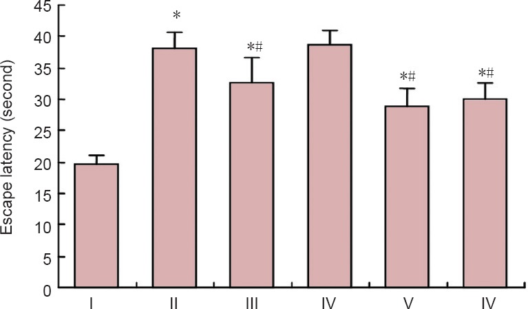
At 4 weeks after chronic cerebral ischemia, the learning and memory abilities of rats were tested by Morris water maze. The results showed that, compared with the model group, the escape latency was significantly shortened in the acupuncture group, medium-, and high-dose Angong Niuhuang sticker point application groups (P < 0.01). There was no statistical difference between the medium-, high-dose groups and acupuncture group (P > 0.05; Figure 1).
Figure 1.

Effects of Angong Niuhuang sticker point application on escape latency of chronic cerebral ischemia model rats in Morris water maze.
A long escape latency indicated worse learning and memory ability. The data are represented as the mean ± SD, and there were eight rats in each group. *P < 0.01, vs. normal group, #P < 0.01, vs. model group (one-way analysis of variance followed Student-Newman-Keuls test). I: Normal group; II: model group; III: acupuncture group; IV: low-dose Angong Niuhuang sticker point application group; V: medium-dose Angong Niuhuang sticker point application group; VI: high-dose Angong Niuhuang-point application group.
Point application with medium- and high-dose Angong Niuhuang sticker improved pathological changes in the hippocampal CA1 region and cortex of chronic cerebral ischemia model rats
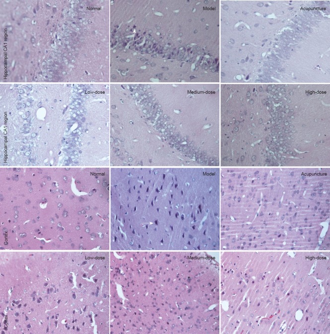
At 4 weeks after modeling, hematoxylin-eosin staining showed no obvious pathological changes in rats of the normal group: the arrangement of pyramidal cells was close and orderly, the morphology of cells was normal, the framework showed integrity with light staining, and the nucleolus was clear in the hippocampus and cortex. In the model group, the arrangement of pyramidal cells in the hippocampal CA1 region and cortex of rats was disordered and sparse, there was nerve cells loss, and edema and deformation were visible with nuclear pyknosis, deep staining and unclear nucleolus. Compared with the model group, the arrangement of pyramidal cells was disordered, but the number was not decreased, an edema, loss and deformation of the nerve cells were alleviated in the hippocampal CA1 region and cortex of rats by varying degrees in point application groups of each-dose and acupuncture group. In addition, there were many nerve cells with complete morphology and a minority had clearly visible nucleolus. This was improved in the medium- and high-dose point application groups as well as the acupuncture group, to a similar degree (Figure 2).
Figure 2.
Effects of point application with Angong Niuhuang sticker on pathological changes in the hippocampal CA1 region and cortex of chronic cerebral ischemia model rats at 4 weeks after modeling (hematoxylin-eosin staining, light microscope, × 400).
In the medium-, high-dose Angong Niuhuang sticker point application groups and acupuncture group, damage to pyramidal cells in the hippocampal CA1 area and cortex of rats was attenuated. Arrows indicate neural cells.
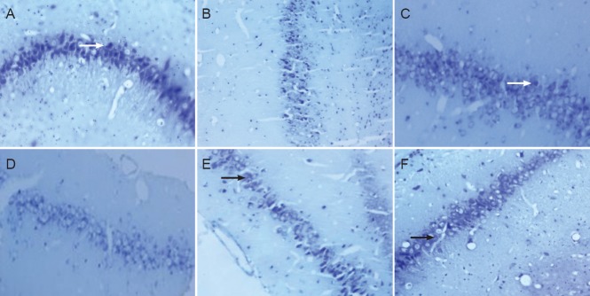
Nissl staining showed as purple blue granular Nissl bodies in neurons in the cytoplasm of hippocampal CA1 areas in rats of the normal group. In the model group, the number of neural cells was significantly reduced in ischemic areas, and the Nissl body color was light in neuron cytoplasm with varying degrees of loss. Compared with the model group, the loss of neural cells was decreased with increased Nissl bodies in neuron cytoplasm in the point application group of each-dose and acupuncture group (Figure 3).
Figure 3.

Effects of point application with Angong Niuhuang sticker on neuronal changes in the hippocampal CA1 region and cortex of chronic cerebral ischemia model rats at 4 weeks after modeling (Nissl staining, light microscope, × 400).
(A) Normal group; (B) model group; (C) acupuncture group; (D–F) low-, medium-, high-dose Angong Niuhuang sticker point application groups. In the medium-, high-dose Angong Niuhuang sticker point application groups and acupuncture group, neuronal injury in the hippocampal CA1 area of rats was attenuated, and Nissl bodies were restored. Arrows indicate Nissl bodies.
Discussion
At present, point application is being researched internationally as a route of administration. Point application is a complex therapy method combining points and meridians with drugs. Point application avoids not only the “peak-valley phenomenon” found with oral administration or injected medications, but also eliminates the first-pass effect in the gastrointestinal tract which reduces side effects. In addition, point application is convenient, easily accepted by patients, and is suitable for long-term administration, especially for the elderly and those who cannot receive oral medication or are afraid of acupuncture (Zhang and Shi, 2013). We extracted and purified components typically found in the prescribed Angong Niuhuang pill. Then we applied transdermal enhancers to Angong Niuhuang stickers. Long-term clinical observations showed that point application with Angong Niuhuang stickers on Dazhui (DU14), Qihai (RN6) and Mingmen (DU4) improved neurological function deficit syndrome of patients suffering from ischemic stroke. There were no adverse reactions or side effects during treatment (Zhang et al., 2008). This study aimed to investigate the protective effect and mechanism against damage of chronic cerebral ischemia in rats with point application.
There have been few research studies regarding the effector mechanisms of acupoint application for the treatment of cerebral ischemia; however, it was confirmed that acupoint application reduced brain edema of ischemic brain injury, improved the activity of plasmin, anti-coagulation, anti-free radical damage and played a role in brain protection in rats (Yan et al., 2004, 2005). Previously, it was shown that acupoint application with Angong Niuhuang stickers can significantly prolong the gasping time after decapitation, survival time after bilateral carotid artery ligation in an acute cerebral ischemia mouse model, reduce stroke index scores, prevent death by pulmonary thrombosis, and prolong the hypoxia tolerance time in mice with similar efficacy to the Angong Niuhuang pill (Chen et al., 2014). Based on previous research, we used a chronic cerebral ischemia model in rats to study the protective effect of acupoint application on neuronal injury in rats with chronic cerebral ischemia.
Chronic cerebral ischemia is the emergence of chronic ischemic damage to the nervous system with long-term low physiological threshold of blood supply to the brain, and it is the main pathological basis of acute and chronic ischemic cerebrovascular disease (Farkas and Luiten, 2001; Wang and Cao, 2013). Permanent ligation of bilateral common carotid artery causes hypoxic-ischemic damage in the brain tissue of rats, especially in vulnerable areas such as the hippocampus and cerebral cortex. A study showed that insufficiency of chronic cerebral blood flow perfusion induced by permanent bilateral common carotid artery ligation was characterized by a reduction of brain blood flow and nerve pathological changes (Zhang et al., 2009). This might explain why hippocampal neuron injury is induced by hypoperfusion in the brain after chronic cerebral ischemia. Another study showed that 3 weeks after ligation, a period of chronic ischemia occurs. Yang et al. (2007) found that chronic hypoxia reduced the spatial memory ability of experimental rats, and had a destructive effect on the morphology and structure of neurons in the hippocampal CA1 area. Thus, the hippocampus is the most sensitive area to cerebral ischemia and an important part of learning and memory with a close relationship with cognitive function. Therefore, this experiment selected the CA1 region of the hippocampus and cortex for observation during treatment.
This study used ligation of the bilateral common carotid artery to induce chronic cerebral ischemia and the Morris water maze to test the ability of learning and memory in rats. The results showed that chronic cerebral ischemia induced significant learning and memory disorders in rats. We used hematoxylin-eosin staining and Nissl staining to observe pathological changes after chronic cerebral ischemia. The normal morphology of cells was altered with a disordered arrangement, loose connections, loss of neurons, and a light color of Nissl bodies indicating varying degrees of loss in the cytoplasm of neurons after chronic cerebral ischemia in the hippocampal CA1 area and cortex. The results of this study showed that point application was effective in improving learning and memory ability after chronic cerebral ischemia in rats, as well as reducing the damage of neurons in the hippocampal CA1 region and cortex by chronic cerebral ischemia. In summary, point application with Angong Niuhuang stickers can improve cognitive function after chronic cerebral ischemia in rats and has a protective effect on neuronal damage. Ischemic stroke has a high incidence, mortality and disability. Acupoint application has shown satisfactory effects on cerebral ischemia in the clinic, and it has the advantages of simple operation, acceptance by patients, long-term use, and non-toxic side effects. In addition, using the transdermal drug delivery method instead of the traditional acupuncture or injection method, will save medical resources, as medical personnel do not require professional training. In addition, there will be reduced environmental pollution and waste caused by discarding disposable acupuncture or injection products, and avoidance of infection caused by needle sharing. Thus, acupoint application is expected to become an effective and economic method for ischemic stroke.
Footnotes
Funding: This study was supported by the National Natural Science Foundation of China, No. 81403458 and a grant from Cultivation Project Foundation for Youth Technological Talents of Southern Medical University, No. B1012015.
Conflicts of interest: None declared.
Copyedited by Croxfor L, Norman C, Yu J, Yang Y, Li CH, Song LP, Zhao M
References
- Bains JS, Shaw CA. Neurodegenerative disorders in humans: the role of glutathione in oxidative stress-mediated neuronal death. Brain Res Rev. 1997;25:335–358. doi: 10.1016/s0165-0173(97)00045-3. [DOI] [PubMed] [Google Scholar]
- Chen J, Zhang DS, Liu YL. The protective effect on neuronal damage after cerebral ischemia in rats with point application. Zhongyiyao Daobao. 2014;3:18–22. [Google Scholar]
- Cui AY. Recent pharmacologic actions and clinical applications of Angong Niuhuang wan. Zhongguo Shiyan Fangji Xue Zazhi. 2012;18:341–343. [Google Scholar]
- Dai FW, Li M, Lei GY, Lei ZB. Analysis of 16 cases of side-effect on Xingnaojing Injection. Haixia Yaoxue. 2009;21:191–193. [Google Scholar]
- Farkas E, Luiten PGM. Cerebral microvascular pathology in aging and Alzheimer's disease. Prog Neurobiol. 2001;64:575–611. doi: 10.1016/s0301-0082(00)00068-x. [DOI] [PubMed] [Google Scholar]
- Feng M, Lu SF, Zhang CS, Yu SG. The analysis and status on domestic experiments of Morris water maze test in rats. Liaoning Zhongyi Zazhi. 2011;38:2170–2172. [Google Scholar]
- Feng SJ, Han JG. Treatment of traumatic brain injury in rats by RhoA gene silencing combinedwith umbilical cord mesenchymal stem cell transplantation. Zhongguo Zuzhi Gongcheng Yanjiu. 2013;17:23–30. [Google Scholar]
- Hua CL, Ye JX. The clinical condition of Xingnaojing injection. Zhongguo Zhonyi Jizheng. 2011;20:626–627. [Google Scholar]
- Kim JH, Choi KH, Jang YJ, Kim HN, Bae SS, Choi BT, Shin HK. Electroacupuncture preconditioning reduces cerebral ischemic injury via BDNF and SDF-1α in mice. BMC Complement Altern Med. 2013;13:22. doi: 10.1186/1472-6882-13-22. [DOI] [PMC free article] [PubMed] [Google Scholar]
- Kong FS. Guangzhou: Guangzhou University of Traditional Chinese Medicine; 2008. Study on the effect and mechanism of acupoint external application on airway inflammation in passive smoking Rat. [Google Scholar]
- Li Z. Beijing: China Medical Science Press; 2010. Experimental Acupuncture Science. [Google Scholar]
- Longa EZ, Weinstein PR, Carlson S, Cummins R. Reversible middle cerebral artery occlusion without craniectomy in rats. Stroke. 1989;20:84–91. doi: 10.1161/01.str.20.1.84. [DOI] [PubMed] [Google Scholar]
- Sarti C, Pantoni L, Bartolini L, Inzitari D. Cognitive impairment and chronic cerebral hypoperfusion: what can be learned from experimental models. J Neurol Sci. 2002:203–204. doi: 10.1016/s0022-510x(02)00302-7. 263-266. [DOI] [PubMed] [Google Scholar]
- Sun L, Zhang Y, Zou XY, Jin T, Wu J, Yao LF. Constract study of cortex and subcortical white matter neuropathology changes of vascular dementia rats after chronic cerebral ischemia. Zhongfeng yu Shenjing Jibing Zazhi. 2004;21:403–405. [Google Scholar]
- Wang M, Cao BZ. Glial cell responses and matrix metalloproteinase-9 expression in the brain of chronic vascular dementia rat models. Zhongguo Zuzhi Gongcheng Yanjiu. 2013;17:1212–1219. [Google Scholar]
- Yan TT, Wang AL, Yang PZ. The suggestions for improvement on preparation of Angong Niuhuang Pill. Zhongguo Yiyuan Yaoxue Zazhi. 1999;19:256. [Google Scholar]
- Yang Y, Tan SY, Zhang XM, Luo YQ. The effect the cognitive function and the ultrastructure of hippocampus CA1 area in rats with chronic intermittent hypoxia. Zhonghua Laonian Yixue Zazhi. 2007;26:145–146. [Google Scholar]
- Yuan Q. Beijing: People's Medical Publishing House; 2006. Academic Features of Jinrui at Acupuncture and Moxibution. [Google Scholar]
- Zhang DS. Guangzhou: Guangzhou University of Chinese Medicine; 2008. The experimental and clinical research of point application on ischemic stroke. [Google Scholar]
- Zhang DS, Shi XM. Beijing: Chemical Industry Press; 2013. 100 Days Master of Acupuncture and Moxibustion. [Google Scholar]
- Zhang DS, Wang SX, Huang Y. Beijing: Chemical Industry Press; 2012a. Jin's Three Needle Therapy. [Google Scholar]
- Zhang DS, Yao LF, Su ZR, Cai DK. Clinical research on SOD and MDA of ishemic stroke patients treated with acupuncture and point application. Zhejiang Zhongyi Zazhi. 2008;43:200–201. [Google Scholar]
- Zhang DS, Li YM, Xu SJ, Wang CY. The study of point application with Angong Niuhuang sticker against cerebral ischemia and hypoxia. Xinzhongyi. 2012b;44:116–118. [Google Scholar]
- Zhang HN, Wu J, Sun L, Wang MY, Tang YY. Ultrastructure of the cerebral cortex and hippocampus of vascular dementia rats following chronic forebrain ischemia at different stage. Zhongfeng yu Shenjing Jibing Zazhi. 2009;26:37–40. [Google Scholar]
- Zhao Y, Cao CY, Wang XR, Cui HF, Wang YS, Wang ZM, Ye ZG, Du GY. Effect of Angong Niuhuang pill containing or not containing cinnaba and realgar on cerebral focal ischemia in rats. Zhongguo Zhongxiyi Jiehe Zazhi. 2002;22:684–686. [Google Scholar]


