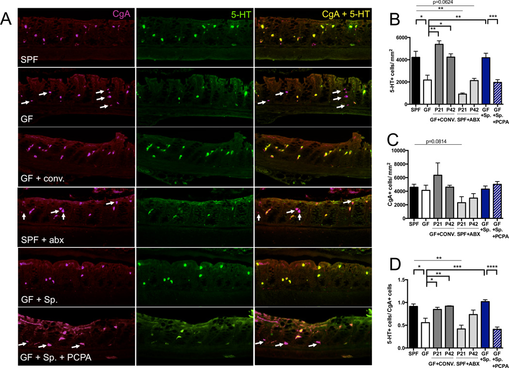
Figure 2. Indigenous Spore-forming Bacteria Increase 5-HT Levels in Colon Enterochromaffin Cells.
(A) Representative images of colons stained for chromagranin A (CgA) (left), 5-HT (center) and merged (right). Arrows indicate CgA-positive cells that lack 5-HT staining. n=3–7 mice/group.
(B) Quantitation of 5-HT+ cell number per area of colonic epithelial tissue. n=3–7 mice/group.
(C) Quantitation of CgA+ cell number per area of colonic epithelial tissue. n=3–7 mice/group.
(D) Ratio of 5-HT+ cells/CgA+ cells per area of colonic epithelial tissue. n=3–7 mice/group.
Data are presented as mean ± SEM. *p < 0.05, **p < 0.01, ***p < 0.001, ****p < 0.0001. SPF=specific pathogen-free (conventionally-colonized), GF=germ-free, CONV.=SPF conventionalized, ABX=antibiotic-treated, Sp=spore-forming bacteria, PCPA=para-chlorophenylalanine. See also Figure S2.

