

Abstract
Parry–Romberg syndrome (PRS) or progressive facial hemiatrophy is a developmental craniofacial disorder of unknown etiology characterized by a slowly progressive unilateral facial atrophy. It is associated with different systemic manifestations particularly, maxillofacial, neurologic and ophthalmologic abnormalities. Dentists must be aware of PRS to identify this invalidating disorder. In this article, we review the etiology, clinical features (especially craniofacial and dental manifestations) and treatment of PRS. We searched in PubMed line using specific words such as PRS from 2008 to 2014 (August). We identify 14 papers have described oral manifestations of this syndrome. We excluded all the article papers that did not indicate to oral manifestations of PRS.
Keywords: Facial hemiatrophy, oral manifestations, Parry–Romberg syndrome
INTRODUCTION
Parry–Romberg syndrome (PRS), also known as progressive hemifacial atrophy was first described by Parry in 1825 and later by Romberg in 1846.[1,2] This syndrome is an uncommon degenerative condition characterized by a slow and progressive but self-limited unilateral atrophy of facial tissues, including muscles, bones, skin and cartilage. It leads to aesthetic troubles as well as functional and psychological problems due to asymmetry of the face.[3]
Etiology is unknown; although trauma, viral infections, genetic factors, autoimmunity, endocrine disturbances, peripheral trigeminal neuritis, increase of cervical sympathetic nerve activity and cerebral disturbance of fat metabolism are possible suggested factors playing a role in pathogenesis of this condition.[4]
Parry–Romberg syndrome is associated with several developmental and congenital deformities, such as neurologic, ophthalmologic, cardiac, endocrine, autoimmune, cranio-maxillofacial and orthodontic abnormalities.[5] Congenital manifestations that might be associated with PRS are contralateral Poland Syndrome,[6] congenital lower limb hypoplasia,[7] congenital ipsilateral cerebral atrophy,[8] microphthalmia[9] and renal malformations.[9] Moreover, hypertrophic cardiomyopathy,[10] hypothyroidism,[11] rheumatoid arthritis,[12] lupus erythematosus[9] and scleroderma[13,14,15] are some developmental conditions reported in PRS.
Parry-Romberg syndrome, although rare, has been reported in the literature quite abundantly, and has been associated with multiple findings. The treatment options offered in the literature are diverse but not curative.[16]
The present review highlights the etiology, clinical oral manifestations and treatment of PRS.
ETIOLOGY
Despite PRS being recognized for >150 years, the exact etiology and pathogenesis of this condition is not well understood and seems to be heterogeneous. Cerebral disturbance on fat metabolism, local facial trauma, endocrine disturbances, autoimmunity, heredity, hyperactivity or hypoactivity of the sympathetic nervous system, abnormality of the trigeminal nerve and viral infections, including Borrelia burgdorferi, are believed to be associated with the pathogenesis of this disease.[3,17,18] Tang et al., 2014 believed that some inherent relationship between PRS and the disorder of neural crest cell migration may exist and that malformation or disturbed migration of neural crest cells might be relevant.[5]
SYSTEMIC AND ORAL MANIFESTATIONS
Parry–Romberg syndrome is a degenerative condition characterized by a slow and progressive atrophy of facial tissues, generally unilateral, including muscles, bones and skin.[3,13,19] Its onset occurs along first two decades of life.[13,20] This syndrome seems to have higher incidence among women and affect most often the left side of the face.[13] The skin may become thin, dry and hyperpigmented.[3] Ocular involvement is common with enophthalmos as a main manifestation.[21] Neurological manifestations such as trigeminal neuralgia, epilepsy, facial paresthesia and migraine may be associated with this condition.[13,22]
Several oral manifestations could be associated with PRS.[5,25,26,27,28,29,30,31,32,33,34,35,36,37] The oral mucosa and tongue can be affected, also jaws, salivary glands and teeth [Table 1]. There is deviation of the mouth and nose toward the affected side.[3] Atrophy of superior lip led to exposure of anterior teeth[3,21] [Figure 1].
Table 1.
Oral manifestations of Parry Romberg syndrome

Figure 1.
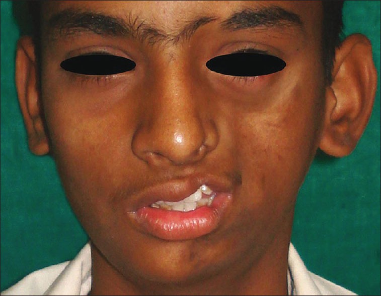
Marked hypoplasia of the left side of the face with deviation of lips and nose toward left side and notching of lips and nose with exposure of teeth. Alopecia in left eyebrow region (Deshingkar et al. 2012)
Intraoral soft tissue and muscles of mastication are also involved in PRS but the normal function like speech, deglution are not disturbed.[20,21] Unilateral atrophy of muscle of the tongue is seen with PRS[3] [Figure 2]. The condition may be associated with deficiencies of the soft and hard palates in all dimensions, shortness and deficiency of the mandibular body and ramus. Delayed tooth eruption, root atrophy and retarded tooth formation may also be observed[23,24] [Figure 3]. However, the affected teeth are normal and vital clinically.[3,23] Frequently, there is unilateral posterior crossbite, as a result of jaw hypoplasia and delayed teeth eruption.[23]
Figure 2.

Unilateral atrophy of tongue papillae of left side (Deshingkar et al. 2012)
Figure 3.

Orthopantomograph showing retarded eruption pattern of teeth on left side compared to that of right side. Decreased vertical height of ramus along with loss of gonial angle prominence on the affected side (Deshingkar et al. 2012)
DENTAL CONSIDERATIONS
Importantly, the dental characteristics in treatment of patients with PRS need to be considered. Correction of malocclusion can be restored by orthodontic movement of teeth. Biomechanical components of functional orthodontic appliance may result in clinically significant morphologic changes in the dentoalveolar and skeletal regions during facial growth. Virtually, the stage of root formation and eruption of permanent teeth is compatible with the active stage of hemifacial atrophy. Accordingly, delayed tooth eruption along with deficient root development appears to occur. Therefore, correcting the eruption problems in patients with PRS can be achieved by application of functional appliance. Prosthetic intervention or additional surgery for the jaws can be used also as a mean to recover the dental occlusion.[36]
TREATMENT AND PROGNOSIS
The disease is self-limiting and has no definite cure. The patients affected should have multidisciplinary attendance, involving experts such as dermatologists, dentists and psychologists.[31] The treatment is usually based on reposition of adipose tissue that was lost due to atrophy. Autogenously fat grafts, cartilage grafts, silicone injections and prostheses, bovine collagen, inorganic implants and recently cell fat mixed with platelet gel are some alternatives to aesthetic correction of the atrophy.[38] Esthetic effects are the usual results from this disorder rather than disability, especially in mild cases. Indeed, the recovery period for overall prognosis of PRS is unpredictable.[28]
CONCLUSION
Parry–Romberg syndrome is an uncommon poorly understood condition characterized by slow progressive atrophy of one side of the face. Its exact etiology is unclear with unknown pathophysiology. More than an aesthetic concern, the condition also causes functional and psychological problems to patients that necessitate a multidisciplinary team approach to identify treatment expectations of these patients.
Footnotes
Source of Support: Nil
Conflict of Interest: None declared.
REFERENCES
- 1.Parry CH. Collections from the Unpublished Medical Writings of the Late Caleb Hillier Parry. London: Underwoods; 1825. pp. 478–80. [Google Scholar]
- 2.Romberg HM. Klinische Ergebnisse. Berlin: Forrtner; 1846. Krankheiten des nervensystems (IV: Trophoneurosen) pp. 75–81. [Google Scholar]
- 3.Pinheiro TP, Silva CC, Silveira CS, Botelho PC, Pinheiro MD, Pinheiro Jde J. Progressive hemifacial atrophy - Case report. Med Oral Patol Oral Cir Bucal. 2006;11:E112–4. [PubMed] [Google Scholar]
- 4.Stone J. Parry–Romberg syndrome. Pract Neurol. 2006;6:185–8. [Google Scholar]
- 5.Tang XJ, Liu W, Yang B, Shi L, Yin L, Zhang ZY. Parry–Romberg syndrome with rare maxillofacial deformities: A report on two cases. J Craniomaxillofac Surg. 2014;42:780–3. doi: 10.1016/j.jcms.2013.11.010. [DOI] [PubMed] [Google Scholar]
- 6.Dintiman BJ, Shapiro RS, Hood AF, Guba AM. Parry–Romberg syndrome in association with contralateral Poland syndrome. J Am Acad Dermatol. 1990;22:371–3. doi: 10.1016/0190-9622(90)70051-i. [DOI] [PubMed] [Google Scholar]
- 7.Zambelis T, Tsivgoulis G, Kokotis P, Spengos K, Karandreas N. Electrophysiological findings in a case of congenital lower limb hypoplasia. Neurol Sci. 2008;29:177–9. doi: 10.1007/s10072-008-0932-3. [DOI] [PubMed] [Google Scholar]
- 8.Catala M. Progressive intracranial aneurysmal disease in a child with progressive hemifacial atrophy (Parry–Romberg disease): Case report. Neurosurgery. 1998;42:1195–6. doi: 10.1097/00006123-199805000-00161. [DOI] [PubMed] [Google Scholar]
- 9.Ruhin B, Bennaceur S, Verecke F, Louafi S, Seddiki B, Ferri J. Progressive hemifacial atrophy in the young patient: Physiopathologic hypotheses, diagnosis and therapy. Rev Stomatol Chir Maxillofac. 2000;101:287–97. [PubMed] [Google Scholar]
- 10.Behera M, Nimkhedkar K, Gupta RS, Hassadi MF, Mousa ME. Facial hemiatrophy (Parry–Romberg syndrome) and hypertrophic cardiomyopathy. J Assoc Physicians India. 1988;36:394–5. [PubMed] [Google Scholar]
- 11.Klene C, Massicot P, Ferrière-Fontan I, Sarlangue J, Fontan D, Guillard JM. “Saber-cut” scleroderma and Parry. Romberg facial hemiatrophy. Nosologic problems. Neurologic complications. Ann Pediatr (Paris) 1989;36:123–5. [PubMed] [Google Scholar]
- 12.Stone J. Parry–Romberg syndrome: A global survey of 205 patients using the Internet. Neurology. 2003;61:674–6. doi: 10.1212/wnl.61.5.674. [DOI] [PubMed] [Google Scholar]
- 13.Tollefson MM, Witman PM. En coup de sabre morphea and Parry–Romberg syndrome: A retrospective review of 54 patients. J Am Acad Dermatol. 2007;56:257–63. doi: 10.1016/j.jaad.2006.10.959. [DOI] [PubMed] [Google Scholar]
- 14.Slimani S, Hounas F, Ladjouze-Rezig A. Multiple linear sclerodermas with a diffuse Parry–Romberg syndrome. Joint Bone Spine. 2009;76:114–6. doi: 10.1016/j.jbspin.2008.07.009. [DOI] [PubMed] [Google Scholar]
- 15.Maletic J, Tsirka V, Ioannides P, Karacostas D, Taskos N. Parry–Romberg Syndrome associated with localized scleroderma. Case Rep Neurol. 2010;2:57–62. doi: 10.1159/000314927. [DOI] [PMC free article] [PubMed] [Google Scholar]
- 16.El-Kehdy J, Abbas O, Rubeiz N. A review of Parry–Romberg syndrome. J Am Acad Dermatol. 2012;67:769–84. doi: 10.1016/j.jaad.2012.01.019. [DOI] [PubMed] [Google Scholar]
- 17.Baskan EB, Kaçar SD, Turan A, Saricaoglu H, Tunali S, Adim SB. Parry–Romberg syndrome associated with borreliosis: Could photochemotherapy halt the progression of the disease? Photodermatol Photoimmunol Photomed. 2006;22:259–61. doi: 10.1111/j.1600-0781.2006.00238.x. [DOI] [PubMed] [Google Scholar]
- 18.Paprocka J, Jamroz E, Adamek D, Marszal E, Mandera M. Difficulties in differentiation of Parry–Romberg syndrome, unilateral facial sclerodermia, and Rasmussen syndrome. Childs Nerv Syst. 2006;22:409–15. doi: 10.1007/s00381-005-1262-x. [DOI] [PubMed] [Google Scholar]
- 19.Gonul M, Dogan B, Izci Y, Varol G. Parry–Romberg syndrome in association with anti-dsDNA antibodies: A case report. J Eur Acad Dermatol Venereol. 2005;19:740–2. doi: 10.1111/j.1468-3083.2005.01290.x. [DOI] [PubMed] [Google Scholar]
- 20.Pensler JM, Murphy GF, Mulliken JB. Clinical and ultrastructural studies of Romberg's hemifacial atrophy. Plast Reconstr Surg. 1990;85:669–74. [PubMed] [Google Scholar]
- 21.Mazzeo N, Fisher JG, Mayer MH, Mathieu GP. Progressive hemifacial atrophy (Parry–Romberg syndrome).Case report. Oral Surg Oral Med Oral Pathol Oral Radiol Endod. 1995;79:30–5. doi: 10.1016/s1079-2104(05)80069-1. [DOI] [PubMed] [Google Scholar]
- 22.Blaszczyk M, Królicki L, Krasu M, Glinska O, Jablonska S. Progressive facial hemiatrophy: Central nervous system involvement and relationship with scleroderma en coup de sabre. J Rheumatol. 2003;30:1997–2004. [PubMed] [Google Scholar]
- 23.Foster TD. The effects of hemifacial atrophy on dental growth. Br Dent J. 1979;146:148–50. doi: 10.1038/sj.bdj.4804213. [DOI] [PubMed] [Google Scholar]
- 24.Glass D. Hemifacial atrophy. Br J Oral Surg. 1964;1:194–9. [PubMed] [Google Scholar]
- 25.Fornwalt BE, Altman JS. Parry–Romberg syndrome in an adult: Report of a case. Ear Nose Throat J. 2013;92:E1–3. doi: 10.1177/014556131309200115. [DOI] [PubMed] [Google Scholar]
- 26.Kaukayil D, Anuradha S, Mukuna A, Basheer AB. Parry Romberg syndrome: A case report and an insight in to the advances in its pathophysiology and treatment. Indian J Oral Sci. 2013;4:130–3. [Google Scholar]
- 27.Khan M, Khan M, Negi R, Gupta N. Parry Romberg syndrome with localized scleroderma: A case report. J Clin Exp Dent. 2014;6:e313–6. doi: 10.4317/jced.51409. [DOI] [PMC free article] [PubMed] [Google Scholar]
- 28.Patel H, Thakkar C, Patel K. Parry–Romberg syndrome: A rare entity. J Maxillofac Oral Surg. 2010;9:247–50. doi: 10.1007/s12663-010-0103-y. [DOI] [PMC free article] [PubMed] [Google Scholar]
- 29.Rangare AL, Babu SG, Thomas PS, Shetty SR. Parry–Romberg syndrome: A rare case report. J Oral Maxillofac Res. 2011;2:e5. doi: 10.5037/jomr.2011.2205. [DOI] [PMC free article] [PubMed] [Google Scholar]
- 30.Reddy SS, Devaraju D, Kunal A. Parry Romberg syndrome: Report of two cases with rare dental and skeletal findings. EJ Dent. 2012;2:212–5. [Google Scholar]
- 31.de Vasconcelos Carvalho M, do Nascimento GJ, Andrade E, Andrade M, Sobral AP. Association of aesthetic and orthodontic treatment in Parry–Romberg syndrome. J Craniofac Surg. 2010;21:436–9. doi: 10.1097/SCS.0b013e3181cfe917. [DOI] [PubMed] [Google Scholar]
- 32.Deshingkar SA, Barpande SR, Bhavthankar JD, Humbe JG. Progressive hemifacial atrophy (Parry–Romberg Syndrome) Contemp Clin Dent. 2012;3:S78–81. doi: 10.4103/0976-237X.95111. [DOI] [PMC free article] [PubMed] [Google Scholar]
- 33.Madasamy R, Jayanandan M, Adhavan UR, Gopalakrishnan S, Mahendra L. Parry Romberg syndrome: A case report and discussion. J Oral Maxillofac Pathol. 2012;16:406–10. doi: 10.4103/0973-029X.102498. [DOI] [PMC free article] [PubMed] [Google Scholar]
- 34.Sande A, Risbud M, Kshar A, Paranjpe AO. Progressive hemifacial atrophy. Dent Res J (Isfahan) 2013;10:108–11. doi: 10.4103/1735-3327.111810. [DOI] [PMC free article] [PubMed] [Google Scholar]
- 35.Tijare MS, Sandhya S, Misurya RV, Lanje AH, Ghom A. Facial hemiatrophy: Review of literature and a case report. J Indian Acad Oral Med Radiol. 2011;23:S478–80. [Google Scholar]
- 36.You KH, Baik HS. Orthopedic and orthodontic treatment of Parry–Romberg syndrome. J Craniofac Surg. 2011;22:970–3. doi: 10.1097/SCS.0b013e31820fe339. [DOI] [PubMed] [Google Scholar]
- 37.Colquhoun AN, Ferguson MM, Doyle TC. Hemifacial atrophy with bilateral short roots. Br J Oral Maxillofac Surg. 2000;38:533–6. doi: 10.1054/bjom.2000.0461. [DOI] [PubMed] [Google Scholar]
- 38.Roddi R, Riggio E, Gilbert PM, Hovius SE, Vaandrager JM, van der Meulen JC. Clinical evaluation of techniques used in the surgical treatment of progressive hemifacial atrophy. J Craniomaxillofac Surg. 1994;22:23–32. doi: 10.1016/s1010-5182(05)80292-6. [DOI] [PubMed] [Google Scholar]