

Abstract
It has become evident that the prion protein (PrP) can form a diverse range of self-replicating structures in addition to bona fide PrPSc or strain-specific PrPSc variants. Some self-replicating states can be only produced in vitro, whereas others can be formed in vivo and in vitro. While transmissible, not all states that replicate in vivo are truly pathogenic. Some of them can replicate silently without causing symptoms or clinical diseases. In the current article we discuss the data on PK-digestion patterns of different self-replicating PrP states in connection with other structural data available to date and assess possible relationships between different self-replicating states. Even though different self-replicating PrP states appear to have significantly different global folding patterns, it seems that the C-terminal region exhibits a cross-β-sheet structure in all self-replicating states, as this region acquires the proteolytically most stable conformation. We also discuss the possibility of the transformation of self-replicating states and triggering of PrPSc formation within the frame of the deformed templating model. The spread of silent self-replicating states is of a particular concern because they can lead to transmissible prion disease. Moreover, examples on how different replication requirements favor different states are discussed. This knowledge can help in designing conditions for selective amplification of a particular PrP state in vitro.
Keywords: prion protein, prion diseases, amyloid fibrils, deformed templating, synthetic prions, protein misfolding cyclic amplification
1. Introduction
According to the protein only hypothesis, the prion protein (PrP) can adopt two alternative structural forms: the normal, cellular form PrP designated as PrPC and the disease-associated, self-replicating form designated as PrPSc (Prusiner, 1982). To explain the diversity of prion disease phenotypes within the same host, the protein only hypothesis proposes that multiple, stable, conformationally distinct self-replicating PrPSc states exist and account for different prion strains. Indeed, FTIR spectroscopy and other biochemical assays revealed differences in PrPSc structure isolated from animals infected with different prion strains (Caughey et al., 1998; Thomzig et al., 2004). It is believed that PrPSc of different strains share a common folding pattern and that strain-specific differences in PrPSc conformation are relatively minor.
In the past decade, a number of studies illustrated that the spectrum of structures acquired by the prion protein is much broader than PrPC, PrPSc or strain-specific PrPSc variants. Some self-replicating states of the prion protein can only be generated in vitro, such as amyloid fibrils produced from recombinant PrP (rPrP) in the absence of cellular cofactors (Baskakov et al., 2002; Bocharova et al., 2005a). Other PrP states, also of amyloid nature, are capable of replicating in the brain and can even be serially transmitted from animal to animal in a laboratory setting. While transmissible, they appear to be neither toxic nor associated with any clinical signs of prion disease. Examples of these include transmissible amyloid plaques in transgenic PrP (P101L) mice induced by material from a Gerstmann-Straussler-Scheinker patient with the P102L mutation (Piccardo et al., 2007); amyloid deposits in transgenic mice expressing GPI- anchorless PrPC (Chesebro et al., 2005); and self-propagating PrP forms characterized by abnormally short, C-terminal PK-resistant fragment (atypical PrPres) observed in animals inoculated with rPrP fibrils (Kovacs et al., 2013; Makarava et al., 2011). In addition, a number of abnormal PK-resistant fragments different from that of bona fide PrPSc or PK- sensitive disease-associated PrP states were observed in human and animals affected by a variety of prion diseases including sporadic CJD (sCJD), iatrogenic CJD, variable protease-sensitive prionopathy and atypical scrapie and BSE (Baron et al., 2008; Biacabe et al., 2007; Satoh et al., 2003; Zou et al., 2003; Zou et al., 2010).
Considering that some abnormal PrP states can replicate silently, i.e. without causing clinical symptoms, their spread within or between individual organisms can occur undetected. Our work on synthetic prions demonstrated that replication of clinically silent PrP states can give rise to PrPSc and transmissible prion diseases (Makarava et al., 2011; Makarava et al., 2012b). In addition, a rapidly growing number of studies suggests that amyloid states of other amyloidogenic proteins associated with neurodegenerative diseases including Alzheimer’s, Parkinson and Huntington’s diseases and Amyotrophic Lateral Sclerosis can also spread from cell to cell in a prion-like manner (reviewed in (Jucker and Walker, 2013)). However, it is not known what self-replicating states are truly pathogenic, what states while capable of replicating in vivo do not cause pathogenic effects, and what structural features separate pathogenic from non-pathogenic self- replicating states.
While the spectrum of self-replicating PrP states continue to expand, our understanding of structural differences and their relationship to bona fide PrPSc remain primitive. With most of the studies focused on elucidating molecular structure of PrPSc, several alternative PrPSc models have been proposed in the past (DeMarco and Daggett, 2004; Govaerts et al., 2004; Langedijk et al., 2006; Stork et al., 2005; Tattum et al., 2006; Wille et al., 2002; Yang et al., 2005). In some aspects the current models contradict each other and are, in part, at odds with growing experimental observations (Requena and Wille, 2014; Shirai et al., 2014). The main challenge of the next decade is to determine the structure of bona fide PrPSc and other self-replicating PrP states. In the current study we discuss the data accumulated up to date on PK-digestion patterns of different self-replicating PrP states and assess possible relationships between these states. We also discuss the possibility of the transformation of self-replicating states within the frame of the deformed templating model and triggering of transmissible prion diseases by silent self-replicating PrP states.
2. Materials and Methods
2.1. Expression and purification of full-length rPrP and formation of rPrP fibrils
Human full-length recombinant PrP encompassing residues 23–231 (variant 129V) was expressed and purified according to previously described procedures (Makarava and Baskakov, 2012). Lyophilized rPrP was dissolved in 5 mM HEPES (pH 7.0) immediately before use. To form fibrils, the rPrP stock solution (0.25 mg/mL) was incubated in 50 mM MES (pH 6.0), 0.5 M GdnHCl at 37°C under continuous shaking at 600 rpm using a Delfia plate shaker (Wallac). Amyloid formation was confirmed by Thioflavin T fluorescence assay (Makarava and Baskakov, 2012). The work on purification and fibrillation of human rPrP was performed in a Biosafety Level 2 laboratory.
2.2. Preparation of normal brain homogenate
Brains of transgenic mice expressing human PrPc (variant 129V) were kindly provided by Dr. Wen-Quan Zou (Case Western Reserve University, Cleveland, OH). 10% NBH (w/v) was prepared using brains of healthy transgenic mice expressing human PrPc (variant 129V) as previously described (Makarava et al., 2011). Briefly, the brains were ground with low speed tissue grinders in ice-cold conversion buffer until homogenous. The conversion buffer consists of Ca2+-free and Mg2+-free PBS, pH 7.5, supplemented with 0.15 M NaCl, 1.0% Triton, and one tablet of Complete protease inhibitors cocktail (Roche, Cat# 1836145) per 50 ml of buffer. The 10% NBH homogenate was pre-cleared by 2 min of centrifugation at 500 g and the supernatant was used for dgPMCAb experiments.
2.3. Protein misfolding cyclic amplification with partially deglycosylated substrate (dgPMCAb)
Protein misfolding cyclic amplification that employs partially deglycosylated PrPC as a substrate is referred to as dgPMCAb. To produce partially de-glycosylated substrate, 10% NBH mice was treated with PNGase F (New England BioLabs, glycerol-free) as follows. After pre-clearance of 10% NBH, 1500 U/ml PNGase F was added to the supernatant, and the reaction was incubated on a rotator at 37°C for 5 hours. The resulting substrate was used in dgPMCAb using the following sonication conditions: the standard sonication program for rounds 0 to 6 consisted of 30 sec sonication pulses delivered at 50% power efficiency applied every 30 min during a 24 hour period, while rounds 7 to 9 consisted of 5 sec sonication every 10 min during a 24 hour period. The dgPMCA reactions were seeded by adding 10 μL of rPrP fibrils (25 μg/mL) to 90 μL of 10% NBH. For rounds 2 to 6, 10 μl of the reaction mixture from the previous round was added to 90 μl of 10% NBH; for rounds 7 to 9, 30 μL of the previous reaction was added to 70 μL of substrate. Each dgPMCAb reaction was carried out in the presence of two 3/32″ Teflon beads (McMaster Carr).
To analyze production of PK-resistant PrP material in dgPMCAb, 10 μL of the sample was supplemented with 5 μL SDS and 5 μl PK, to a final concentration of SDS and PK of 0.25% and 50 μg/ml respectively, followed by incubation at 37°C for 1 hour. The digestion was terminated by addition of SDS-sample buffer and heating the samples for 10 min in a boiling water bath. Samples were loaded onto NuPAGE 12% BisTris gels, transferred to PVDF membrane, and probed with anti-C human PrP antibody (gift from Dr. Wen-Quan Zou, the Case Western Reserve University) that recognizes the epitope 220–231.
3. Results and Discussion
3.1. Insight into PrPSc structure from limited proteolysis
Limited proteolysis with PK produces a truncated form of PrPSc referred to as PrP27–30 that spans residues ~90 to 231 for most prion strains (Basler et al., 1986; Oesch et al., 1985). Detailed analysis by mass spectroscopy revealed that each prion strain or isolate is characterized by multiple PK-cleavage sites that are distributed in a strain-specific manner along the region spanning from amino acid residue 74 to 102 (Fig 1A) (Parchi et al., 2000; Sajnani et al., 2008). In addition to strain-dependent conformations, the distribution of PK-cleavage sites also depends on PK-digestion conditions including pH, presence of detergents and PK concentration (Notari et al., 2004). Harsher digestion conditions shift the PK sites in the direction of the C-terminus. For instance, upon treatment with high concentrations of PK, additional PK cleavage sites were identified within the central region, including residues 117, 119, 135, 139, 142, and 154 (residue numbers are given for hamster PrP) using limited proteolysis and mass spectroscopy (Fig. 1A) (Sajnani et al., 2008). Subsequent study of PrPSc devoid of its glycosylphosphatidylinositol anchor and carbohydrates also showed that in addition to the conventional PK-cleavage sites around the residue 90, the following residues within the central region were accessible to PK: 116, 118, 133, 134, 141, 152, 153, 162, 169 and 179 (residue numbers are given for mouse PrP) (Vazquez-Fernandez et al., 2012). In agreement with the data obtained by limited proteolysis and mass spectroscopy, the studies on chemical modifications of tyrosine or lysine residues suggested that in PrPSc the C-terminal region is considerably less accessible than the central region (residues 90–162) (Gong et al., 2011).
Figure 1.

Schematic diagram showing PK-resistant fragments and PK cleavage sites in PrPSc (A), PrPSc subjected to partial denaturation (B) (Kocisko et al., 1996), atypical PrPres found in sCJD (C) (Zou et al., 2003), and rPrP fibrils generated in vitro (D) (Bocharova et al., 2005b). Epitopes for anti-PrP antinodies D13, D18, AH6 and R1 used in the immunoconformational assay are shown in panel D. PK-resistant regions are indicated by black bars, partially PK-resistant regions – by gray bars, and PK cleavage sites by arrows. (E), 3D AFM image of a partially fractured amyloid fibril formed using full-length rPrP. Location of epitopes of D13, D18, AH6 and R1 antibody is indicated as assigned using results of the immunoconformational microscopy assay (Novitskaya et al., 2006). (F), Fluorescence microscopy images of rPrP fibrils kept under native conditions (top panel) or exposed to 4 M GdnHCl (middle panel) or 6 M GdnHCl (bottom panel) and double stained with R1 and reference AG4 (epitope 37–50) antibody labeled with Alexa-488 (green) and Alexa-546 (red), respectively, as previously described (Novitskaya et al., 2006).
In a manner similar to harsh proteolytic treatment, pretreatment of PrPSc under partially denaturing conditions exposed new PK-cleavage sites. Under reversible denaturing conditions the central region (up to a.a. residue 143) was found to acquire a PK-sensitive conformation, whereas the C-terminal region remained PK resistant (Fig. 1B) (Kocisko et al., 1996).
Considering the data on limited proteolysis it is reasonable to speculate that as the most resistant to denaturation and proteolytic digestion, the C-terminal region (residues ~160–231) adapts a cross-β-sheet structure in PrPSc. One can argue that proteolytic resistance of this region could be attributed to bulky carbohydrates attached to asparagine residues at positions 181 and 197 (numbers are given for hamster sequence), but not due to β-sheet structure. To test this possibility, we produced PrPSc using dgPMCA, a protein misfolding cyclic amplification reaction that employs partially deglycosylated PrPC as a substrate. While dgPMCA-derived PrPSc had considerably higher proportions of un- and mono-glycosylated glycoforms compared to those in brain- or PMCA-derived PrPSc, the C-terminal region (residues ~150–231) was the most resistant to PK in all three sources of PrPSc (Makarava et al., 2013). Furthermore, new PK-cleavage sites appeared in the central but not C-terminal region of dgPMCA-derived PrPSc (Makarava et al., 2013).
3.2. Atypical disease-associated PrP forms
In addition to authentic PrPSc that produces PrP27–30 upon proteolytic treatment, a number of atypical or short PK-resistant fragments were identified in human and animal prion diseases. C-terminal PK-resistant fragments encompassing residues 154/156–231 and 162/167–231 were found in the majority of patients with sCJD (Fig. 1C) (Zou et al., 2003). C-terminal PK-resistant products of similar size and location were also found in iatrogenic CJD (Satoh et al., 2003) and in mice infected with mouse-passaged hamster scrapie (Lawson et al., 2004). Furthermore, similar C-terminal PK-resistant fragments were found in atypical bovine spongiform encephalopathy (H-BSE), which is believed to be sporadic in origin (Biacabe et al., 2007), and in certain types of ovine scrapie (Baron et al., 2008). The role of the C-terminal fragments found in sCJD, iatrogenic CJD or in association with H-BSE in prion diseases and their relationship to PrPSc are currently uncertain. The hypotheses under consideration are the following: (i) atypical PrP forms are by-products of or on-pathway intermediates toward bona fide PrPSc; (ii) PrPSc and atypical PrPs are produced via independent pathways; and (iii) atypical PrP and PrPSc forms are not separate states, instead atypical PrP represents a semi-stable intermediate of PrPSc degradation in a cell. According to the last hypothesis, cellular clearance of PrPSc leads to accumulation of atypical PrP states that consists of the most proteolytically resistant regions that are the most difficult to clear.
3.3. Insight into the structure of rPrP amyloid fibrils
In the absence of feasible approaches for elucidating the molecular structure of PrPSc, amyloid fibrils produced in vitro using recombinant PrP have been employed in the past for gaining insight into structures of self-propagating PrP states. Our studies revealed that upon treatment with PK, fibrils generated from full-length rPrP produced progressively smaller PK resistant fragments encompassing residues 138/141–231, 152/153–231 and 162–231 (Fig. 1D) (Bocharova et al., 2005b). Imaging of individual fibrils using an immunoconformational microscopy assay revealed that the epitopes within the C-terminal region (residues 159–174 and 225–231) were the most resistant to GdnHCl-induced denaturation and became exposed to a solvent only upon treatment with 6 M GdnHCl, arguing that the C-terminal region acquires a cross-β structure (Fig. 1E, F) (Novitskaya et al., 2006). The epitope 96–106 was fully accessible to a solvent under native conditions, whereas the epitope 133–157 was secluded but became solvent accessible under partially denaturing conditions, suggesting that this epitope contributes to a lateral association between filaments (Fig. 1F) (Novitskaya et al., 2006). Notably, upon PK treatment that digests the C-terminal region, the fibrils split apart into individual filaments (Anderson et al., 2006), yet the PK-resistant regions 152/153–231 and 162–231 preserved high β-sheet rich structure, filament morphology and seeding activity as judged from the fibrillation assay (Bocharova et al., 2005b). Taken together, these results argue that the cross-β core consists of residues 152–231 or 162–231, whereas the central region between residues 106 and 152 is presumably responsible for lateral association between filaments. In agreement with the microscopy assay of single fibrils and PK digestion assay, site-specific conformational stability studies revealed that in the amyloid state the C-terminal region exhibited higher thermodynamic stability than that of the central region, whereas the unfolding of the N-terminal region was non-cooperative (Sun et al., 2007). Interestingly, brief heating of rPrP fibrils in the presence of Triton X-100 or normal brain homogenate resulted in an extension of the PK-resistant region into the central region in a process referred to as annealing (Bocharova et al., 2006). However, annealing does not significantly increase fibril’s infectivity (Makarava et al., 2010; Makarava et al., 2011; Makarava et al., 2012b) arguing that the structure of annealed or not annealed fibrils is substantially different from that of bona fide PrPSc.
In agreement with the results on limited proteolysis, solid-state NMR studies of fibrils formed using full-length rPrP demonstrated that the structurally ordered cross-β core can be assigned to the C-terminal region that forms in-register parallel β-structures (Tycko et al., 2010). Analysis using a Monte Carlo/simulated annealing algorithm suggests that the core is comprised primarily of the residues in the 173–224 range (Tycko et al., 2010). An EPR spectroscopy study of spin-labeled fibrils prepared from truncated PrP 90–231 also revealed in-register parallel β-sheet structures within residues 160–220 (Cobb et al., 2007).
While the above studies provide the first insight into the molecular structure of PrP amyloids, subsequent studies suggest that the molecular structure of rPrP fibrils is fundamentally different from that of authentic PrPSc. While both rPrP fibrils and PrPSc showed 4.8 A meridional diffraction, a characteristic of cross-β structure, X-ray diffraction studies showed that infectious PrP27–30 produce X-ray diffraction patterns different from those of rPrP fibrils (Wille et al., 2009). Moreover, when subjected to amplification in vitro, rPrP fibrils and PrPSc displayed different amplification requirements (Makarava et al., 2012b; Miller et al., 2011). When added to PMCA reactions, rPrP did not support but interfered with amplification of PrPSc (Gonzalez- Montalban et al., 2011a; Nishina et al., 2006; Yuan et al., 2013). Nevertheless, while global folding patterns in rPrP fibrils and PrPSc appear to be different, they might share common structural elements, as evident from partial overlap of PK cleavage sites (Fig. 1).
3.4. Transformation of PrP self-replicating states
While the structures of rPrP fibrils and PrPSc are believed to be substantially different, they can give rise to each other upon changes in replication environment and exposure to an appropriate substrate (Fig. 2). For instance, when exposed to rPrP in vitro, brain-derived PrPSc triggers formation of amyloid fibrils in so-called quaking or amyloid seeding assays (Fig. 2A) (Atarashi et al., 2007; Colby et al., 2007). rPrP fibrils induced by PrPSc displayed minimal infectivity and have PK resistant products similar to that of rPrP fibrils formed in the absence of PrPSc seeds (Atarashi et al., 2007). At the same time, serial passaging in animals of amyloid fibrils formed in non-seeded reactions in vitro gave rise to bona fide PrPSc and transmissible prion disease (Fig. 2B) (Makarava et al., 2010; Makarava et al., 2011; Makarava et al., 2012b). A transformation of one self- replicating state into an alternative state is described by the deformed templating model (Makarava and Baskakov, 2013). According to this model, if a self-replicating state does not fit well into a new environment, multiple new self-replicating variants are generated via deformed templating. A variant that is the best fit to a new substrate or environment receives selective advantage and surpasses alternative structures (Fig. 2). Direct support for the deformed templating model was provided by observation of hybrid fibrils using a novel imaging approach that combines atomic force microscopy with immunofluorescence microscopy on one platform (Makarava et al., 2009).
Figure 2.

Schematic diagram illustrating the deformed templating mechanism. The deformed templating model postulates that diverse structural variants are generated as a result of changes in replication environment via numerous trial-and-error seeding events. A newly generated variant that is the best fit to an altered environment not only replaces the original variant but also surpasses newly generated competing variants. Examples of deformed templating are (A) PrPSc-seeded fibrillation of rPrP in vitro or (B) generation of synthetic strains upon serial passaging of rPrP fibrils in animals.
Detailed analysis of serial transmission of rPrP fibrils provides mechanistic insight into the evolution of PrP self-replicating states in vivo. The first product of PrPC misfolding triggered by rPrP fibrils in animals was so-called atypical PrPres (Fig. 3) (Makarava et al., 2011; Makarava et al., 2012b). Atypical PrPres had a PK-resistant core similar to that of rPrP fibrils and displayed in vitro replication requirements different from that of authentic PrPSc. In contrast to PrPSc, replication of atypical PrPres in PMCA was RNA independent. Atypical PrPres prefers un- and monoglycosylated PrPC as a substrate, whereas PrPSc predominantly recruits diglycosylated PrPC (Kovacs et al., 2013). Moreover, seeding of dgPMCA with rPrP fibrils resulted in atypical PrPres with properties very similar to those of atypical PrPres formed in animal brains inoculated with rPrP fibrils (Fig. 3) (Makarava et al., 2012b). Accumulation of atypical PrPres in animal brains did not lead to any notable clinical signs of prion diseases and was associated only with minor lesions (Kovacs et al., 2013). Atypical PrPres, while capable of replicating in a brain using PrPC as a substrate, was a clinically silent abnormal PrP state that appeared to be structurally different from PrPSc but similar to rPrP fibrils. During two or three serial passages, atypical PrPres was fully replaced by PrPSc (Makarava et al., 2011; Makarava et al., 2012b). While the appearance of PrPSc was stochastic, it always followed accumulation of atypical PrPres. The dynamics between the two states suggests that appearance of PrPSc is a result of a series of deformed templating events and a selection of the most favorable structural variants that were best suited for replication in animal brains (Makarava et al., 2011; Makarava et al., 2012b). While animals infected with strains of synthetic origin (SSLOW, LOTSS and S05) display all the key characteristics that define transmissible prion disease, these strains show a number of remarkable features with respect to disease phenotype, cellular localization, their deposition pattern and behavior in PMCA, as described in details elsewhere (Gonzalez-Montalban and Baskakov, 2012; Gonzalez-Montalban et al., 2011b; Jeffrey et al., 2014; Makarava et al., 2012a; Makarava et al., 2012c). Such peculiar features places them into a group separate from that of hamster-adapted strains of natural origin.
Figure 3.
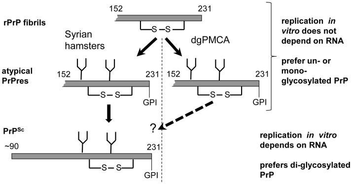
Schematic diagram illustrating that in animals or in dgPMCAb rPrP fibrils trigger atypical PrPres, in which the same region acquires the same PK-resistant conformation as in rPrP fibrils. Atypical PrPres formed in animals then gave rise to PrPSc. It remains to be shown whether atypical PrPres triggered by rPrP fibrils in dgPMCAb can induce transmissible prion disease in animals and whether strain-specific properties are similar to that of previously produced synthetic strains. Only PK-resistant regions of rPrP fibrils, atypical PrPres and PrPSc are shown (gray bars).
Not all types of rPrP amyloid fibrils produced in vitro can give rise to PrPSc. For instance, in contrast to R fibrils, S fibrils which were formed using the same stocks of full-length rPrP and under the same solvent conditions as R fibrils but using a different shaking mode, failed to induce PrPSc or prion diseases in animals (Makarava et al., 2012b). Such opposite outcomes were presumably due do fundamental differences between S- and R-specific folding patterns, as their secondary, tertiary and quaternary structures were found to be considerably different (Makarava and Baskakov, 2008; Ostapchenko et al., 2010). Not every preparation of R fibrils can induce prion disease either (Makarava et al., 2012b). The incubation time to disease and the features of the resulting PrPSc were found to be a function of conformational stability of the fibrils (Colby et al., 2009). The high concentrations of denaturants (urea or GdnHCl), while accelerating the reactions of rPrP fibrillation, give selective advantages to the thermodynamically more stable conformations of R-fibrils (Baskakov et al., 2004; Sun et al., 2008). The higher stability of R-fibrils produces a longer incubation time to disease when inoculated into transgenic mice with high PrP expression (Colby et al., 2009). When inoculated into wild type animals, highly stable R-fibril preparations failed to induce clinical or subclinical disease (Makarava et al., 2012b). These results illustrate that the spectrum of possible self-propagating variants generated by a specific amyloid template in a new environment is not endless. Perhaps, the transition from rPrP fibrils to bona fide PrPSc can occur successfully only if the two states have a reasonable overlap in their folding patterns.
3.5. PMCA and selective amplification of atypical PrPres or PrPSc in vitro
As discussed above, atypical PrPres of synthetic origin and PrPSc exhibit different in vitro replications requirements. This raises the possibility that different self-replicating PrP states could be selectively amplified if one design sets of conditions that provide selective advantages to a particular state. Indeed, we found that under a modified PMCAb format that employs partially deglycosylated PrPC (dgPMCAb), atypical PrPres can be selectively amplified from a mixture with PrPSc (Makarava et al., 2013). Moreover, depletion of RNA gives further selective advantages to atypical PrPres, since amplification of PrPSc is RNA-dependent, whereas amplification of atypical PrPres is RNA-independent (Makarava et al., 2012b; Makarava et al., 2013). In vitro amplification requirements were only tested so far for atypical PrPres of synthetic origin. It is not known whether the same rules apply to amplification of atypical PrPres observed in sCJD or animal prion diseases. While atypical PrPres of synthetic origin and those found in sCJD and iatrogenic CJD display very similar if not identical PK-resistant fragments (Fig. 1), it is not known whether these forms are structurally similar.
3.6. Testing whether atypical PrPres can be generated in vitro using human rPrP fibrils
The previous chapter discussed the relationship between atypical PrPres of synthetic origin and PrPSc. The majority of sCJD patients accumulate atypical PrPres, which appears to be very similar to that of synthetic origin, as judged from their PK-digestion profile (Zou et al., 2003) (Fig. 1C). However, the relationship of atypical PrPres and PrPSc in sCJD is not clear. Could atypical PrPres be an intermediate to PrPSc? If so, could it be produced in vitro in a similar manner to that of hamster atypical PrPres formed in dgPMCA seeded with hamster rPrP fibrils? To address this question, we tested whether human atypical PrPres could be produced de novo in dgPMCA upon seeding with human rPrP fibrils.
Full-length human (hu) rPrP (129V variant) was expressed in E. coli and purified using the same protocol as previously used for purification of hamster full-length rPrP (Makarava and Baskakov, 2012). This purification procedure yields only ~1 mg of hu rPrP per 1 L of culture, which is approximately 10-fold less than the yield routinely found for hamster or mouse rPrP. Such low yield was due to the high tendency of hu rPrP to aggregate. Hu rPrP was converted into amyloid fibrils under solvent conditions (50 mM MES, pH 6.0, 0.5 M GdnHCl) that were previously used for producing fibrils from hamster rPrP, the inoculation of which gave rise to atypical PrPres and synthetic strain S05 (Makarava et al., 2012b). Formation of amyloid fibrils was confirmed by Tioflavin T assay. While seeding of dgPMCA with hamster rPrP fibrils consistently triggered atypical PrPres, seeding with hu rPrP fibrils failed to do so (Fig. 4A, C). Barely detectible PK resistant bands with patterns of atypical PrPres were observed in the 5th and 6th dgPMCAb rounds in overexposed blots (Fig. 4A). However, further efforts to amplify this material using the dgPMCAb protocol and low dilution factors between rounds were not successful (Fig. 4B).
Figure 4.

dgPMCAb reactions seeded with two preparations of hu rPrP fibrils and conducted with 10-fold dilutions between serial rounds (A); subsequent dgPMCAb reactions seeded with a mixture of reaction products from the 5th and 6th rounds and conducted with 3.3-fold dilutions between rounds (B). dgPMCAb reactions seeded with two preparations of hamster rPrP fibrils and conducted with 10-fold dilutions between serial rounds (C). Western blots were stained with anti-C antibody in panels A and B and SAF-84 antibody in panel C. Atypical PrPres shows characteristic PK-resistant bands at 23, 16 and 13 kDa that correspond to di-, mono- and un glycosylated PK-resistant fragment 152–231, respectively (arrows).
In summary, we were not able to produce hu atypical PrPres in vitro using an approach that worked well for generating hamster atypical PrPres. Currently we are testing whether hamster atypical PrPres generated in vitro induces transmissible prion disease. The failure to form hu atypical PrPres suggests that while showing the same PK-resistant products as hamster, atypical PrPres of synthetic origin might not be related to the hu atypical PrPres found in sCJD. Alternatively, low conversion efficiency and/or differences in replication environment might account for the negative result.
Conclusions
The prion protein is able to form several distinct self-replicating states characterized by different global folding patterns. Some self-replicating states can only be formed in vitro, while other states can replicate in vivo and in vitro. While transmissible, not all states that replicate in vivo are truly pathogenic; some of them can replicate silently without causing clinical diseases. However, regardless of a specific folding pattern, the C-terminal PrP region acquires the most proteolytically and thermodynamically stable conformation suggesting that the cross-β-sheet structure involves the C-terminal region in all self-replicating states. The specific replication environment, including cellular cofactors and PrP posttranslational modifications, guide PrP to acquire and maintain a particular self-replicating state. Upon changes in replication environment, one state might give rise to an alternative state that fits better into the new environment. When undetected, a spread of silent self-replicating states is of particular concern because they can produce a truly pathogenic transmissible state or PrPSc via a deformed templating mechanism. Knowledge about replication requirements can help with designing PMCA conditions for selective amplification of a particular state in vitro.
Highlights.
Prion protein can form a diverse range of self-replicating states
Distinct states exhibit significantly different global folding patterns
The C-terminal region adapt a cross-β-sheet structure in all self-replicating states
Different self-replicating states can trigger each other via deformed templating
Acknowledgments
We thank Dr. Wen-Quan Zou (the Case Western Reserve University) for providing brains of transgenic mice expressing human PrPC and anti-PrP antibody, and Pamela Wright for editing the manuscript. This work was supported by a NIH grants NS045585 and NS074998 to IVB.
Footnotes
Publisher's Disclaimer: This is a PDF file of an unedited manuscript that has been accepted for publication. As a service to our customers we are providing this early version of the manuscript. The manuscript will undergo copyediting, typesetting, and review of the resulting proof before it is published in its final citable form. Please note that during the production process errors may be discovered which could affect the content, and all legal disclaimers that apply to the journal pertain.
References
- Anderson M, Bocharova OV, Makarava N, Breydo L, Salnikov VV, Baskakov IV. Polymorphysm and Ultrastructural Organization of Prion Protein Amyloid Fibrils: An Insight from High Resolution Atomic Force Microscopy. J Mol Biol. 2006;358:580–596. doi: 10.1016/j.jmb.2006.02.007. [DOI] [PubMed] [Google Scholar]
- Atarashi R, Moore RA, Sim VL, Hughson AG, Dorward DW, Onwubiko HA, Priola SA, Caughey B. Ultrasensitive detection of scrapie prion protein using seeded conversion of recombinant prion protein. Nature Methods. 2007;4:645–650. doi: 10.1038/nmeth1066. [DOI] [PubMed] [Google Scholar]
- Baron T, Bencsik A, Vulin J, Biacabe AG, Morignat E, Verchere J, Betemps D. A C-terminal protease-resistant prion fragment distinguishes ovine “CH1641-like” scrapie from bovine classical and L-Type BSE in ovine transgenic mice. PLOS Pathog. 2008;4:e1000137. doi: 10.1371/journal.ppat.1000137. [DOI] [PMC free article] [PubMed] [Google Scholar]
- Baskakov IV, Legname G, Baldwin MA, Prusiner SB, Cohen FE. Pathway Complexity of Prion Protein Assembly into Amyloid. J Biol Chem. 2002;277:21140–21148. doi: 10.1074/jbc.M111402200. [DOI] [PubMed] [Google Scholar]
- Baskakov IV, Legname G, Gryczynski Z, Prusiner SB. The peculiar nature of unfolding of human prion protein. Protein Sci. 2004;13:586–595. doi: 10.1110/ps.03457204. [DOI] [PMC free article] [PubMed] [Google Scholar]
- Basler K, Oesch B, Scott M, Westaway D, Wälchli M, Groth DF, McKinley MP, Prusiner SB, Weissmann C. Scrapie and cellular PrP isoforms are encoded by the same chromosomal gene. Cell. 1986;46:417–428. doi: 10.1016/0092-8674(86)90662-8. [DOI] [PubMed] [Google Scholar]
- Biacabe AG, Jacobs JG, Bencsik A, Langeveld JP, Baron TG. H-type bovine spongiform encephalopathy: complex molecular features and similarities with human prion diseases. Prion. 2007;1:61–68. doi: 10.4161/pri.1.1.3828. [DOI] [PMC free article] [PubMed] [Google Scholar]
- Bocharova OV, Breydo L, Parfenov AS, Salnikov VV, Baskakov IV. In vitro conversion of full length mammalian prion protein produces amyloid form with physical property of PrPSc. J Mol Biol. 2005a;346:645–659. doi: 10.1016/j.jmb.2004.11.068. [DOI] [PubMed] [Google Scholar]
- Bocharova OV, Breydo L, Salnikov VV, Gill AC, Baskakov IV. Synthetic prions generated in vitro are similar to a newly identified subpopulation of PrPSc from sporadic Creutzfeldt-Jakob Disease PrP Sc Prot. Science. 2005b;14:1222–1232. doi: 10.1110/ps.041186605. [DOI] [PMC free article] [PubMed] [Google Scholar]
- Bocharova OV, Makarava N, Breydo L, Anderson M, Salnikov VV, Baskakov IV. Annealing PrP amyloid firbils at high temperature results in extension of a proteinase K resistant core. J Biol Chem. 2006;281:2373–2379. doi: 10.1074/jbc.M510840200. [DOI] [PubMed] [Google Scholar]
- Caughey B, Raymond GJ, Bessen RA. Strain-dependent differences in β-sheet conformations of abnormal prion protein. J Biol Chem. 1998;273:32230–32235. doi: 10.1074/jbc.273.48.32230. [DOI] [PubMed] [Google Scholar]
- Chesebro B, Trifilo M, Race M, Meade-White K, Teng C, LaCasse R, Raymond L, Favara C, Baron G, Priola S, Caughey B, Masliah E, Oldstone M. Anchorless prion protein result in infectious amyloid disease without clinical scrapie. Science. 2005;308:1435–1439. doi: 10.1126/science.1110837. [DOI] [PubMed] [Google Scholar]
- Cobb NJ, Sonnichsen FD, McHaourab H, Surewicz W. Molecular architecture of human prion protein amyloid: A parallel, in-register b-structure. Proc Acad Natl Sci USA. 2007;104:18946–18951. doi: 10.1073/pnas.0706522104. [DOI] [PMC free article] [PubMed] [Google Scholar]
- Colby DW, Giles K, Legname G, Wille H, Baskakov IV, DeArmond SJ, Prusiner SB. Design and construction of diverse mammalian prion strains. Proc Acad Natl Sci USA. 2009;106:20417–20422. doi: 10.1073/pnas.0910350106. [DOI] [PMC free article] [PubMed] [Google Scholar]
- Colby DW, Zhang Q, Wang S, Groth D, Legname G, Riesner D, Prusiner SB. Prion detection by an amyloid seeding assay. Proc Acad Natl Sci USA. 2007;104:20914–20919. doi: 10.1073/pnas.0710152105. [DOI] [PMC free article] [PubMed] [Google Scholar]
- DeMarco ML, Daggett V. From conversion to aggregation: protofibrils formation of the prion protein. Proc Acad Natl Sci USA. 2004;101:2293–2298. doi: 10.1073/pnas.0307178101. [DOI] [PMC free article] [PubMed] [Google Scholar]
- Gong B, Ramos A, Vazquez-Fernandez E, Silva CJ, Alonso J, Liu Z, Requena J. Probing structural differences between PrPC and PrPSc by surface nitration and acetylation: evidence of conformational change in the C-terminus. Biochemistry. 2011;50:4963–4972. doi: 10.1021/bi102073j. [DOI] [PubMed] [Google Scholar]
- Gonzalez-Montalban N, Baskakov IV. Assessment of strain-specific PrPSc elongation rates revealed a transformation of PrPSc properties during Protein Misfolding Cyclic Amplification. PLoS One. 2012;7:0041210. doi: 10.1371/journal.pone.0041210. [DOI] [PMC free article] [PubMed] [Google Scholar]
- Gonzalez-Montalban N, Makarava N, Ostapchenko VG, Savtchenko R, Alexeeva I, Rohwer RG, Baskakov IV. Highly Efficient Protein Misfolding Cyclic Amplification. PLoS Pathogen. 2011a;7:e1001277. doi: 10.1371/journal.ppat.1001277. [DOI] [PMC free article] [PubMed] [Google Scholar]
- Gonzalez-Montalban N, Makarava N, Savtchenko R, Baskakov IV. Relationship between Conformational Stability and Amplification Efficiency of Prions. Biochemistry. 2011b;50:7933–7940. doi: 10.1021/bi200950v. [DOI] [PMC free article] [PubMed] [Google Scholar]
- Govaerts C, Wille H, Prusiner SB, Cohen FE. Evidance for assembly of prions with left-handed b-helices into trimers. Proc Acad Natl Sci USA. 2004;101:8342–8347. doi: 10.1073/pnas.0402254101. [DOI] [PMC free article] [PubMed] [Google Scholar]
- Jeffrey M, McGovern G, Makarava N, Gonzalez L, Kim YS, Rohwer RG, Baskakov IV. Pathology of SSLOW, a transmissible and fatal synthetic prion protein disorder, and comparison with naturally occurring classical transmissible spongoform encephalopathies. Neuropath Appl Neurobiol. 2014;40:296–310. doi: 10.1111/nan.12053. [DOI] [PMC free article] [PubMed] [Google Scholar]
- Jucker M, Walker LC. Self-propagation of pathogenic protein aggregates in neurodegenerative diseases. Nature. 2013;501:45–51. doi: 10.1038/nature12481. [DOI] [PMC free article] [PubMed] [Google Scholar]
- Kocisko DA, Lansbury JPT, Caughey B. Partial unfolding and refolding of scrapie-associated prion protein: evidence for a critical 16-kDa C-terminal domain. Biochemistry. 1996;35:13434–13442. doi: 10.1021/bi9610562. [DOI] [PubMed] [Google Scholar]
- Kovacs GG, Makarava N, Savtchenko R, Baskakov IV. Atypical and classical forms of the disease-associated state of the prion protein exhibit distinct neuronal tropism, deposition patterns, and lesion profiles. Am J Pathol. 2013;183:1539–1547. doi: 10.1016/j.ajpath.2013.07.024. [DOI] [PMC free article] [PubMed] [Google Scholar]
- Langedijk JPM, Fuentes G, Boshuizen R, Bonvin AMJJ. Two-rung Model of a Left-handed b-helix for Prions Explains Species Barrier and Strain Variation in Transmissible Spongiform Encephalopathies. J Mol Biol. 2006;360:907–920. doi: 10.1016/j.jmb.2006.05.042. [DOI] [PubMed] [Google Scholar]
- Lawson VA, Priola SA, Meade-White K, Lawton M, Chesebro B. Flexible N-terminal region of prion protein influences conformation of protease resistant prion protein isoforms associated with cross-species scrapie infection in vivo and in vitro. J Biol Chem. 2004;279:13689–13695. doi: 10.1074/jbc.M303697200. [DOI] [PubMed] [Google Scholar]
- Makarava N, Baskakov IV. The same primary structure of the prion protein yields two distinct self-propagating states. J Biol Chem. 2008;283:15988–15996. doi: 10.1074/jbc.M800562200. [DOI] [PMC free article] [PubMed] [Google Scholar]
- Makarava N, Baskakov IV. Purification and fibrillation of full-length recombinant PrP. Methods Mol Biol. 2012;849:33–52. doi: 10.1007/978-1-61779-551-0_4. [DOI] [PMC free article] [PubMed] [Google Scholar]
- Makarava N, Baskakov IV. The Evolution of Transmissible Prions: The Role of Deformed Templating. PLOS Pathog. 2013;9:e1003759. doi: 10.1371/journal.ppat.1003759. [DOI] [PMC free article] [PubMed] [Google Scholar]
- Makarava N, Kovacs GG, Bocharova OV, Savtchenko R, Alexeeva I, Budka H, Rohwer RG, Baskakov IV. Recombinant prion protein induces a new transmissible prion disease in wild type animals. Acta Neuropathol. 2010;119:177–187. doi: 10.1007/s00401-009-0633-x. [DOI] [PMC free article] [PubMed] [Google Scholar]
- Makarava N, Kovacs GG, Savtchenko R, Alexeeva I, Budka H, Rohwer RG, Baskakov IV. Genesis of mammalian prions: from non-infectious amyloid fibrils to a transmissible prion disease. PLoS Pathogen. 2011;7:e1002419. doi: 10.1371/journal.ppat.1002419. [DOI] [PMC free article] [PubMed] [Google Scholar]
- Makarava N, Kovacs GG, Savtchenko R, Alexeeva I, Budka H, Rohwer RG, Baskakov IV. Stabilization of a prion strain of synthetic origin requires multiple serial passages. J Biol Chem. 2012a;287:30205–30214. doi: 10.1074/jbc.M112.392985. [DOI] [PMC free article] [PubMed] [Google Scholar]
- Makarava N, Kovacs GG, Savtchenko R, Alexeeva I, Ostapchenko VG, Budka H, Rohwer RG, Baskakov IV. A New Mechanism for Transmissible Prion Diseases. J Neurosci. 2012b;32:7345–7355. doi: 10.1523/JNEUROSCI.6351-11.2012. [DOI] [PMC free article] [PubMed] [Google Scholar]
- Makarava N, Ostapchenko VG, Savtchenko R, Baskakov IV. Conformational switching within individual amyloid fibrils. J Biol Chem. 2009;284:14386–14395. doi: 10.1074/jbc.M900533200. [DOI] [PMC free article] [PubMed] [Google Scholar]
- Makarava N, Savtchenko R, Alexeeva I, Rohwer RG, Baskakov IV. Fast and ultrasensitive method for quantitating prion infectivity titer. Nature Commun. 2012c;3:741. doi: 10.1038/ncomms1730. [DOI] [PMC free article] [PubMed] [Google Scholar]
- Makarava N, Savtchenko R, Baskakov IV. Selective amplification of classical and atypical prions using modified protein misfolding cyclic amplification. J Biol Chem. 2013;288:33–41. doi: 10.1074/jbc.M112.419531. [DOI] [PMC free article] [PubMed] [Google Scholar]
- Miller MB, Geoghegan JC, Supattapone S. Dissociation of infectivity from seeding ability in prions with alternate docking mechanism. PLoS Pathogen. 2011;7:e1002128. doi: 10.1371/journal.ppat.1002128. [DOI] [PMC free article] [PubMed] [Google Scholar]
- Nishina K, Deleault NR, Mahal S, Baskakov I, Luhrs T, Riek R, Supattapone S. The Stoichiometry of Host PrPC Glycoforms Modulates the Efficiency of PrPSc formation in vitro. Biochemistry. 2006;45:14129–14139. doi: 10.1021/bi061526k. [DOI] [PubMed] [Google Scholar]
- Notari S, Capellari S, Giese A, Westner I, Baruzzi A, Ghetti B, Gambetti P, Kretzschmar HA, Parchi P. Effects of Different Experimental Conditions on the PrPSc Core Generated by Protease Digestion. J Biol Chem. 2004;279:16797–16804. doi: 10.1074/jbc.M313220200. [DOI] [PubMed] [Google Scholar]
- Novitskaya V, Makarava N, Bellon A, Bocharova OV, Bronstein IB, Williamson RA, Baskakov IV. Probing the conformation of the prion protein within a single amyloid fibril using a novel immunoconformational assay. J Biol Chem. 2006;281:15536–15545. doi: 10.1074/jbc.M601349200. [DOI] [PubMed] [Google Scholar]
- Oesch B, Westaway D, Wälchli M, McKinley MP, Kent SBH, Aebersold R, Barry RA, Tempst P, Teplow DB, Hood LE, Prusiner SB, Weissmann C. A cellular gene encodes scrapie PrP 27–30 protein. Cell. 1985;40:735–746. doi: 10.1016/0092-8674(85)90333-2. [DOI] [PubMed] [Google Scholar]
- Ostapchenko VG, Sawaya MR, Makarava N, Savtchenko R, Nilsson KP, Eisenberg D, Baskakov IV. Two amyloid states of the prion protein display significantly different folding patterns. J Mol Biol. 2010;400:908–921. doi: 10.1016/j.jmb.2010.05.051. [DOI] [PMC free article] [PubMed] [Google Scholar]
- Parchi P, Zou W, Wang W, Brown P, Capellari S, Ghetti B, Kopp N, Schulz-Schaeffer WJ, Kretzschmar HA, Head MW, Ironside JW, Gambetti P, Chen SG. Genetic Influence on the structural variations of the abnormal prion protein. Proc Acad Natl Sci USA. 2000;97:10168–10172. doi: 10.1073/pnas.97.18.10168. [DOI] [PMC free article] [PubMed] [Google Scholar]
- Piccardo P, Manson JC, King D, Ghetti B, Barron RM. Accumulation of prion protein in the brain that is not associated with transmissible disease. Proc Acad Natl Sci USA. 2007;104:4712–4717. doi: 10.1073/pnas.0609241104. [DOI] [PMC free article] [PubMed] [Google Scholar]
- Prusiner SB. Novel proteinaceous infectious particles cause scrapie. Science. 1982;216:136–144. doi: 10.1126/science.6801762. [DOI] [PubMed] [Google Scholar]
- Requena JR, Wille H. The Structure of the infectious prion protein: Experimental data and molecular models. Prion. 2014;8:60–66. doi: 10.4161/pri.28368. [DOI] [PMC free article] [PubMed] [Google Scholar]
- Sajnani G, Pastrana MA, Dynin I, Onisko B, Requena JR. Scrapie prion protein structural constraints obtained by limited proteolysis and mass spectrometry. J Mol Biol. 2008;382:88–98. doi: 10.1016/j.jmb.2008.06.070. [DOI] [PubMed] [Google Scholar]
- Satoh K, Muramoto T, Tanaka T, Kitamoto N, Ironside JW, Nagashima K, Yamada M, Sato T, Mohri S, Kitamoto T. Association of an 11–12 kDa protease-resistant prion protein fragment with subtypes of dura graft-associated Creutzfeldt-Jakob disease and other prion diseases. J Gen Virol. 2003;84:2885–2893. doi: 10.1099/vir.0.19236-0. [DOI] [PubMed] [Google Scholar]
- Shirai T, Saito M, Kobayashi A, Asano M, Hizume M, Ikeda S, Teruya K, Morita M, Kitamoto T. Evaluating prion models based on comprehensive mutation data of mouse PrP. Structure. 2014;8:560–571. doi: 10.1016/j.str.2013.12.019. [DOI] [PubMed] [Google Scholar]
- Stork M, Giese A, Kretzchmar HA, Tavan P. Molecular Dynamics Simulations Indicate a Possible Role of Parallel b-Helices in Seeded Aggregation of Poly-Gln. Biophys J. 2005;88:2442–2451. doi: 10.1529/biophysj.104.052415. [DOI] [PMC free article] [PubMed] [Google Scholar]
- Sun Y, Breydo L, Makarava N, Yang Q, Bocharova OV, Baskakov IV. Site-specific conformational studies of PrP amyloid fibrils revealed two cooperative folding domain within amyloid structure. J Biol Chem. 2007;282:9090–9097. doi: 10.1074/jbc.M608623200. [DOI] [PubMed] [Google Scholar]
- Sun Y, Makarava N, Lee CI, Laksanalamai P, Robb FT, Baskakov IV. Conformational stability of PrP amyloid firbils controls their smallest possible fragment size. J Mol Biol. 2008;376:1155–1167. doi: 10.1016/j.jmb.2007.12.053. [DOI] [PMC free article] [PubMed] [Google Scholar]
- Tattum MH, Cohen-Krausz S, Thumanu K, Wharton CW, Khalili-Shirazi A, Jackson GS, Orlova EV, Collinge J, Clarke AR, Saibil HR. Elongated oligomers assemble into mammalian PrP amyloid fibrils. J Mol Biol. 2006;357:975–985. doi: 10.1016/j.jmb.2006.01.052. [DOI] [PubMed] [Google Scholar]
- Thomzig A, Spassov S, Friedrich M, Naumann D, Beekes M. Discriminating Scrapie and Bovine Spongiform Encephalopathy Isolates by Infrared Spectroscopy of Pathological Prion Protein. J Biol Chem. 2004;279:33854. doi: 10.1074/jbc.M403730200. [DOI] [PubMed] [Google Scholar]
- Tycko R, Savtchenko R, Ostapchenko VG, Makarava N, Baskakov IV. The a-Helical C-Terminal Domain of Full-Length Recombinant PrP Converts to an In-Register Parallel β-Sheet Structure in PrP Fibrils: Evidence from Solid State Nuclear Magnetic Resonance. Biochemistry. 2010;49:9488–9497. doi: 10.1021/bi1013134. [DOI] [PMC free article] [PubMed] [Google Scholar]
- Vazquez-Fernandez E, Alonso J, Pastrana MA, Ramos A, Stitz L, Vidal E, Dynin I, Petsch B, Silva CJ, Requena JR. Structural organization of mammalian prions as probed by limited proteolysis. Plos ONE. 2012;7:e50111. doi: 10.1371/journal.pone.0050111. [DOI] [PMC free article] [PubMed] [Google Scholar]
- Wille H, Bian W, McDonald M, Kendall A, Colby DW, Bloch L, Ollesh J, Borovinskiy AL, Cohen FE, Prusiner SB, Stubbs G. Natural and synthetic prion structure from X-ray fiber diffraction. Proc Acad Natl Sci USA. 2009;106:16990–16995. doi: 10.1073/pnas.0909006106. [DOI] [PMC free article] [PubMed] [Google Scholar]
- Wille H, Michelitsch MD, Guenebaut V, Supattapone S, Serban A, Cohen FE, Agard DA, Prusiner SB. Structural studies of the scrapie prion protein by electron crystallography. Proc Acad Natl Sci USA. 2002;99:3563–3568. doi: 10.1073/pnas.052703499. [DOI] [PMC free article] [PubMed] [Google Scholar]
- Yang S, LeVine H, Onuchic JN, Cox DL. Structure of infectious prions: stabilization by domain swapping. Faseb J. 2005;19:1778–1782. doi: 10.1096/fj.05-4067hyp. [DOI] [PubMed] [Google Scholar]
- Yuan J, Zhan YA, Abskharon R, Xiao X, Martinez MC, Zhou X, Kneale G, Mikol J, Lehmann S, Surewicz WK, Castilla J, Steyaert J, Zhang S, Kong Q, Petersen RB, Wohlkonig A, Zou WQ. Recombinant human prion protein inhibits prion propagation in vitro. Sci Rep. 2013;3:2911. doi: 10.1038/srep02911. [DOI] [PMC free article] [PubMed] [Google Scholar]
- Zou WQ, Capellari S, Parchi P, Sy MS, Gambetti P, Chen SG. Identification of Novel Proteinase K-resistant C-terminal Fragments of PrP in Creutzfeldt-Jakob Disease. J Biol Chem. 2003;278:40429–40436. doi: 10.1074/jbc.M308550200. [DOI] [PubMed] [Google Scholar]
- Zou WQ, Puoti G, Xiao X, Yuan J, Qing L, Cali I, Shimoji M, Langeveld JP, Castellani R, Notari S, Crain B, Schmidt RE, Geschwind M, DeArmond SJ, Cairns NJ, Dickson D, Honig L, Torres JM, Mastrianni J, Capellari S, Giaccone G, Belay ED, Schonberger LB, Cohen M, Perry G, Kong Q, Parchi P, Tagliavini F, Gambetti P. Variably protease-sensitive prionopathy: a new sporadic disease of the prion protein. Ann Neurol. 2010;68:162–172. doi: 10.1002/ana.22094. [DOI] [PMC free article] [PubMed] [Google Scholar]