Abstract
We present a four-year-old wth ethylmalonic encephalopathy who presented with delayed walking. She had bilateral hyperintense lesions in the basal ganglia. Molecular analysis revealed a homozygous c.3G>T mutation in the ETHE1 gene. She did not have typical findings of the disease including recurrent petechia, chronic diarrhea and acrocyanosis was very subtle and orthostatic. She benefited from riboflavine and Q10 treatments. We suggest that acrocyanosis should be questioned and examined in patients with motor delay.
Keywords: Acrocyanosis, ethylmalonic encephalopathy, motor delay
Introduction
Ethylmalonic encephalopathy is a rare neurometabolic disorder caused by mutations in the ETHE1 gene. We present a four-year-old girl who presented with delayed walking. Acrocyanosis was very subtle and orthostatic in her. We suggest that acrocyanosis should be questioned and examined in patients with motor delay.
Case Report
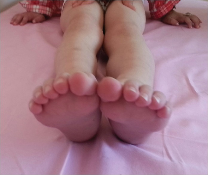
A 4-year-old girl was admitted for the investigation for delayed walking. She started to walk at the age of 2.5 years. Since she started to walk, she had tip toe walking, clumsy feet, and easy fatigability while climbing stairs. She was easily falling when she attempted to perform fast run. The symptoms increased at times when she had an infection. She was born at term with a birth weight of 3150 g. The parents were consanguineous. Neurologic examination revealed esotropia in the left eye, mild spasticity in lower extremities with increased deep tendon reflexes, bilateral clonus, and Babinski sign. Brain magnetic resonance imaging revealed bilateral punctuate hyperintense T2 signals in the basal ganglia with minor lactate peak on spectroscopy. Plasma lactic acid level was minimally elevated (2.92 [0.5–2.2] mmol/L). Plasma acylcarnitine analysis was normal and urine organic acid analyses revealed a slight increase in excretion of ethylmalonic acid (41 [1.7–14.6] mmol/mol creatinine). There was no history of diarrhea, petechiae, ecchimoses and acute metabolic decompensation. Molecular analysis revealed a homozygous c. 3 G >T mutation in the ETHE1 gene, previously reported in association with ethylmalonic encephalopathy, which alters the initiator methionine codon. After the genetic diagnosis, we observed that the patient had mild orthostatic acrocyanosis in feet [Figure 1]. Riboflavine (100 mg/day) and coenzyme Q10 (20 mg/kg/day) were started.
Figure 1.

Acrocyanosis of the feet
Discussion
Ethylmalonic encephalopathy is a rare neurometabolic disorder caused by mutations in the ETHE1 gene. Neurodevelopmental delay and regression, pyramidal and extrapyramidal involvement, episodes of acrocyanosis, recurrent petechiae and chronic diarrhea are cardinal features of the disease.[1] Characteristic metabolic findings include lactic acidemia, elevated plasma C4 and C5 acylcarnitines, C4-5 acyglycines and remarkable ethylmalonic aciduria. Brain magnetic resonance findings are not considered specific for the disorder and include frontotemporal atrophy and areas of high signal in the caudate nuclei, putamina and posterior fossa. Fatal neurologic deterioration due to intercurrent infection frequently occurs in infancy or early childhood, but a few children with a milder, chronic form of this disorder have been reported.[2] Our patient had milder form of the disease with prominent pyramidal tract signs, bilateral punctuate hyperintense T2 signals in the basal ganglia and mildly elevated serum lactic acid and urinary ethylmalonic acid. Not all patients with ethylmalonic encephalopathy have classical findings like recurrent petechia, orthostatic acrocyanosis and chronic diarrhea leading to diagnosis. We observed mild orthostatic acrocyanosis just after the genetic confirmation of the disease.
Acrocyanosis is a peripheral vascular disorder characterized by bluish discoloration of the skin and mucous membrane due to diminished oxyhemoglobin. It is caused by chronic vasospasm of small cutaneous arteries and arterioles along with compensatory dilatation in the capillary and postcapillary venules. It can be both primary and secondary to psychiatric, neurologic, autoimmune, infective and metabolic causes. Inherited metabolic disorders causing acrocyanosis include fucosidosis, hyperoxaluria type I and mitochondrial disorders with onset in early infancy and multisystem involvement. Orthostatic acrocyanosis is one of the important signs of ethylmalonic encephalopathy.[3] ETHE1 encodes a mitochondrial sulfur dioxygenase that takes part in aerobic energetic exploitation of and detoxification from sulfide. Sulfide can act as a vasodilator thus explaining the acrocyanosis and can be toxic for the microvessels thus explaining the petechiae.[4]
In conclusion, acrocyanosis may be subtle in cases with ethylmalonic encephalopathy, it should be questioned and examined in patients presenting with motor delay and bilateral hyperintense lesions in the basal ganglia.
Footnotes
Source of Support: Nil.
Conflict of Interest: None declared.
References
- 1.Ozand PT, Rashed M, Millington DS, Sakati N, Hazzaa S, Rahbeeni Z, et al. Ethylmalonic aciduria: An organic acidemia with CNS involvement and vasculopathy. Brain Dev. 1994;16(Suppl):12–22. doi: 10.1016/0387-7604(94)90092-2. [DOI] [PubMed] [Google Scholar]
- 2.Grosso S, Mostardini R, Farnetani MA, Molinelli M, Berardi R, Dionisi-Vici C, et al. Ethylmalonic encephalopathy: Further clinical and neuroradiological characterization. J Neurol. 2002;249:1446–50. doi: 10.1007/s00415-002-0880-4. [DOI] [PubMed] [Google Scholar]
- 3.Das S, Maiti A. Acrocyanosis: An overview. Indian J Dermatol. 2013;58:417–20. doi: 10.4103/0019-5154.119946. [DOI] [PMC free article] [PubMed] [Google Scholar]
- 4.Tiranti V, Zeviani M. Altered sulfide (H (2) S) metabolism in ethylmalonic encephalopathy. Cold Spring Harb Perspect Biol. 2013;5:a011437. doi: 10.1101/cshperspect.a011437. [DOI] [PMC free article] [PubMed] [Google Scholar]


