Abstract
Primary chondroid tumors of the larynx represent less than 1% of all laryngeal tumors. Most of them are chondromas and they often involve to the cricoid cartilage. They are characterized by a low tendency to metastatic diffusion (low grade). The treatment of choice is surgery, which may be endoscopic or “open partial surgery”, if extension of the cancer is limited. Prognosis is generally good. In this report, two cases of low grade chondrosarcoma of the larynx are presented, one was treated surgically with cricoidectomy and partial laryngectomy, and another was treated surgically with hemicricoidectomy. Laryngoscopy reveals tumefaction of the larynx, covered by intact mucosa. Computerized tomography imaging with contrast and magnetic resonance imaging defines not only coarse calcifications, pathognomonic of chondromatous neoformations but also the relationship of the neoformation with the surrounding tissues. Treatment is essentially surgical, given the importance of preserving the larynx to patients’ quality of life, the only risk is recurrence, which is treated by a second surgery.
Keywords: Chondrosarcoma, larynx, laryngectomy
Introduction
Chondrosarcomas are slow-growing invasive tumors. They represent approximately 11% of all primary malignant bone tumors. Only 1 to 12% of chondrosarcomas occur in the head and neck region representing 0.1% of its neoplasms [1]. Chondroma and low-grade chondrosarcoma are most common types [2]. These tumors are localized in posterior lamina of cricoids cartilage [1]. Malignforms generally are seen in old and male patients [2]. The most common symptoms are dyspnea, hoarseness, and compression to the neighbor tissues.
Herein we report two cases of low grade chondrosarcoma of the larynx, one was treated surgically with cricoidectomy and partial laryngectomy, another one was treated surgically with hemicricoidectomy.
Case report
Case 1
A 59-years-old female presented with a history of progressive hoarseness of voice for 3 months. Enquiry revealed that the patient had earlier sought medical intervention about 58 months ago for gradually increasing hoarseness of voice, and underwent hemicricoidectomy and partial laryngectomy, the histopathological report suggested chondroma of the larynx. The patient was followed up for 55 months. He presently is symptom free without any sign of recurrence. Three months before attending our Department with gradually increasing hoarseness of voice.
A smooth swelling involving the left arytenoid had been found by direct laryngoscopy. Computerized tomography (CT) scan of larynx done subsequently showed an isodense soft tissue mass arising from the left arytenoid cartilage and involving the cricoid cartilage with evidence of erosion and areas of mottled calcification (Figure 1). There was no evidence of enlarged cervical lymph nodes. Magnetic resonance imaging (MRI) was then performed, at arytenoid cartilage level, showed a low signal in T1 and T2 dependent sections, but appeared globally located within a thickening of solid tissue with an overall diameter of 2 cm, the formation showed clear margins and no infiltrations of the adjacent surgical area was observed. The latero-cervical soft parts were normal and no adenopathy was observed (Figure 2).
Figure 1.
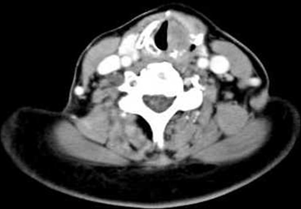
Computerized tomography (CT) scan showing mass arising from the left arytenoid with areas of mottled calcification and erosion.
Figure 2.
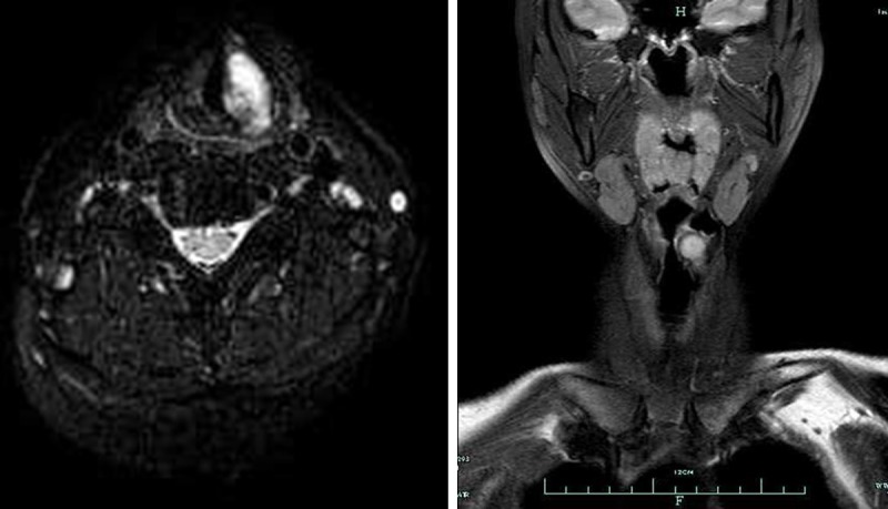
Magnetic resonance imaging (MRI) scanning showed mass arising from the left arytenoid cartilage, and showed clear margins and no infiltrations of the adjacent surgical area.
Biopsy was taken and the histopathological report now came as chondrosarcoma of the larynx (Low grade, well differentiated) (Figure 3). Metastatic work up was done, which revealed no evidence of metastasis.
Figure 3.
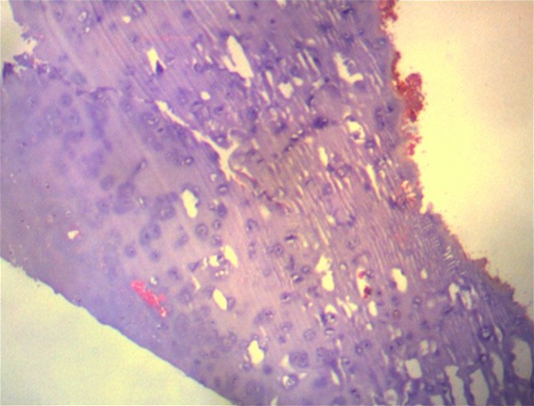
Histopathology showing lobular and patchily hypercellular proliferation of atypical chondrocytes. Magnification × 100.
As less than 50% of the cricoid cartilage was involved, hemicricoidectomy, partial laryngectomy, and a permanent tracheostomy was done under general anesthesia. The strap muscles of the neck were opened to reconstruction laryngeal functions. A smooth, mucus membrane covered 2 cm × 2 cm swelling arising from the left arytenoid was found extending towards the glottis, causing narrowing of the airway with slight distortion of the laryngeal anatomy. Post-operative period was uneventful. The patient attended a follow-up CT one year after surgery. CT showed no evidence of recurrence (Figure 4).
Figure 4.
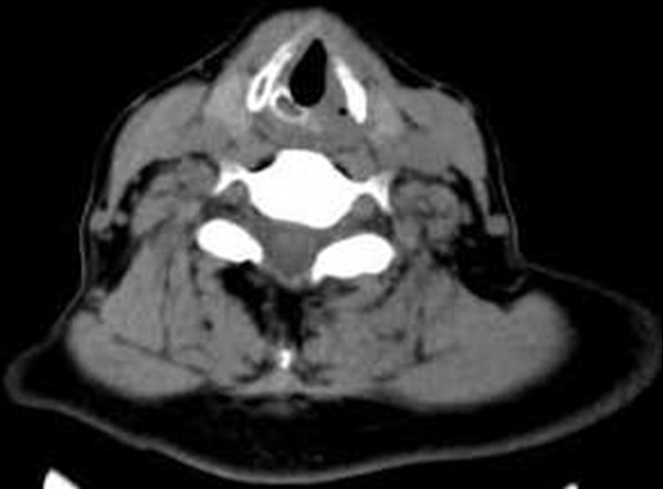
CT scan showed no evidence of recurrence after one year postoperation.
Case 2
A 34-year-old male, non-smoker, arrived at our Department with Pharyngeal foreign body sensation which had been slowly increasing for about two weeks. The indirect laryngoscopic test with flexible fibre optics showed a posterior left paramedian subglottic tumefaction at cricoid cartilage level, surrounded by intact mucosa (Figure 5). Cordal mobility was preserved and the laryngeal respiratory space was good. No adenopathy was found upon palpation at laterocervical level.
Figure 5.
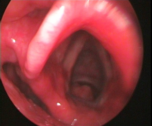
Endoscopic examination shown a mass was covered by intact mucosa. The lesion was scraped from posterior wall of cricoid cartilage.
The patient underwent neck computerized tomography (CT) with contrast medium using the spiral technique with 3 mm scans which showed: “caudally at glottis plan, at cricoid cartilage level, a coarse calcification, behind the aerial canal and at the larynx-trachea junction, diameter approximately 2 cm, in a left para-median location, with slight tumefaction of the adjacent soft tissue. No infiltrations of adjacent surgical plans were detected. More caudally, the trachea seemed in axis and with normal para-tracheal adipose tissue. At the latero-cervical location, the structures related to vascular-nervous bundles were normal and no adenopathies were observed” (Figure 6).
Figure 6.
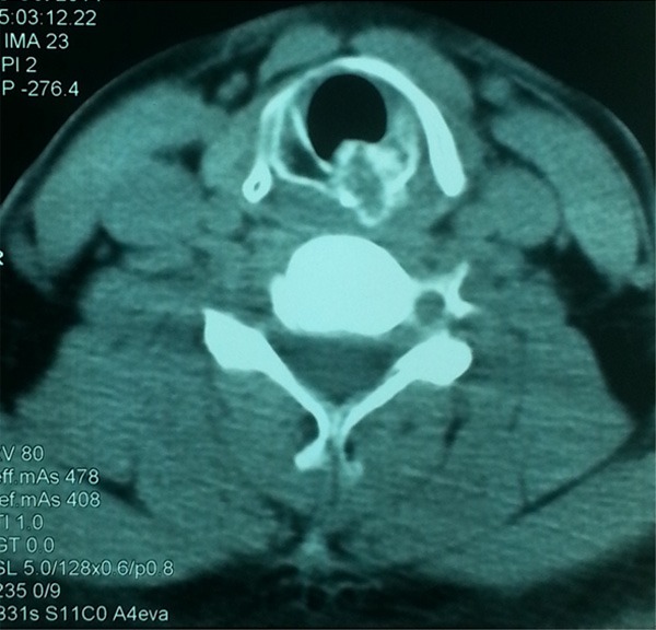
Computerized tomography (CT) scan showing mass arising from the left cricoid with areas of mottled calcification and erosion.
Magnetic resonance imaging (MRI) was then performed, with thin multiplane sections T1 and T2 dependent: “the coarse calcification, already highlighted at CT, at cricoid cartilage level, showed a low signal in T1 and T2 dependent sections, but appeared globally located within a thickening of solid tissue with an overall diameter of 2 cm, which clearly filled the back-cricoid space, with consequent reduction of the lumen of the anterior aerial canal and with compression of the soft parts of the pre-cervical area. The formation showed clear margins and no infiltrations of the adjacent surgical area was observed. The coarse calcification was, therefore, likely part of a larger solid structure, similar to a chondromatous neoformation. The latero-cervical soft parts were normal and no adenopathy was observed” (Figure 7).
Figure 7.
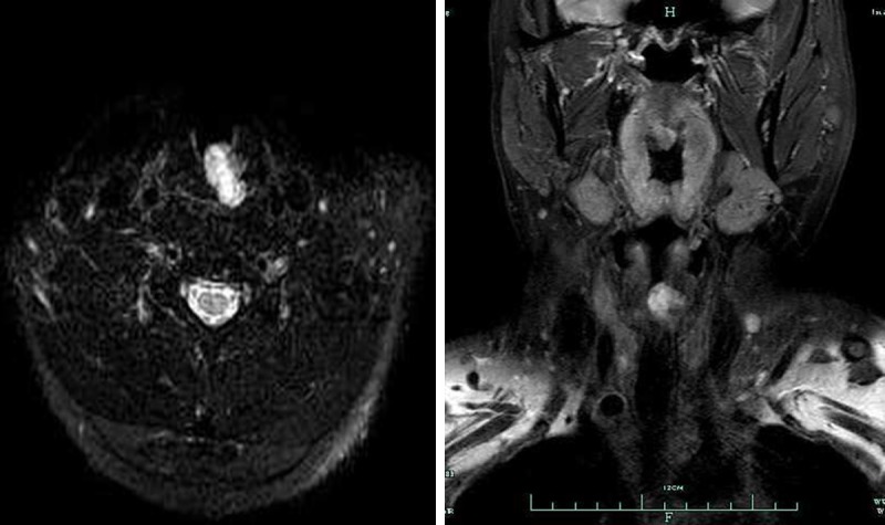
Magnetic resonance imaging (MRI) scanning showed a high signal intensity mass indicating chondroid matrix arising from the left cricoid cartilage.
During direct microlaryngoscopy, a biopsy was collected, which is fundamental for low malignancy chondrosarcoma.
On the basis of the histological and radiological examinations performed, conservative functional surgery was planned in “open surgery”. The patient, under general anaesthesia, after tracheotomy, underwent hemicricoidectomy.
The final histological examination confirmed the diagnosis of well-differentiated I grade chondrosarcoma of the larynx (Figure 8).
Figure 8.
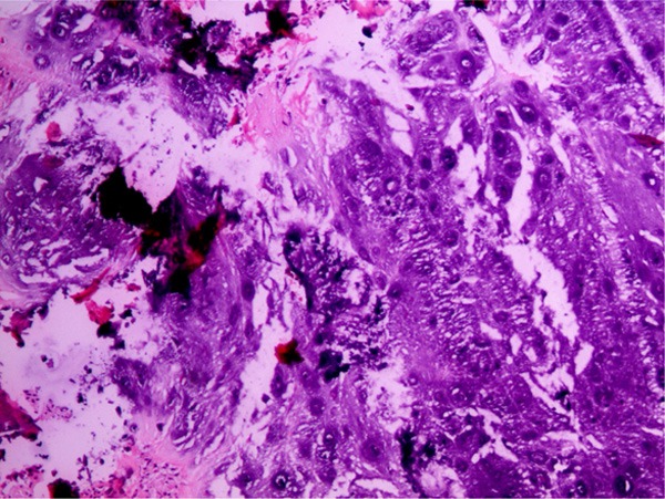
Histopathology showing hypocellular and homogenous appearance of tumor. Tumor cells are small, oval-round shaped (no mitosis and atypia). Magnification × 200.
The patient was followed up for 30 months. He presently is symptom free without any sign of recurrence.
Discussion
chondrosarcoma of the larynx is an uncommon tumor, accounting for approximately 1% of all laryngeal neoplasms [2]. The etiology of laryngeal chondrosarcoma is unknown, although it is usually assumed to derive from disordered ossification of the laryngeal cartilage [3,4]. No relationship to tobacco use or alcohol consumption has been proved [2].
The mean age at diagnosis is 60 to 64 years, with a male predominance [5,6]. Symptoms vary, depending on the location of the mass, and include hoarseness (most patients) caused by narrowing of the glottic plane and compression of the inferior laryngeal nerves; dyspnea and airway obstruction due to endolaryngeal and subglottic growth; dysphagia due to extralaryngeal growth, originating in the posterior cricoid; and painless neck mass due to tumor involvement of the thyroid cartilage (when present) [2-4].
The tumor arises from hyaline-and not elastic-cartilage: cricoid cartilage in 75% of patients, thyroid cartilage in 17%, arytenoid cartilage in 5%, and epiglottis and accessory cartilages in 3% [4]. The tumor in our case originated from the left arytenoid cartilage and left cricoid cartilage, respectively.
Imaging studies have some diagnostic value, although it is impossible to distinguish chondromas from chondrosarcomas [7]. The imaging modality of choice is computed tomography. Typical findings are a hypodense, well-circumscribed. Magnetic resonance imaging has the added advantage of superior contrast resolution of the tumor and paralaryngeal tissues [8].
Chondroid tumors of larynx can be easily recognized histologically, because of their characteristic features. But morphologic features must be well known for differential diagnosis between subtypes. Histological findings of chondrosarcomas demonstrate hyaline cartilage of variable degrees of cellularity, anaplasia, and mitotic index [9]. Cellularity, mitotic activity, and nuclear size are key features in grading and distinguishing benign from malignant tumors. Microscopically chondromas are hypocellular (30-40 nuclei/HPF) lesions with homogenous lobular growing pattern. They have no nuclear atypia and mitosis. Increased cellularity (more than 40 nuclei/HPF), nuclear atypia, binucleation, mitosis, and pleomorphism are variable in low- and high-grade chondrosarcomas. Our lesion was characterized by hypocellularity, no nuclear atypia, mitosis, binucleation, and pleomorphism. We diagnosed this lesion as “laryngeal chondroma” with these morphologic findings.
The main treatment of laryngeal cartilaginous tumors is surgical excision. Surgery that protects laryngeal functions is recommended if possible [10,11]. But 75% of laryngeal CS develops from cricoid cartilage which is considered crucial for normal laryngeal function and to protect laryngeal functions is not possiblemostly [12]. Total laryngectomy is recommended for CS destructing more than half of the cricoid cartilage. Total cricoidectomy protecting laryngeal functions is also defined by de Vincentiis et al. [13]. They mentioned 3 patients who underwent total cricoidectomy with an end to-end an astomosis between the remaining larynx and the trachea for the treatment of low-grade CS. Our study confirms other observations in the literature that radical surgery is usually unnecessary for laryngeal chondrosarcoma because of the relatively benign course of the disease [9,10]. In our series, the disease recurred in the patient with 58 months postoperative, and second surgery was performed by hemicricoidectomy and partial laryngectomy. None of our patients had distant metastases.
Anastomosis or reconstruction by free flap is used to close the defect. In our cases, the strap muscles of the neck were opened to reconstruction laryngeal functions. Recurrence is common, with rates of 35-40%. Nevertheless, the long-term prognosis of laryngeal chondrosarcoma is good (95%, 10 year survival) and metastasis is rare (up to 10%) [10]. We recommend long term follow-up, as one recurrence in our series developed 58 months after the initial diagnosis.
In conclusion, histopathologically, differential diagnosis of laryngeal chondromas should be planned very carefully. Especially differential diagnosis between chondromas and low-grade chondrosarcomas is important for planning of treatment. Given the importance of preserving the larynx to patients’ quality of life, the only risk is recurrence, which is treated by a second surgery. In our series, there were no metastases, and no patient died from the disease. We suggested that the differential diagnosis of chondromas must be done carefully and the follows-up of patients must be planned more frequently. Radical surgery is usually unnecessary for laryngeal chondrosarcoma because of the relatively benign course of the disease.
Disclosure of conflict of interest
None.
References
- 1.Coca-Pelaz A, Rodrigo JP, Triantafyllou A, Hunt JL, Fernández-Miranda JC, Strojan P, de Bree R, Rinaldo A, Takes RP, Ferlito A. Chondrosarcomas of the head and neck. Eur Arch Otorhinolaryngol. 2014;270:2601–9. doi: 10.1007/s00405-013-2807-3. [DOI] [PubMed] [Google Scholar]
- 2.Thompson LD, Gannon FH. Chondrosarcoma of the larynx: a clinicopathologic study of 111 cases with a review of the literature. Am J Surg Pathol. 2002;26:836–51. doi: 10.1097/00000478-200207000-00002. [DOI] [PubMed] [Google Scholar]
- 3.Brandwein M, Moore S, Som P, Biller H. Laryngeal chondrosarcomas: a clinicopathologic study of 11 cases, including two “dedifferentiated” chondrosarcomas. Laryngoscope. 1992;102:858–67. doi: 10.1288/00005537-199208000-00004. [DOI] [PubMed] [Google Scholar]
- 4.Casiraghi O, Martinez-Madrigal F, Pineda-Daboin K, Mamelle G, Resta L, Luna MA. Chondroid tumors of the larynx: a clinicopathologic study of 19 cases, including two dedifferentiated chondrosarcomas. Ann Diagn Pathol. 2004;8:189–97. doi: 10.1053/j.anndiagpath.2004.04.001. [DOI] [PubMed] [Google Scholar]
- 5.Thompson LD. Chondrosarcoma of the larynx. Ear Nose Throat J. 2004;83:609. [PubMed] [Google Scholar]
- 6.Cohen JT, Postma GN, Gupta S, Koufman JA. Hemicricoidectomy as the primary diagnosis and treatment for cricoid chondrosarcomas. Laryngoscope. 2003;113:1817–19. doi: 10.1097/00005537-200310000-00029. [DOI] [PubMed] [Google Scholar]
- 7.Gil Z, Fliss DM. Contemporary management of head and neck cancers. IMAJ Isr Med Assoc J. 2009;11:296–300. [PubMed] [Google Scholar]
- 8.Mishell JH, Schild JA, Mafee MF. Chondrosarcoma of the larynx. Diagnosis with magnetic resonance imaging and computed tomography. Arch Otolaryngol Head Neck Surg. 1990;116:1338–41. doi: 10.1001/archotol.1990.01870110110016. [DOI] [PubMed] [Google Scholar]
- 9.Buda I, Hod R, Feinmesser R, Shvero J. Chondrosarcoma of the larynx. Isr Med Assoc J. 2012;14:681–4. [PubMed] [Google Scholar]
- 10.Thompson LD, Gannon FH. Chondrosarcoma of the larynx: a clinicopathologic study of 111 cases with a review of the literature. Am J Surg Pathol. 2002;26:836–851. doi: 10.1097/00000478-200207000-00002. [DOI] [PubMed] [Google Scholar]
- 11.Oliveira JF, Branquinho FA, Monteiro AR, Portugal ME, Guimarães AM. Laryngeal chondrosarcoma-ten years of experience. Braz J Otorhinolaryngol. 2014;80:354–8. doi: 10.1016/j.bjorl.2014.05.004. [DOI] [PMC free article] [PubMed] [Google Scholar]
- 12.Sauter A, Bersch C, Lambert KL, Hörmann K, Naim R. Chondrosarcoma of the larynx and review of the literature. Anticancer Res. 2007;27:2925–2929. [PubMed] [Google Scholar]
- 13.de Vincentiis M, Greco A, Fusconi M, Pagliuca G, Martellucci S, Gallo A. Total cricoidectomy in the treatment of laryngeal chondrosarcomas. Laryngoscope. 2011;121:2375–2380. doi: 10.1002/lary.22337. [DOI] [PubMed] [Google Scholar]


