Abstract
Background
Advanced laparoscopy for pancreatic cancer surgery should include laparoscopic ultrasound (LUS), in order to accurately evaluate resectability and rule out the presence of undetected metastases and/or vascular infiltration. LUS should be done as a preliminary step whenever pre-operative imaging casts doubts on resectability.
Patients and methods
We hereby report our experience of 18 consecutive patients, aged 43–76, coming to our attention during a six months period (Jan–Jun 2013), with a diagnosis of pancreas head or body cancer.
Results
LUS allowed to rule out undetected metastases or mesenteric vessels infiltration in 11 patients (61.1%), who were submitted, as previously scheduled, to radical duodeno-pancreatectomy (9 cases) and spleno-caudal pancreatectomy (2 cases). Among the remaining patients, three had been correctly evaluated as non resectable radically at pre-operative work out, and confirmed at LUS, while LUS detected non resectable disease in further 4 patients (22.2%), who underwent palliative procedures. In 2 patients of this group liver micro-metastases were found, while 2 were excluded because of mesenteric vessels infiltration.
Conclusions
LUS provided a higher level of diagnostic accuracy, allowing in our experience to exclude 4 patients from radical surgery (22.2%). The evaluation of surgical resectability is an issue of crucial importance to decide surgical strategy in pancreas tumor surgery. In our opinion LUS should be considered a mandatory step in laparoscopic approach to pancreatic tumors, to better define disease staging and evaluate resectability.
Keywords: Ultrasonography (laparoscopic), Pancreatic neoplasms, Pancreas surgery
Introduction
The evaluation of pancreatic cancer resectability is an issue, when considering a patient for radical surgery.
Although advances in radiologic imaging techniques have determined better definition and staging of disease, still doubts remain on undetected peritoneal dissemination, liver micro-metastases, mesenteric vessels infiltration, lymphnodes involvement. It has been suggested that staging laparoscopy (1–4) may be useful in patients with locally advanced disease, in order to better plan surgical procedures.
The aim of our study was to retrospectively assess the effectiveness and contribution of laparoscopy and laparoscopic ultrasound, to adjust surgical approach in these patients.
Patients and methods
Eighteen consecutive patients, aged 43–76, came to our observation in the period January–June 2013, with a diagnosis of pancreatic head or body cancer.
Complete pre-operative imaging work out was performed, including contrast CT and ultrasound. In all patients laparoscopic ultrasonography was performed at the beginning of surgical procedure, by means of an Aloka ProSound Alpha7 device (Hitachi Aloka Medical Ltd. Tokyo, Japan), equipped with an electronic intra-operative flexible laparoscopic probe with a 38 mm field side view, with a frequency range of 4–11 Mhz, sterilized by means of 58% density hydrogen peroxide (Sterrad®). Adherence to the pancreatic surface was provided by tilting the flexible tip of the probe.
11 patients were judged to be radically resectable at pre-operative work out, laparoscopy and LUS confirmed this finding, and the patients underwent duodeno-pancreatectomy (9 cases) or caudal spleno-pancreatectomy (2 cases).
All the intra-operative examinations were performed by two surgeons of the surgical team (D.P. and A.S.), specially trained in intra-operative ultrasound.
3 patients were deemed non resectable at pre-operative study, laparoscopy and LUS confirmed non resectability.
4 further patients, in whom resectability was questionable, were excluded by means of laparoscopy and LUS, two because of undetected liver micro-metastases, two because of infiltration of mesenteric vessels. In all these patients palliative surgery was performed, consisting of hepatico-jejunostomy and gastro-jejunostomy.
Results
Laparoscopy and LUS confirmed pre-operative results in 14 patients (77.7%), 11 of whom were radically resected, while three were submitted to palliative procedures (Tab. 1).
Table 1.
RESECTABILITY IN 18 PANCREATIC CANCER (14 HEAD + 4 BODY NEOPLASMS).
| Resectable | Unresectable | Doubt | |
|---|---|---|---|
| Contrast CT +US | 11 | 3 | 4 |
| LUS | 11 | 7 | |
| Surgery | 11 | 7 | |
| Concordance LUS/surgery | 100% | 100% |
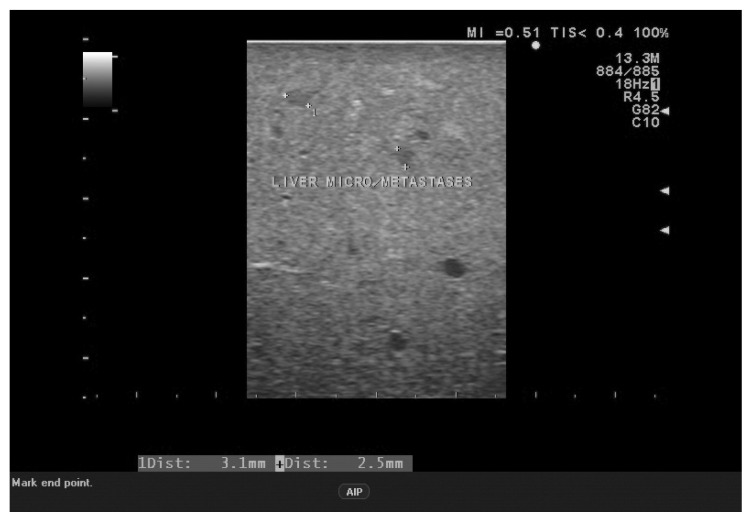
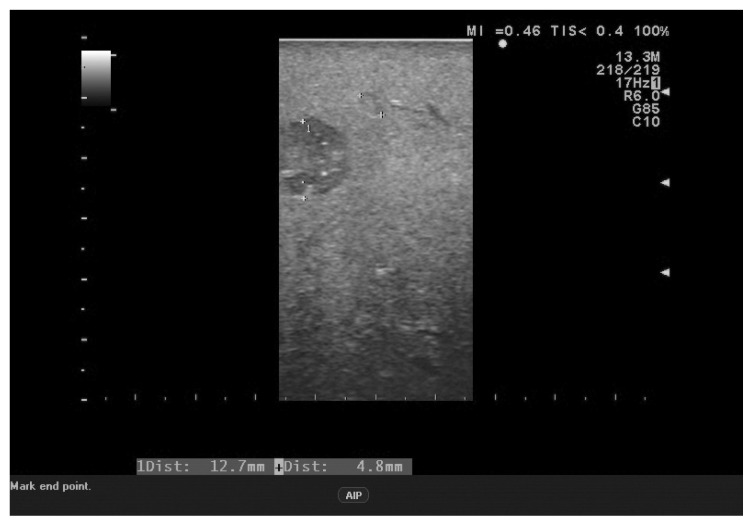
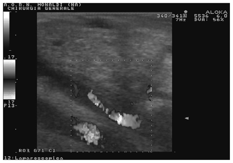
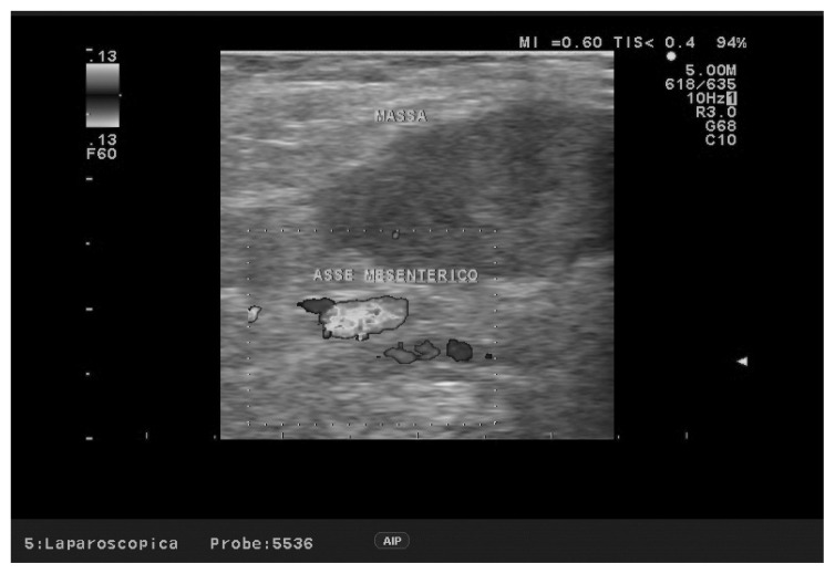
In four patients, in whom resectability was questionable, laparoscopy and LUS allowed to rule out the possibility of radical surgery (22.2%), showing the presence of non detected intra-hepatic micro-metastases in 2 cases (Figures 1, 2), and infiltration of mesenteric vein in two cases (Figures 3, 4).
Fig. 1.
LUS: undetected hepatic micro-metastases.
Fig. 2.
LUS: hypoechoic hepatic nodules suspect for metastases. Intra-operative ultrasonic-guided biopsy was performed.
Fig. 3.
LUS with duplex Doppler showing impingement of pancreatic tumor into the mesenteric vein.
Fig. 4.
LUS with duplex Doppler showing stenosis of the superior mesenteric vein due to tumor compression.
Discussion
Although the routine use of laparoscopy and LUS in pancreatic cancer surgery is not universally supported (5, 6), in our experience these techniques allowed for better definition of pathology and decision of surgical strategy in almost one fourth of the patients.
For these reasons we strongly recommend to perform laparoscopy and LUS as a first step in all these patients. This attitude is supported by the experience of Long (7) who reported a sensitivity of 78% for LUS and 93% for open ultrasound, in determining pancreas tumor resectability, thus facilitating intra-operative decision making.
Doucas (1) and Norton (8) reported 44% change of surgical management with laparoscopy and ultrasound in patients with pancreatic malignancy.
Liu and Mayo (3, 4) reported the finding of occult tumor during staging laparoscopy in 34% and 27.6% of the patients, respectively.
Menack (9) reported that in 26% of the patients laparoscopy defined unresectable disease and LUS further identified three non resectable patients (portal vein occlusion and non detected liver or lymphnodes metastases).
Cirimbei (10) reported change in therapeutic attitude with intra-operative ultrasound (open and laparoscopic) in more than 12% of the patients.
Zhao (11) reported avoidance of laparotomy in 5 patients, out of 22, with laparoscopy and LUS.
Laparoscopy is able to detect peritoneal carcinomatosis, while the addition of LUS allows definition of extent of the primary tumor, involvement of liver parenchyma and surrounding organs and infiltration (encasement or impingement) of vascular structures (12, 13).
Moreover the low costs of these techniques, which require the insertion of just two 10mm. trocars, laparoscopic optics and a dedicated ultrasound linear flexible probe, compare favourably with the costs of useless laparotomies.
Although the infiltration of the mesenteric vein is not considered an absolute contra-indication to resection, we recommend, in accordance with other groups (14) that LUS finding of vein involvement should suggest a variable surgical approach, which can be either palliative or radical, including vascular resection, according to patient’s life expectancy.
Intra-operative ultrasound can be repeated, if necessary, after conversion to open procedure, but in our experience it gave no further information as compared to LUS.
In our experience laparoscopy and laparoscopic ultrasound prevented a significant number of patients from undergoing open surgery, thus giving an essential support to the staging of pancreatic tumors.
Footnotes
Disclosures
The Authors: Domenico Piccolboni, Anna Settembre, Pierluigi Angelini, Francesco Esposito, Simona Palladino, Francesco Corcione have no conflicts of interest and received no benefits related to the present manuscript. No specific funding resource was utilized for this paper.
References
- 1.Doucas H, Sutton CD, Zimmerman A, Dennison AR, Berry DP. Assessment of pancreatic malignancy with laparoscopy and intraoperative ultrasound. Surg Endosc. 2007;21(7):1147–1152. doi: 10.1007/s00464-006-9093-8. [DOI] [PubMed] [Google Scholar]
- 2.Shoup MG, Winston C, Brennan MF, Bassman D, Conlon KC. Is there a role for staging laparoscopy in patients with locally advanced, unresectable pancreatic adenocarcinoma? J Gastrointest Surg. 2004;8:1068–1071. doi: 10.1016/j.gassur.2004.09.026. [DOI] [PubMed] [Google Scholar]
- 3.Liu RC, Traverso LW. Diagnostic laparoscopy improves staging of pancreatic cancer deemed locally unresectable by computed tomography. Surg Endosc. 2005;19(5):638–642. doi: 10.1007/s00464-004-8165-x. [DOI] [PubMed] [Google Scholar]
- 4.Mayo SC, Austin DF, Sheppard BC, Mori M, Shipley DK, Billingsley KG. Evolving preoperative evaluation of patients with pancreatic cancer: does laparoscopy have a role in the current era? J Am Coll Surg. 2009;208(1):87–95. doi: 10.1016/j.jamcollsurg.2008.10.014. [DOI] [PubMed] [Google Scholar]
- 5.Ni Mhuircheartaigh JM, Sun MRM, Callery MP, Siewert B, Vollmer CM, Kane RA. Pancreatic Surgery: a multidisciplinary assessment of the value of intra-operative ultrasound. Radiology. 2013;266(3):945–955. doi: 10.1148/radiol.12120201. [DOI] [PubMed] [Google Scholar]
- 6.Barabino M, Santambrogio R, Pisani Ceretti A, Scalzone R, Montorsi M, Opocher E. Is there still a role for laparoscopy combined with laparoscopic ultrasonography in the staging of pancreatic cancer? Surg Endosc. 2011;25(1):160–165. doi: 10.1007/s00464-010-1150-7. [DOI] [PubMed] [Google Scholar]
- 7.Long EE, Van Dam J, Weinstein S, Jeffrey B, Desser T, Norton JA. Computed tomography, endoscopic, laparoscopic, and intra-operative sonography for assessing resectability of pancreatic cancer. Surg Oncol. 2005;14(2):105–113. doi: 10.1016/j.suronc.2005.07.001. [DOI] [PubMed] [Google Scholar]
- 8.Norton JA. Intraoperative methods to stage and localize pancreatic and duodenal tumors. Ann Oncol. 1999;10(suppl 4):S182–S184. [PubMed] [Google Scholar]
- 9.Menack MJ, Spitz JD, Arregui ME. Staging of pancreatic and ampullary cancers for resectability using laparoscopy with laparoscopic ultrasound. Surg Endosc. 2001;15(10):1129–1134. doi: 10.1007/s00464-001-0030-6. [DOI] [PubMed] [Google Scholar]
- 10.Cirimbei S, Puscu C, Lucenco L, Bratucu E. The role of intra-operative ultrasound in establishing the surgical strategy regarding hepato-bilio-pancreatic pathology. Chirurgia. 2013;108:643–651. [PubMed] [Google Scholar]
- 11.Zhao ZW, He JY, Tan G, Wang HJ, Li KJ. Laparoscopy and laparoscopic ultrasonography in judging resectability of pancreatic head cancer. Hepatobiliary Pancreat Dis Int. 2003;2(4):609–611. [PubMed] [Google Scholar]
- 12.Piccolboni D, Ciccone F, Settembre A, Corcione F. The role of echo-laparoscopy in abdominal surgery: five years’ experience in a dedicated center. Surg Endosc. 2008;22(1):112–117. doi: 10.1007/s00464-007-9382-x. [DOI] [PubMed] [Google Scholar]
- 13.Piccolboni D, Ciccone F, Settembre A, Corcione F. Laparoscopic intra-operative ultrasound in liver and pancreas resection: analysis of 93 cases. J Ultrasound. 2010;13:3–8. doi: 10.1016/j.jus.2010.06.001. [DOI] [PMC free article] [PubMed] [Google Scholar]
- 14.Sun MRM, Brennan DD, Kruskal JB, Kane RA. Intraoperative ultrasonography of the pancreas. RadioGraphics. 2010;30:1935–1953. doi: 10.1148/rg.307105051. [DOI] [PubMed] [Google Scholar]


