

Abstract
Objectives
This study presents the successful posterior surgical reduction and fusion on a 26-month-old child with chronic unilateral locked facet joint and spinal cord injury (SCI).
Methods
A 26-month-old child with chronic unilateral locked facet joint and SCI treated by posterior surgical reduction and fusion. Plaster external fixation was applied and rehabilitation exercise was trained post-operatively.
Results
Chronic unilateral locked facet joint was reduced successfully and bone fusion of C4/5 was achieved 3 months after surgery. The function of both lower limbs was improved 1 year after surgery, aided with physical rehabilitation.
Conclusion
Unilateral locked facet joint in pediatric population is rare. Few clinical experiences were found in the literature. Non-surgical treatment has advantages of not being invasive and is preferred for acute patients; however, it may not be suitable for chronic unilateral locked facet joint with SCI, in which surgical intervention is needed.
Keywords: Unilateral locked facet joint, Pediatric population, Case report
Introduction
Unilateral locked facet joint in the pediatric population is rare.1 Asymptomatic unilateral locked facet joint of the pediatric population may be difficult to diagnose, however, neurological symptoms will indicate to doctors the possibility of spinal cord injury (SCI).
Therefore, for most unilateral locked facet joint with SCI, accurate diagnosis can be established at the acute phase. Non-surgical treatment may be enough for these patients, thus surgical intervention can be avoided.1
In some rare conditions, like our present case, combined unilateral locked facet joint with lower limb injury, the reduced activities of lower limbs was misdiagnosed and was considered as caused by lower limb injury.
The unilateral locked facet joint with SCI was not diagnosed until many weeks after injury, and the optimal time for treatment was missed. If pediatric unilateral locked facet joint lasts more than 3 weeks, it is defined as chronic unilateral locked facet joint.
Lack of clinical experience and few available descriptions in the literature leave unclear as how to manage chronic pediatric unilateral locked facet joint. Here, we present a 26-month-old child with unilateral C4/5 chronic unilateral locked facet joint and treated by posterior surgical reduction and fusion.
Case report
A 26-month-old girl experienced high frequency of crying, reduced activities of both lower limbs, and pain of left lower limb after motor vehicle accident. The X-ray at another hospital showed fracture of left femoral shaft, therefore the reduced activities of the other lower limb was ignored. She was treated only for left femoral shaft fracture in the other hospital. Twenty-five days after the accident, the parents of the child found that the reduced activities of both lower limbs had still not significantly improved; at that time, the new symptom of frequent micturition appeared in the patient, and she was transported to the Department of Pediatrics of our hospital, where we were invited for consultation to discover the reasons for the reduced activities of both lower limbs.
Normal muscle strength was found in both upper limbs, and graded 1 of both lower limbs by physical examination. Magnetic resonance imaging (MRI) of the whole spinal cord was examined; the MRI showed the dislocation of C4/5 and high signal of spinal cord from C6 to T2 region on T2-weight image (Fig. 1). Two-dimensional computed tomography (CT) showed left unilateral locked facet joint of C4/5 (Fig. 2A and B).
Figure 1 .

T2-weight MRI image showed unilateral locked facet joint of C4/5 and high signal of spinal cord from C6 to T2 region.
Figure 2 .

(A/B) Pre-operative 2D-CT showed left lateral locked facet joint of C4/5.
Closed reduction was applied under general anesthesia and monitored somatosensory-evoked potential and motor-evoked potential, however, reduction could not be achieved. At last, posterior surgery was performed; reduction still could not be achieved by leverage. Part of the C4 inferior articular process was removed, and reduction was assisted by titanium mini-plate and lateral mass screws. Decortications and autogenously bone graft were performed on lamina of C4/5.
After the wound was checked for hemostasis and closure completed, the patient was immobilized by plaster external fixation (Fig. 3), and rehabilitation exercise was begun under a rehabilitative therapist.
Figure 3 .
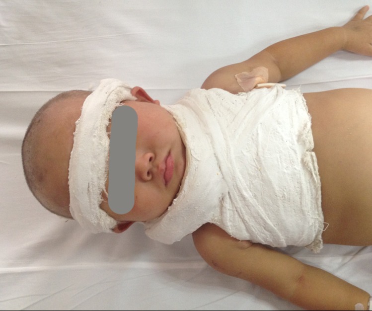
Patient was immobilized by plaster external fixation after operation.
Post-operative lateral film showed that the screws were placed well (Fig. 4). Bone fusion of C4/5 was achieved at 3 months after the operation (Fig. 5). One year after surgery, the muscle strength of both lower limbs was improved to grade 3–4, without complications of screws loosening or breaking (Fig. 6).
Figure 4 .

Post-operative lateral radiograph showed that the screws were placed well.
Figure 5 .

Bone fusion was achieved of C4/5 at 3D-CT image of 3 months after operation.
Figure 6 .

No complication of screws loosening or breakage could be observed on the lateral radiograph of 1 year after surgery.
Discussion
Because of the unique anatomic and biomechanical features of the pediatric spine, cervical spine injuries in pediatric population are not common,2 the major pediatric cervical injuries occurring in the upper cervical spine,3,4 while lower cervical spine injuries are rare.1
Hott et al.1 reported a 10-month-old boy with unilateral locked facet joint, who, fortunately, did not present any neurological symptoms. Manual reduction was applied unsuccessfully; therefore, the patient was placed in a specially designed, full-body (papoose) external orthosis, and discharged from the hospital without event. Flexion–extension radiograph obtained 2 months after injury of this patient revealed normal osseous alignment and no abnormal motion; dynamic cervical radiographs taken 18 months after injury demonstrated no instability. Therefore, external orthosis may be enough for the child with acute unilateral locked facet joint.
However, for the present case, with a patient with SCI, manual reduction having been applied unsuccessfully, we thought that unilateral locked facet joint of C4/5 was the reason for the high signal of C6-T2 spinal cord, because no other abnormality was found. We felt, therefore, that to discharge her from the hospital without any further treatment was improper. Recognizing that non-surgical treatment may be not suitable for chronic unilateral locked facet joint patient, especially one with SCI, we at last decided that surgical intervention should be performed.
For adult patients, anterior discectomy and fusion may be recommended,5 however, we lack successful experience of management of unilateral locked facet joint for children. As is well-known, the bone growth of children is very fast; because the diagnosis was made 25 days after the accident, the possibility of union between C4 inferior articular process and C5 superior articular process was considered pre-operation. Thus, posterior surgical reduction was performed.
At the time of surgery, we found that some connective tissue had grown between C4 inferior articular process and C5 superior articular process, and the reduction could not be achieved by leverage; at last, we removed part of the C4 inferior articular process, and the reduction was finally achieved with the assistance of a titanium mini-plate and lateral mass screws.
Although there are many advantages to non-surgical treatment, the articular capsule, posterior longitudinal and interspinous ligaments were damaged and the disk was torn in the patient's locked facet joint6; due to lack of the long-term follow-up evidence, the efficacy and safety of non-surgical treatment without fusion is still uncertain. For patients with SCI, especially for chronic unilateral facet dislocation lasting longer than 3 weeks, the enduring locked facet joint stiffens the soft tissue and bone construction, and reduction may not be achieved even of using the leverage by open posterior approach. In the present case, removing part of the C4 inferior articular process decreased the risk of the iatrogenic neurological injury as low as possible.
Although the preliminary results of posterior fixation and fusion were successful, the future possibility of cervical kyphosis by asymmetry of anterior and posterior spinal growth (crankshaft phenomenon) and accelerated degeneration of adjacent segment is a concern, and long-term follow-up should be pursued.
Conclusion
Unilateral locked facet joint in pediatric population is rare, and few clinical experiences could be found in the literature. Despite the advantages of non-surgical treatment for acute patients, closed reduction may be unsuccessful for patients with chronic unilateral locked facet joint, especially for those with SCI. Posterior surgery of fixation and fusion may be more suitable for them; however, long-term complications are still unpredictable.
Disclaimer statements
Contributors AMW, XYW and PL contribute to the concept and design. AMW, XYW and HZX acquisition of data or analysis and interpretation of data. AMW, XYW and YLC drafted the article. All authors revised it critically for important intellectual content and approved the version to be published.
Conflicts of interest The authors declare that they have no conflict of interest.
Ethics approval This paper has received ethical approval from the Institutional Review Board of Second Affiliated Hospital & Yuying Pediatric Hospital of Wenzhou Medical University.
Funding XYW was supported by Qianjiang Talents Project of Technology Office of Zhejiang Province (2010R10075), Natural Science Foundation of Zhejiang Province for Distinguished Young Scholars (LR12H06001) and National Nature Foundation of China (81371988).
References
- 1.Hott JS, Feiz-Erfan I, Kim LJ, Rekate HL, Sonntag VK. Nonsurgical treatment of a C6-7 unilateral locked facet joint in an infant. Case report. J Neurosurg 2004;1002 Suppl Pediatrics:220–2. [DOI] [PubMed] [Google Scholar]
- 2.Klimo P Jr, Ware ML, Gupta N, Brockmeyer D. Cervical spine trauma in the pediatric patient. Neurosurg Clin N Am 2007;18(4):599–620. [DOI] [PubMed] [Google Scholar]
- 3.Lowry DW, Pollack IF, Clyde B, Albright AL, Adelson PD. Upper cervical spine fusion in the pediatric population. J Neurosurg 1997;87(5):671–6. [DOI] [PubMed] [Google Scholar]
- 4.Eleraky MA, Theodore N, Adams M, Rekate HL, Sonntag VK. Pediatric cervical spine injuries: report of 102 cases and review of the literature. J Neurosurg 2000;92(1 Suppl):12–7. [DOI] [PubMed] [Google Scholar]
- 5.Schwarz N, Sim E. Treatment problems in unilateral locked facet syndrome of the cervical spine. Eur Spine J 1993;2(2):65–71. [DOI] [PubMed] [Google Scholar]
- 6.Crawford NR, Duggal N, Chamberlain RH, Park SC, Sonntag VK, Dickman CA. Unilateral cervical facet dislocation: injury mechanism and biomechanical consequences. Spine 2002;27(17):1858–64; discussion 64. [DOI] [PubMed] [Google Scholar]