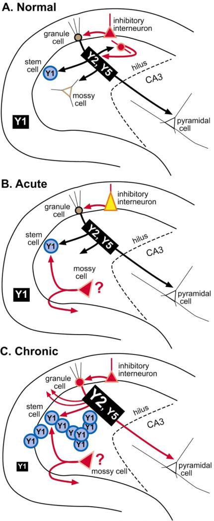
Figure 7.
Summary of NPY plasticity in the rat dentate gyrus after seizures. A summary of the changes that occur in protein and receptor expression after acute seizures and further changes after chronic seizures. A. Normal condition B. After acute seizures, NPY protein increases in inhibitory neurons, although some may also be lost due to seizure-induced neuronal death, and some may sprout collaterals, making the new NPY intemeurons network potentially novel. In addition, some hilar cells that do not normally express NPY protein may begin to do so, such as surviving mossy cells. C. After chronic seizures, granule cells and their axons, the mossy fibers, express NPY and sprout into the inner molecular layer. Y2, and possibly Y5 receptors, increase in mossy fibers, and Y1 receptors in the molecular layer appear to decrease. In addition, seizures increase the proliferation of granule cells from progenitors in the subgranular zone which express the Y1 receptor; this is likely to occur during a window between day 3–4 and 30 after status epilepticus, at least in the case of pilocarpine-induced status epilepticus [32, 34]. This coincides with the period when spontaneous seizures are beginning to occur, suggesting a role for seizure-induced neurogenesis in epileptogenesis [33].

