Abstract
We report multimodal imaging findings, including enhanced depth imaging-optical coherence tomography, in an affected child with choroidal neurofibromatosis. We identify novel features such as choroidal vessel compression from NF1-related choroidal nodules and an increased subfoveal choroidal thickness. This is the first report to use EDI-OCT to analyze choroidal features in neurofibromatosis type-1.
INTRODUCTION
With an incidence of 1 in 3000, neurofibromatosis type-1 (NF1) is the most common single-gene disorder in which affected individuals display central nervous system (CNS) abnormalities. The ophthalmic examination is a sensitive and specific screen allowing NF1 to be distinguished from other overlapping disorders. In particular, optic nerve gliomas occur in ~15% of NF1 patients,1 and Lisch nodules in ~70%.2, 3 Using invasive and non-invasive tools such as indocyanine-green (ICG) angiography and near-infrared reflectance (NIR) imaging, respectively choroidal abnormalities are observed in 80–100% of NF1-affected individuals.2, 3 Enhanced depth imaging spectral-domain optical coherence tomography (EDI-OCT), has emerged as a non-invasive technique to visualize cross-sectional architecture of the choroid in a variety of neoplastic, neovascular, and degenerative retinochoroidal disorders,4 but has not yet been applied to imaging choroidal neurofibromatosis.
We report multimodal imaging findings, including for the first time EDI-OCT, in an affected child with choroidal neurofibromatosis.
CASE REPORT
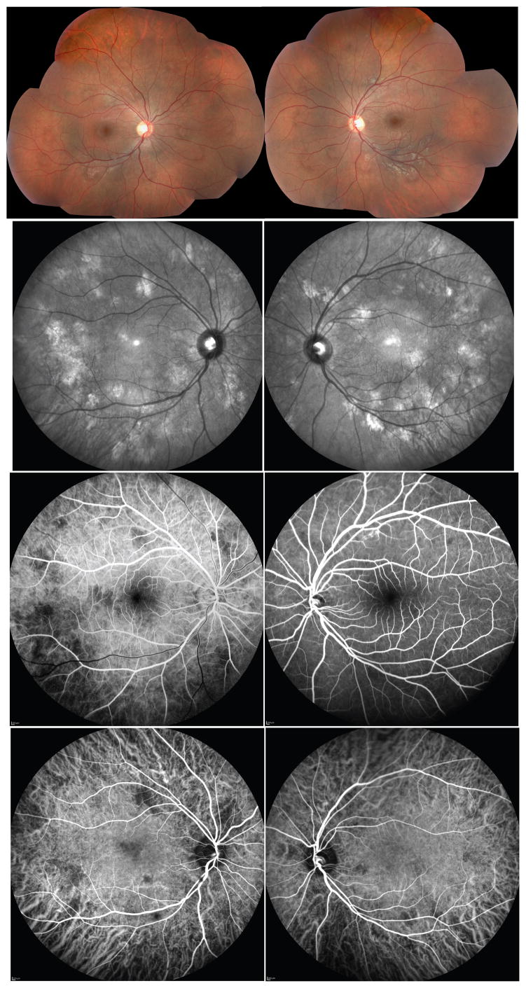
A 7-year-old girl, with a known diagnosis of NF1, presented for an ophthalmic examination. Lisch nodules (iris hamartomas) were present (data not shown). The visual acuities were 20/20 OD and 20/25 OS. There was no afferent pupillary defect. Color photos (Fig 1. A, B), fundus autofluorescence (FAF) and red-free imaging (data not shown) were unremarkable. NIR imaging demonstrated gray-white patches throughout the posterior pole (Fig 1. C, D, arrowheads). These patches corresponded to multifocal areas of hypofluorescence in the venous laminar phase of the fluorescein angiogram (FA) but were not as obvious in the later phases of the angiogram (Fig 1. E vs F, arrowheads). In contrast to FA, ICG angiography highlighted the NIR findings as corresponding patches of hypofluorescence (Fig 1. G, H, arrowheads).
Figure 1. Multimodal Imaging Findings in Choroidal Neurofibromatosis.
(A,B) Color fundus photos in the right (A) and left (B) eyes of a 7-year-old girl with NF1. (B,C) NIR imaging demonstrates multiple grey-white patches throughout the posterior poles of both eyes (arrowheads). (D) Multiple hypofluorescent foci in the venous laminar phase of FA correspond to the NIR patches in the right eye but in the late phase of FA of the left eye (E), only a few hypofluorescent foci are visible (arrowheads). (F, G) ICG angiography highlights the NIR features as corresponding patches of hypofluorescence (arrowheads).
EDI-OCT imaging through the NIR lesions (Fig 2. A, B, inset arrowheads) demonstrated hyperreflective choroidal nodules. The nodules appeared to abut the overlying choroidal and choriocapillaris tissue, with loss of choroidal lucency, suggestive of choroidal vessel compression (Fig 2. A, B, double arrowheads). However, no overlying retinal abnormalities were observed. The subfoveal choroidal thickness was 403 and 492 microns in the right and left eyes, respectively (Fig 2. C, D).
Figure 2. EDI-OCT Features in Choroidal Neurofibromatosis.
(A,B) EDI-OCT imaging through the NIR lesions (green line, insets and arrowheads) demonstrates hyperreflective choroidal nodules (arrowheads). The nodules compress the overlying choroidal vessels and choriocapillaris. (C,D, double arrowheads) The subfoveal choroidal thickness was 403 and 492 microns in the right (C) and left (D) eyes.
DISCUSSION
The patchy NIR changes and corresponding ICG hypofluorescence—in conjunction with less obvious or negative findings with dilated fundus examination, FA, FAF and red-free imaging—appear sensitive and specific for choroidal neurofibromatosis.3 For this reason, these multimodal imaging findings have been suggested as new diagnostic criteria for NF1 diagnosis.3 Our report confirms these features in an affected child. When analyzed with EDI-OCT, the high-signal NIR patches correspond to choroidal nodules that compress overlying choroidal vessels. The compression, while not significant enough to cause outer retinal thinning, may indicate ischemic choroidal foci visible with ICG angiography as punctate hypofluorescent regions. These nodules may correspond to ovoid bodies of proliferating, neoplastic Schwann cells arranged in concentric rings around axons,5 or choroidal melanocytic proliferations. Interestingly, the subfoveal choroidal thickness in this patient is greater than the reported mean choroidal thickness in healthy children.6 Perhaps the posterior pole choroidal nodules contribute to this apparent difference. However, a larger EDI-OCT study comparing subfoveal and eccentric choroidal thicknesses of NF1-affected children to healthy controls would be needed to confirm whether there is an actual difference.
Combining EDI-OCT, ICG angiography, and microperimetry would provide a unique structural and functional tool to follow these nodules as they increase in size or number with time,3 and potentially impair choroidal blood flow and compromise overlying retinal sensitivity. Together, these modalities pave the way toward a new understanding of the natural history of retinochoroidal neurofibromatosis. To our knowledge, this is the first report to use EDI-OCT to describe choroidal changes and subfoveal choroidal thickness in NF1.
Acknowledgments
R.C.R. is supported by NEI K12EY022299. The funding source had no role in the development or publication of this work.
Footnotes
CONFLICT OF INTEREST: NONE.
Contributor Information
Rajesh C. Rao, Email: rajeshcrao@gmail.com, Department of Ophthalmology & Visual Sciences, W.K. Kellogg Eye Center, Department of Pathology, University of Michigan Medical School, 1000 Wall St., Brehm Rm 8326, Ann Arbor, MI 48105 USA.
Netan Choudhry, Herzig Eye Institute, Toronto, Ontario, M5S 1R1 Canada.
References
- 1.Avery RA, Fisher MJ, Liu GT. Optic pathway gliomas. J Neuroophthalmol. 2011;31:269–278. doi: 10.1097/WNO.0b013e31822aef82. [DOI] [PubMed] [Google Scholar]
- 2.Yasunari T, Shiraki K, Hattori H, et al. Frequency of choroidal abnormalities in neurofibromatosis type 1. Lancet. 2000;356:988–992. doi: 10.1016/S0140-6736(00)02716-1. [DOI] [PubMed] [Google Scholar]
- 3.Viola F, Villani E, Natacci F, et al. Choroidal abnormalities detected by near-infrared reflectance imaging as a new diagnostic criterion for neurofibromatosis 1. Ophthalmology. 2012;119:369–375. doi: 10.1016/j.ophtha.2011.07.046. [DOI] [PubMed] [Google Scholar]
- 4.Rao RC, Choudhry N, Gragoudas ES. Enhanced depth imaging spectral-domain optical coherence tomography findings in sclerochoroidal calcification. Retina. 2012;32:1226–1227. doi: 10.1097/IAE.0b013e3182576e50. [DOI] [PubMed] [Google Scholar]
- 5.Kurosawa A, Kurosawa H. Ovoid bodies in choroidal neurofibromatosis. Arch Ophthalmol. 1982;100:1939–1941. doi: 10.1001/archopht.1982.01030040919010. [DOI] [PubMed] [Google Scholar]
- 6.Park KA, Oh SY. Choroidal thickness in healthy children. Retina. 2013;33:1971–1976. doi: 10.1097/IAE.0b013e3182923477. [DOI] [PubMed] [Google Scholar]


