Abstract
The identification of new virulence factors in Yersinia pestis and understanding their molecular mechanisms during an infection process are necessary in designing a better vaccine or to formulate an appropriate therapeutic intervention. By using a high-throughput, signature-tagged mutagenic approach, we created 5,088 mutants of Y. pestis strain CO92 and screened them in a mouse model of pneumonic plague at a dose equivalent to 5 50% lethal doses (LD50) of wild-type (WT) CO92. From this screen, we obtained 118 clones showing impairment in disseminating to the spleen, based on hybridization of input versus output DNA from mutant pools with 53 unique signature tags. In the subsequent screen, 20/118 mutants exhibited attenuation at 8 LD50 when tested in a mouse model of bubonic plague, with infection by 10/20 of the aforementioned mutants resulting in 40% or higher survival rates at an infectious dose of 40 LD50. Upon sequencing, six of the attenuated mutants were found to carry interruptions in genes encoding hypothetical proteins or proteins with putative functions. Mutants with in-frame deletion mutations of two of the genes identified from the screen, namely, rbsA, which codes for a putative sugar transport system ATP-binding protein, and vasK, a component of the type VI secretion system, were also found to exhibit some attenuation at 11 or 12 LD50 in a mouse model of pneumonic plague. Likewise, among the remaining 18 signature-tagged mutants, 9 were also attenuated (40 to 100%) at 12 LD50 in a pneumonic plague mouse model. Previously, we found that deleting genes encoding Braun lipoprotein (Lpp) and acyltransferase (MsbB), the latter of which modifies lipopolysaccharide function, reduced the virulence of Y. pestis CO92 in mouse models of bubonic and pneumonic plague. Deletion of rbsA and vasK genes from either the Δlpp single or the Δlpp ΔmsbB double mutant augmented the attenuation to provide 90 to 100% survivability to mice in a pneumonic plague model at 20 to 50 LD50. The mice infected with the Δlpp ΔmsbB ΔrbsA triple mutant at 50 LD50 were 90% protected upon subsequent challenge with 12 LD50 of WT CO92, suggesting that this mutant or others carrying combinational deletions of genes identified through our screen could potentially be further tested and developed into a live attenuated plague vaccine(s).
INTRODUCTION
Yersinia pestis is a tier 1 select agent that leads to three pathodynamic manifestations in humans, namely, bubonic, septicemic, and pneumonic plague (1). Although the disease is endemic in certain regions of the globe (2), the potential use of this organism as a biological warfare agent is a significant worldwide concern. In particular, aerosolized droplets charged with Y. pestis can lead to primary pneumonic plague and subsequent person-to-person spread, with a narrow window for antibiotic intervention (3–5). Consequently, an ideal strategy to combat the disease is to have a vaccine offering long-lasting immunity.
Until 1999, a heat-killed plague vaccine composed of Y. pestis 195/P strain was available for use in the United States; however, the production of this vaccine was discontinued because of its reactogenicity and effectiveness only against bubonic and not pneumonic plague (6, 7). As a live attenuated vaccine, a strain of Y. pestis in which the pigmentation locus (required for iron acquisition) is deleted, designated EV76, is currently used in China and the states of the former Soviet Union where plague is endemic. Although protective against both bubonic and pneumonic plague, its reactogenicity and the possibility that EV76 could behave like a virulent wild-type (WT) strain in individuals with underlying diseases, e.g., hemochromatosis (8), precludes Food and Drug Administration (FDA) approval of such vaccines for human use.
Consequently, significant efforts have been made in recent years to formulate recombinant subunit and DNA-based vaccines to combat Y. pestis infections (7, 9). However, a majority of these vaccines are composed of two dominant antigens of Y. pestis: F1 capsular antigen and V antigen (a structural component of the type 3 secretion system [T3SS]) (7). One of the concerns associated with such vaccines is that F1 capsular antigen is dispensable for the bacterium's virulence (10), and another is that the gene encoding V antigen is not fully conserved among various virulent strains of Y. pestis (11). Thus, F1-V antigen-based vaccines may provide minimal cross protection. Furthermore, both humoral and cell-mediated immune responses play roles during protection of the host from plague, and hence, subunit vaccines may not be optimal (12, 13). Consequently, serious consideration should be given to developing live attenuated plague vaccines.
The identification and characterization of novel virulence factors of Y. pestis to rationally design a better live attenuated vaccine and, also, to formulate effective new therapeutics are of significant importance. Various virulence factors of Y. pestis have been identified and are primarily of plasmid origin, e.g., T3SS pathway genes are carried by the pCD1 plasmid, the plasminogen activator (Pla) protease and pesticin genes are harbored on the pPCP1 plasmid, and the F1 capsular antigen-encoding gene is located on the pMT1 plasmid (14–18). Apart from these well-known virulence factors of Y. pestis, very limited information is available on other virulence factors/mechanisms that contribute to the extremely virulent phenotype of the plague bacterium. More recently, Braun lipoprotein (Lpp) and an acyltransferase (MsbB) that modifies the lipid A moiety of lipopolysaccharide (LPS) were shown to contribute to Y. pestis virulence during both bubonic and pneumonic plague, and currently, mutants devoid of these genes are being exploited for developing live attenuated plague vaccines (18–20). Similarly, an outer membrane protein, Ail (attachment invasion locus), that provides serum resistance to Y. pestis plays an important role during septicemic plague, allowing the plague bacterium to resist host complement-mediated killing (21). Since Y. pestis is a facultative intracellular pathogen, the bacterium upregulates the expression of various virulence genes during its intracellular life cycle, including those that code for F1 capsular antigen and pH 6 antigen (PsaA), the latter of which is an adherence factor (22, 23).
Recently, a number of genome-wide functional studies have been performed, mainly utilizing array-based approaches, to identify other possible virulence factors of Y. pestis. During mammalian host infection, Y. pestis increases its expression of genes associated with insecticidal toxin synthesis, iron acquisition and storage, metabolite transportation, amino acid biosynthesis, and proteins that provide Y. pestis a survival advantage against neutrophil-generated reactive nitrogen species (24–27). Although efforts have been made to further explore these targets to comprehend their underlying pathophysiological mechanisms in the disease process, the knowledge accumulated in this area is still limited (1, 28). In the same vein, we performed these studies to identify novel virulence factors that are critical during the infection and dissemination of Y. pestis in a mouse model. We employed a high-throughput signature-tagged mutagenesis (STM) approach and subsequently screened the mutants for in vivo attenuation models of bubonic and pneumonic plague.
STM is a powerful genome manipulation technique in both prokaryotes and eukaryotes and has been successfully used to identify virulence factors of many pathogens, such as Salmonella enterica serovar Typhimurium, Mycobacterium tuberculosis, Vibrio cholerae, and Yersinia enterocolitica (29). In this approach, multiple mutants can be combined and subjected to a screening process to determine the competitive value of each of the mutants. A recent study by Palace et al. that focused on factors essential for deep tissue growth revealed that various amino acid and sugar transporters are necessary during the deep tissue survival of Y. pestis (30). Notably, a branched-chain amino acid importer gene (brnQ) was identified as essential in evoking bubonic plague in a mouse model (30). The use of this approach in other Yersinia species helped in identifying genes related to the biosynthesis of LPS, T3SS, and other metabolic pathways as necessary virulence factors during infection of the host (31–33).
In this study, by using the STM approach with 53 unique signature tags, 5,088 mutants of Y. pestis CO92 were created and screened for impairment in disseminating to the spleen in a mouse model of pneumonic plague. Among 118 clones that failed to disseminate to the spleen, 15 mutants were attenuated either in a mouse model of bubonic plague at a higher infectious dose or/and in a pneumonic mouse model with an infectious dose equivalent to 12 50% lethal doses (LD50) of WT CO92. Subsequently, the pathogenic roles in Y. pestis infection of rbsA, which codes for a putative sugar transport system ATP-binding protein, vasK, a component of the type VI secretion system, and ypo0498, a gene within another T6SS cluster encoding a protein with a putative function, were studied by in-frame deletion of these genes from the WT or the Δlpp single and Δlpp ΔmsbB double mutant background strains of CO92.
MATERIALS AND METHODS
Bacterial strains, plasmids, and culture conditions.
The bacterial strains and plasmids used in this study are provided in Table 1. Escherichia coli cultures were grown overnight at 37°C with 180 rpm shaking in Luria-Bertani (LB) broth or on LB agar plates for 18 to 20 h. Y. pestis strains were cultured overnight at 28°C, unless specifically noted otherwise, with shaking at 180 rpm in heart infusion broth (HIB) (Difco; Voigt Global Distribution, Inc., Lawrence, KS) or grown for 48 h on 5% sheep blood agar (SBA) (Teknova, Hollister, CA) or HIB agar plates. As appropriate, the organisms were cultivated in the presence of antibiotics, such as ampicillin, kanamycin, and polymyxin B at concentrations of 100, 50, and 35 μg/ml, respectively. All of the experiments with Y. pestis were performed in the Centers for Disease Control and Prevention (CDC)-approved select agent laboratory in the Galveston National Laboratory (GNL), UTMB.
TABLE 1.
Bacterial strains and plasmids used in this studya
| Strain or plasmid | Genotype and/or relevant characteristics | Reference or Source |
|---|---|---|
| Y. pestis CO92 strains | ||
| WT CO92 | Virulent Y. pestis biovar Orientalis strain isolated in 1992 from a fatal human pneumonic plague case and naturally resistant to polymyxin B | CDC |
| WT CO92(pBR322) | WT Y. pestis CO92 transformed with pBR322 (Tcs) | 18 |
| WT CO92 luc2 | WT Y. pestis CO92 integrated with the luciferase gene (luc), used as a reporter strain | 39 |
| miniTn5Km2STM mutants | Random transposon insertion mutants of Y. pestis CO92 | This study |
| WT CO92(pKD46) | WT Y. pestis CO92 transformed with plasmid encoding phage λ recombination system | This study |
| Δypo0498 strain | ypo0498 deletion mutant of Y. pestis CO92 | This study |
| ΔrbsA strain | rbsA deletion mutant of Y. pestis CO92 | This study |
| ΔrbsA(pBR322) strain | ΔrbsA strain transformed with pBR322 (Tcs) | This study |
| ΔrbsA(pBR322-rbsA) strain | ΔrbsA strain complemented with pBR322-rbsA (Tcs) | This study |
| ΔvasK strain | vasK deletion mutant of Y. pestis CO92 | This study |
| ΔvasK(pBR322) strain | ΔvasK strain transformed with pBR322 (Tcs) | This study |
| ΔvasK(pBR322-vasK) strain | ΔvasK strain complemented with pBR322-vasK (Tcs) | This study |
| Δlpp strain | lpp deletion mutant of Y. pestis CO92 | 20 |
| Δlpp(pKD46) strain | Δlpp strain transformed with plasmid encoding phage λ recombination system | This study |
| Δlpp(pBR322) strain | Δlpp strain transformed with pBR322 (Tcs) | This study |
| Δlpp ΔrbsA strain | lpp rbsA double deletion mutant of Y. pestis CO92 | This study |
| Δlpp ΔrbsA(pBR322) strain | Δlpp ΔrbsA double mutant transformed with pBR322 (Tcs) | This study |
| Δlpp ΔrbsA(pBR322-rbsA) strain | Δlpp ΔrbsA double mutant complemented with pBR322-rbsA (Tcs) | This study |
| Δlpp ΔmsbB strain | lpp msbB double deletion mutant of Y. pestis CO92 | 19 |
| Δlpp ΔmsbB(pKD46) strain | Δlpp ΔrbsA double mutant transformed with plasmid encoding phage λ recombination system | This study |
| Δlpp ΔmsbB ΔrbsA strain | lpp msbB rbsA triple deletion mutant of Y. pestis CO92 | This study |
| Δlpp ΔmsbB ΔrbsA(pBR322) strain | Δlpp ΔmsbB ΔrbsA trible mutant transformed with pBR322 (Tcs) | This study |
| Δlpp ΔvasK strain | lpp vasK double deletion mutant of Y. pestis CO92 | This study |
| Δlpp ΔvasK(pBR322) strain | Δlpp ΔvasK double mutant transformed with pBR322 (Tcs) | This study |
| A. hydrophila strainsb | ||
| SSU | Aeromonas hydrophila human diarrheal isolate | 43 |
| ΔvasK strain | vasK deletion mutant of A. hydrophila SSU | 43 |
| E. coli S17-1(pUTminiTn5Km2STM) | E. coli S17-1, with chromosomally integrated recA pro hsdR RP4-2-Tc::Mu-Km::Tn7 λpir, carrying a pUTminiTn5Km2STM plasmid | 35 |
| Plasmids | ||
| pUTminiTn5Km2STM | Minitransposon plasmid carrying 1 of 53 unique STM tags | 35 |
| pKD46 | Plasmid for phage λ recombination system under arabinose-inducible promoter | 37 |
| pKD13 | Template plasmid for PCR amplification of the Kmr gene cassette flanked by FRT sites | 37 |
| pFlp2 | Plasmid for FLP enzyme under constitutively expressed lac promoter (Apr) | 38 |
| pBR322 | A variant of pBR322 (Tcs) | 70 |
| pBR322-rbsA | Plasmid containing the rbsA coding region and its putative promoter inserted in the Tcr cassette of vector pBR322 | This study |
| pBR322-vasK | Plasmid containing the vasK coding region and its putative promoter inserted in the Tcr cassette of vector pBR322 | This study |
CDC, Centers for Disease Control and Prevention; Tcr, tetracycline resistance; Tcs, tetracycline susceptibility; Apr, ampicillin resistance; FLP, flippase; FRT, flippase recognition target.
A. hydrophila SSU has now been reclassified as A. dhakensis SSU (40).
Construction of Y. pestis CO92 signature-tagged transposon mutant library.
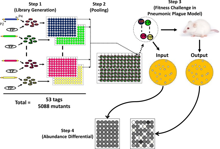
A total of 5,088 transposon mutants of WT CO92 were created, which included 96 mutants for each of the 53 unique 40-bp signature tags (34). As a source of the tags, 53 E. coli S-17 strains, each harboring the plasmid pUTminiTn5Km2STM with a unique tag, were used as donor strains and conjugated with WT CO92 (35). Initially, 56 tags were chosen, as previously described (35), and were tested for their cross-hybridization. Three of the 56 tags showed a cross-reaction under our tested conditions and, therefore, were excluded from the study. For each of the 53 signature tags used, the following procedures were carried out. The E. coli S17-1 strain (Table 1) carrying the transposon with a unique signature tag was grown overnight, subcultured, and then further grown for 4 h (optical density at 600 nm [OD600] of ∼0.6). Separately, WT CO92 was grown overnight and mixed in a 4-to-1 ratio with each of the above-described donor E. coli strains. An aliquot of each mixture was spread on LB agar plates and incubated at 30°C for 24 h. Subsequently, the cultures from the LB plates were collected in sterile phosphate-buffered saline (PBS), and a portion of each mixture was spread on HIB agar plates containing polymyxin B and kanamycin for 48 h at 28°C. Following the incubation period, separate transconjugant colonies were tested for resistance to polymyxin B (WT CO92 is naturally resistant to this antibiotic) (Table 1) and kanamycin but sensitivity to ampicillin. Finally, 96 of the transconjugants carrying each tag that did not show any obvious growth defects were randomly picked and individually inoculated into the wells of a 96-well microtiter plate. After 24 h of growth, glycerol was added to a final concentration of 15% and the plates were stored at −80°C (Fig. 1). From the 53 96-well-plate stocks, 96 mutant pools were prepared by combining 20 μl of stock cultures from the same respective positions of 96-well microtiter plates (Fig. 1). Thus, each mutant pool represented a collection of 53 transposon mutants, each with a unique signature tag.
FIG 1.
Schematic illustration of the signature-tagged mutagenesis (STM) approach. Step 1, transposon mutants with 53 unique signature tags were generated through a mini-Tn5 transposon system to create a library of 96 representative Y. pestis (YP) mutants for each tag, totaling 5,088 mutants. Step 2, one mutant for each tag was combined to generate 96 pools of 53 uniquely tagged clones. Step 3, each pool was used to generate an input pool of bacterial DNA and to infect mice by the i.n. route (5 LD50). At 3 days p.i., disseminated mutant bacteria were isolated from the spleens and their DNA extracted, providing the output pool of bacterial DNA. Step 4, signature tag probes were generated by PCR with primer pair P2-P4 for input and output pools of bacterial DNA, with primers binding to common sequences adjacent to the signature tags. Then, each pool of the DNA probes (input and out) was separately hybridized to membranes spotted with an array of the 53 unique signature tags. After hybridization, the membranes were developed and the input and output pool membranes compared for changes in the abundance of corresponding signatures.
Preparation of input mutant pools of Y. pestis CO92 and collection of corresponding output mutant pools from the spleen in a mouse model of pneumonic plague.
Each of the 96 mutant pools prepared as described above was individually tested in female Swiss-Webster mice (Taconic Biosciences, Inc., Hudson, NY) that were infected via the intranasal (i.n.) route. The animal experiments were conducted in accordance with the Institutional Animal Care and Use Committee (IACUC)-approved protocol in the animal biosafety level 3 (ABSL-3) facility located in the GNL. Figure 1 shows the approach used for screening the mutants. For each of the mutant pools with 53 unique DNA tags, three animals were infected. Before infection of the mice, a portion of the bacterial inoculum was subjected to genomic DNA isolation using a DNeasy blood and tissue kit (Qiagen, Inc., Valencia, CA), and the resulting DNA was referred to as the input DNA pool, or input pool. The remaining inoculum was used to infect mice at a dose of 2,500 CFU, representing the equivalent of 5 LD50 (1 LD50 = 500 CFU) of WT CO92 (18). Three days postinfection (p.i.), the spleens were excised to recover the output mutants. Briefly, the spleens were homogenized in sterile PBS and aliquots of the homogenates was spread on LB agar plates containing kanamycin. After 24 h of incubation, the bacterial colonies were collected for genomic DNA isolation; the resulting DNA is referred to as the output DNA pool or output pool.
DNA hybridization-based screening of input and output mutant pools of Y. pestis CO92.
The DNA hybridizations were performed separately for each of the input and output pools, as previously described (35). Briefly, 53 signature tags were each PCR amplified (Phusion high-fidelity PCR kit; New England BioLabs, Inc., Ipswich, MA), using primers P2 and P4, from transposon plasmid pUTminiTn5Km2STM carrying the respective tag (Fig. 1 and Tables 1 and 2). The PCR products were digested with HindIII restriction enzyme (New England BioLabs, Inc.) to remove primer sequences and gel purified using a QIAquick gel extraction kit (Qiagen, Inc.), and then 15-ng amounts of the tag DNA were spotted individually on a positively charged nylon membrane (Amersham Hybond-N+; GE Healthcare Life Sciences, Pittsburgh, PA). The DNA probes on the membranes were sequentially subjected to denaturation in 1.5 M NaCl–0.5 M NaOH solution for 3 min, neutralization in 1.5 M NaCl–0.5 M Tris-HCl (pH 7.4) solution for 5 min and again for 1 min in the same but fresh solution, and washing in 2× SSC (1× SSC is 0.15 M NaCl plus 0.015 M sodium citrate [7.0]) for 2 min. Finally, the DNA was UV cross-linked to the membranes.
TABLE 2.
Sequences of primers used in this study
| Primer or primer pair | Primer sequences (5′–3′; forward, reverse) | Purpose |
|---|---|---|
| P2-P4 | TACCTACAACCTCAAGCT, TACCCATTCTAACCAAGC | PCR amplification of STM tags |
| P1-P3 | GCGCAACGGAACATTCATC, GCAAGCTTCGGCCGCCTAGG | Identification of mini-Tn5 insertion sites |
| P5-P6 | AGGGTCAGCCTGAATACGCG, CTGACTCTTATACACAAGTGC | Identification of mini-Tn5 insertion sites |
| Kmypo2500 | TTAGCTGGTAAGCGTGTCAATTCTCGCTCTGCTCAGGCAGAGCGATAACCGTGTAGGCTGGAGCTGCTTC (FRT sequence), TAATGCACTCCCTGTTGCGTGAAGCATGATGTTATTAGATTCAATTTCATATTCCGGGGATCCGTCGACC (FRT sequence) | Construction of a DNA fragment with Kmr gene cassette and FRT sequence for the rbsA mutation |
| ypo2500V | CGTATTGCACTGGGTATCGCGTTGG, GTCATTTAACCCGCTCATTAAGACA | PCR verification of rbsA deletion |
| ypo2500C | CGGGATCCGGTTAGCGTAGACGGCCAACCA (BamHI), ACGCGTCGACTCATAATGCACTCCCTGTTG (SalI) | Cloning of rbsA in plasmid pBR322 |
| Kmypo3603 | GACAACTCAAACCATGATCGCCATGGAATGCCACGGAGCGTTAGCGCATGTGTAGGCTGGAGCTGCTTC (FRT sequence), TGCGATCTGTCTCAATACAATCGTGTGTCTCAATACAGAGTGTCTGGCAGATTCCGGGGATCCGTCGACC (FRT sequence) | Construction of a DNA fragment with Kmr gene cassette and FRT sequence for the vasK mutation |
| ypo3603V | TAAACCGGCAACCACAGCAATCCGA, TTGACCTCTGGCCGTGCCGGGTGGT | PCR verification of vasK deletion |
| ypo3603C | CTAGCTAGCCTACAGATGATAAACCGGCAA (NheI), ACGCGTCGACTCAATACAGAGTGTCTGGCA (SalI) | Cloning of vasK into plasmid pBR322 |
| Kmypo0498 | ACCATTAGCACGATGACGTGGATGAATAGCCAAAATAAGAGGACATAGATGTGTAGGCTGGAGCTGCTTC (FRT sequence), TACCTCTTAATCTCCAGAGATTTTAGATCCTTTGCGTGTCAGATAGGACAATTCCGGGGATCCGTCGACC (FRT sequence) | Construction of a DNA fragment with Kmr gene cassette and FRT sequence for the ypo0498 mutation |
| ypo0498V | GCTATTCGCTGGTTGAGGCT, AACGCTGGCAGAGAGATGAG | PCR verification of the ypo0498 deletion |
With the P2 and P4 primers (Table 2), the tag sequences from each of the input and corresponding output DNA pools were PCR amplified and gel purified as described above and were digoxigenin (DIG) (Roche Applied Science, Indianapolis, IN) labeled by PCR using the P2 and P4 primers as described previously (35). The labeled tags were digested with HindIII restriction enzyme to remove the primer sequences and denatured at 95°C for 5 min before proceeding to hybridization. The membranes prepared as described above were prehybridized with the DIG hybridization solution (Roche Applied Science), and finally, the labeled tags were added to the membrane in a fresh hybridization solution and incubated overnight at 42°C.
Following hybridization, the membranes were subjected to washing, blocking, and developing at room temperature, unless otherwise stated, as follows: (i) twice for 5 min each in 2× SSC plus 0.1% sodium dodecyl sulfate (SDS), (ii) twice for 5 min each in 0.1× SSC plus 0.1% SDS at 65°C, and once for 5 min in 0.1 M maleic acid, 0.15 M NaCl (pH 7.5), 0.3% (wt/vol) Tween 20 (MNT) buffer. Then, the membranes were placed in 1× blocking solution (Roche Applied Science) for 30 min and were probed with monoclonal anti-DIG antibody in 1× blocking solution for 30 min. The membranes were washed twice for 5 min each in MNT solution and equilibrated for 5 min in 0.1 M Tris-HCl, 0.1 M NaCl (pH 9.5) solution. Finally, ready-to-use CDP-Star solution (Roche Applied Science) was applied to each membrane and incubated for 5 min, and the positive hybridization signals were visualized on a luminescent image analyzer (ImageQuant LAS4000; GE Healthcare Life Sciences). All of the hybridization steps were performed in a hybridization oven.
Testing individual signature-tagged transposon mutants of Y. pestis CO92 in bubonic and pneumonic plague mouse models.
Mutant clones that exhibited either complete or partial loss in virulence in terms of their ability to disseminate to the spleen, as determined by the hybridization reactions, were selected for further study. Each of the mutants was individually used to infect a group of five Swiss-Webster mice via the subcutaneous (s.c.) route at a dose equivalent to 8 LD50 of WT CO92 (1 LD50 by the s.c. route is 50 CFU) (18). The mutants found to be attenuated for bubonic infection after the first screen were subjected to a stringent second screen in mice (n = 10 to 20) at a higher infectious dose of 40 LD50. The animals were observed for mortality over a period of 21 to 28 days. The mutant clones that were sufficiently attenuated to show at least 40% animal survival were selected for genomic characterization of the transposon insertion sites.
Transposon mutants that showed promising results during the first s.c. screening were further tested for their level of attenuation in a pneumonic plague mouse model. Each selected mutant was used to infect a group of five Swiss-Webster mice at an infection dose equivalent to 12 LD50 of WT CO92. The animals were observed for mortality over a period of 9 days.
Genomic characterization of transposon insertion sites in the signature-tagged mutants of Y. pestis CO92.
Inverse PCR was used to amplify the DNA fragment flanking the mini-Tn5 insertion site as described previously (35, 36). Briefly, genomic DNA from the selected signature-tagged transposon mutants described above was extracted by using a DNeasy blood and tissue kit (Qiagen, Inc.). An aliquot (2 μg) of the genomic DNA was digested with BamHI, MluI, PstI, SalI, or XbaI restriction enzyme (New England BioLabs), and the resulting fragments were ligated using T4 DNA ligase (Promega, Madison, WI). Inverse PCR was performed to anneal outward-facing primers P1 and P3 to the mini-Tn5 sequence (Table 2). Subsequently, a nested PCR amplification was carried out on each of the inverse PCR products using primers P5 and P6 (Table 2). Primers P5 and P6 annealed downstream to the primer pair P1 and P3. Then, the resulting PCR products were gel purified and sequenced using primer P6 (Table 2). Based on the sequence information, transposon insertion sites in the genome of Y. pestis CO92 were identified (15).
Construction of in-frame deletion mutants and testing in mouse models of bubonic and pneumonic plague.
To construct in-frame deletion mutants of Y. pestis CO92, the phage λ recombination system was used (37). Initially, the WT CO92 strain was transformed with plasmid pKD46 (Table 1) and grown in the presence of 1 mM l-arabinose to induce the expression of the phage λ recombination system. The above-described Y. pestis culture was processed for the preparation of electroporation-competent cells (10, 37). The latter were then transformed with 0.5 to 1.0 μg of the linear double-stranded DNA constructs carrying the kanamycin resistance (Kmr) gene cassette that was immediately flanked by a bacterial flippase (FLP) recombination target (FRT) sequence and followed on either side by 50 bp of DNA sequences homologous to the 5′ and 3′ ends of the gene to be deleted from WT CO92. Plasmid pKD46 was cured from the mutants that had successful Kmr gene cassette integration at the correct location by growing the bacteria at 37°C. The latter mutants were transformed with plasmid pFlp2 (Table 1) to excise the Kmr gene cassette (38). Eventually, plasmid pFlp2 was also cured from the kanamycin-sensitive (Kms) clones by growing them in a medium containing 5% sucrose (38). To confirm the in-frame deletion, mutants showing sensitivity to kanamycin and ampicillin were tested by PCR using appropriate primer pairs (Table 2) and sequencing of the PCR products.
To construct double or triple in-frame deletion mutants of CO92, a similar procedure was followed using selected single (Δlpp) or double (Δlpp ΔmsbB) in-frame deletion mutants that existed in the laboratory (Table 1). To construct a recombinant plasmid for complementation studies, the complete open reading frame of the gene of interest, along with 200 bp of the upstream DNA sequence corresponding to the promoter region of that gene from WT CO92, was PCR amplified using a Phusion high-fidelity PCR kit (New England Biolabs). Then, the DNA construct was cloned in plasmid pBR322 in place of the tetracycline resistance (Tcr)-conferring gene cassette (Table 1).
Single, double, and triple isogenic mutants and their complemented strains were then tested in both bubonic and pneumonic plague mouse models along with the WT CO92 strain as a control. For rechallenge experiments, after 28 days p.i. with the selected mutants, bioluminescent WT CO92, carrying the luciferase gene operon luxCDABE (Table 1), was used to infect mice as described previously (39). Also, in vivo imaging was performed on rechallenged animals using an IVIS 200 bioluminescence and fluorescence whole-body-imaging workstation (Caliper Corp., Alameda, CA).
Western blot analysis for detecting a T6SS effector, hemolysin-coregulated protein (Hcp), in the isogenic mutants of Y. pestis CO92.
Overnight-grown cultures of various Y. pestis and Aeromonas hydrophila strains (the latter was reclassified as Aeromonas dhakensis) (40) were harvested, and the supernatants mixed with 20% trichloroacetic acid (vol/vol). The resulting precipitates were dissolved in SDS-PAGE buffer by boiling and subjected to SDS–4 to 15% polyacrylamide gel electrophoresis. The proteins from the gel were then transferred to a Hybond ECL nitrocellulose membrane (GE Healthcare) by following the standard procedure (41). The membrane was blocked with 1% bovine serum albumin (BSA) or 5% skim milk and then incubated with anti-Hcp antibody specific for Y. pestis (1:1,000), followed by incubation with the secondary antibody (goat anti-mouse IgG [1:10,000]; Southern Biotechnology Associates, Inc., Birmingham, AL). The membrane was washed with Tris-buffered saline (TBS; 20 mM Tris base, 136 mM NaCl [pH 7.4])–0.05% Tween 20, and the blot was developed using SuperSignal west Dura extended-duration substrate (Pierce, Rockford, IL). Finally, the positive signal was detected by using the ImageQuant LAS4000 platform (GE Healthcare). Polyclonal antibody raised in mice against Hcp of Y. pestis was used for immunoblot analysis. The hcp gene (YPO3708) of Y. pestis CO92 was overexpressed in E. coli as a His-tagged recombinant protein using the pET30a vector system and purified by using Ni2+ chromatography (41). As a loading control for immunoblot analysis, we used monoclonal antibody against DnaK (Enzo, Farmingdale, NY), a member of the conserved Hsp70 chaperone family.
Growth kinetics of WT Y. pestis CO92, its ΔrbsA mutant, and the complemented strain.
Overnight cultures of various Y. pestis strains were washed in PBS and normalized to the same absorbance by measuring the optical density at 600 nm (OD600). These bacterial cultures were then inoculated separately (at approximately 1 × 10⁷ CFU) into 20 ml of modified M9 medium (1× M9 salts [22 mM KH2PO4, 33.7 mM Na2HPO4, 8.55 mM NaCl, 9.35 mM NH4Cl], 1 mM MgSO4, 2.5 mM CaCl2, 0.001 mg/ml FeSO4, 0.0001% thiamine, 0.1% Casamino Acids) (all chemicals were obtained from Sigma-Aldrich, St. Louis, MO) contained in 125-ml polycarbonate Erlenmeyer flasks with HEPA-filtered tops. The medium either did not contain any sugar or was supplemented with 0.4% glucose or 0.4% ribose. The cultures were incubated at 28°C with shaking at 180 rpm. Samples were taken by removing 100 μl of the culture from each of the flasks at the time points indicated in Fig. 6B. Each of the samples was serially diluted, plated on SBA agar plates, and incubated at 28°C for 48 h to determine CFU/ml.
FIG 6.
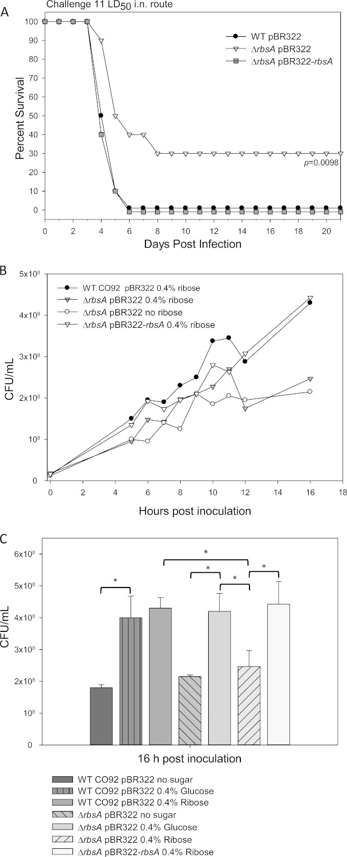
Survival of mice challenged with the ΔrbsA isogenic mutant of Y. pestis CO92 in a pneumonic plague model, and ribose utilization. (A) For each of the strains tested, 10 adult Swiss-Webster mice were challenged by the i.n. route with 11 LD50 and then observed for mortality for 21 days. Survival data were analyzed for significance by Kaplan-Meier survival estimates with the Bonferroni post hoc test. The P values are for comparison of the results for each of the strains to the results for WT CO92. (B) Growth of mutants and WT CO92 in a modified M9 minimal medium with or without supplementation with 0.4% ribose. Samples were taken at the time points indicated and plated for CFU counts. (C) At 16 h postinoculation, culture titrations were determined for the WT CO92(pBR322), ΔrbsA(pBR322), and ΔrbsA(pBR322-rbsA) strains grown in a modified M9 minimal medium with or without supplementation with 0.4% glucose or 0.4% ribose. Statistical significance was analyzed by one-way ANOVA with the Tukey post hoc test. The comparisons between groups indicated by asterisks and brackets are significant at a P value of <0.001.
Statistical procedures.
Animal survival rates were statistically analyzed using Kaplan-Meier survival estimates with the Bonferroni post hoc test. The in vitro growth of WT CO92, its ΔrbsA mutant, or the complemented strain under different nutritional conditions was analyzed by one-way analysis of variance (ANOVA) followed by the Tukey post hoc test. Wherever applicable, the P values are reported, and a P value of ≤0.05 is considered significant.
RESULTS
Y. pestis CO92 signature-tagged transposon mutant library and its primary screen in a mouse model of pneumonic plague.
We generated a library of mutants with 53 unique DNA tags from wild-type (WT) strain CO92 by using a transposon Tn5-based system (Fig. 1). For each signature tag, 96 mutants potentially representing Tn5 hits at different locations on the chromosome or the plasmids of WT CO92 were randomly picked from the HIB agar plates, thus resulting in a library consisting of 5,088 mutants. During this selection process, any mutant clones that exhibited visual growth defects, such as a smaller colony size, were not included as they would be outcompeted in a mixed culture infection, resulting in false positives. We observed a transfer efficiency of 1.5 × 10⁻⁴ transposon mutants per donor colony when the Tn5-harboring E. coli strains were mixed with the recipient WT CO92 strain at a ratio of 4:1 for the conjugation process. Finally, 96 input mutant pools, each containing a collection of 53 mutants (one clone for each one of the signature tags), were generated to screen for attenuation in terms of dissemination to the internal organs, i.e., the spleen, in a mouse model of pneumonic plague (Fig. 1).
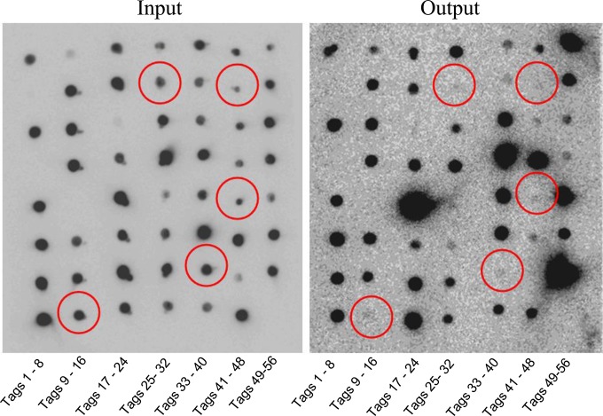
The infectious dose of the 96 input mutant pools given by the i.n. route in mice was the equivalent of 5 LD50 of WT CO92 (18). Three days p.i., ∼60% of the animals died due to developing pneumonic plague and the remaining animals had clinical symptoms of plague, such as lethargy and ruffled fur. The excised spleens from these mice, irrespective of their survival status, had high bacterial counts, in the range of 1 × 10⁷ to 1 × 10⁸ CFU per organ. Under the conditions of hybridization and washing optimized for the study, the nylon membranes harboring the purified 53 unique signature tags were hybridized with the 96 input pool DNA probes (obtained from each transposon mutant pool with 53 signature tags) and showed a clear pattern of positive reactions without any background signal (a representative blot is shown in Fig. 2).
FIG 2.
Representative STM hybridization reactions. Bacterial DNA was isolated from input and output mutant pools and signature tags amplified, labeled with DIG, and then used to hybridize membranes spotted with a signature tag array. Out of 56 initial signature tags, 3 were found to be cross-reactive (4, 13, and 56) and were subsequently excluded. Each array was composed of 53 signature tags that were shown to be specific. Red circles highlight those mutants that show a reduction in abundance between the input and the output pools. The tag numbers (1 to 56) are indicated.
By this hybridization approach, we identified a total of 118 potential mutant candidates; among these, 108 had no detectable signal on the output DNA pool membranes, and the remainder had very weak signals compared to those of the corresponding input DNA pool membranes (a representative blot is shown in Fig. 2).
Second screen of selected signature-tagged transposon mutants of Y. pestis CO92 in a mouse model of bubonic plague.
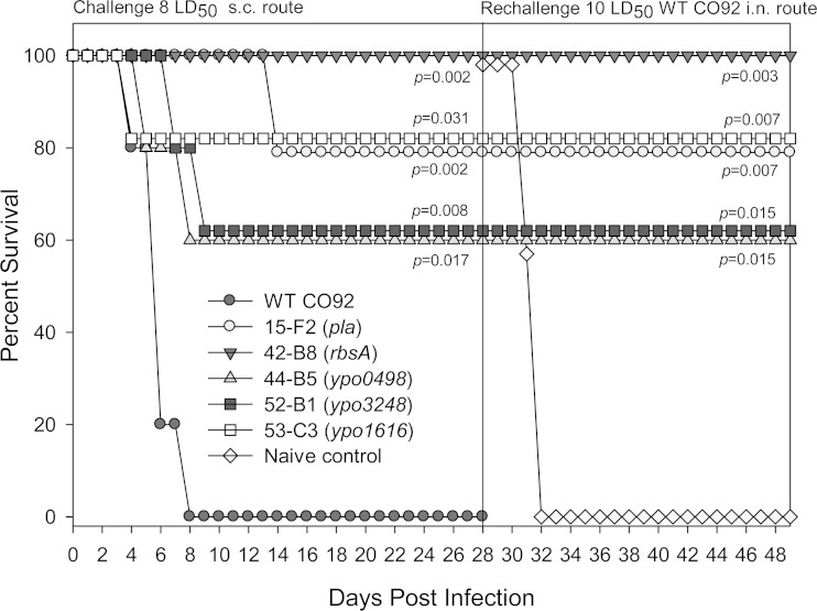
We performed a second screen with the 118 mutant clones generated as described above by injecting mice with individual mutants via the s.c. route with doses aimed at achieving the equivalent of 8 LD50 of WT CO92 (18). Of the 118 mutant clones tested, 20 (∼17%) showed attenuation to the level of at least 20% or more of the animals surviving (up to 100%) compared to the survival rate with WT CO92 on day 21 p.i. (Table 3). One of our long-term goals is to identify candidate genes that could be deleted from the WT CO92 to develop a novel live attenuated plague vaccine. Therefore, we also tested some of the animals that survived infection with representative transposon mutants for the ability to withstand rechallenge with the WT CO92. The rechallenge occurred by a more stringent i.n. route, which evokes pneumonic plague, to gauge the immunogenicity of the transposon mutant clones. As shown by the results in Fig. 3, 100% of the animals that were initially infected with the transposon mutants (15-F2, 42-B8, 44-B5, 52-B1, and 53-C3) (Table 3) and were rechallenged with the WT CO92 strain (10 LD50) were protected over a period of 21 days. All of the naive control mice died by day 4. These second-screen mutant candidates were then subjected to a higher-stringency screen in a bubonic plague mouse model by increasing the challenge dose to 40 LD50 (Table 4). As noted from the data in Table 4, mice infected with 10 of the 20 mutants showed 40% or greater rates of survival on day 21 p.i. Following this final high-stringency screen, we identified the insertion sites within the disrupted genes for each of the mutant candidate strains.
TABLE 3.
Survival patterns for selected transposon mutants in mouse models of bubonic and pneumonic plague
| Clonea | Gene locus | % of mice surviving postinfection withb: |
||||
|---|---|---|---|---|---|---|
| ∼8 LD50 s.c. on day: |
12 LD50 i.n. on day: |
|||||
| 7 | 14 | 21 | 3 | 9 | ||
| WT CO92 | 40 | 0 | 0 | 0 | 0 | |
| 15-F2 | YPPCP1.07 | 100 | 100 | 80 | 60 | 0 |
| 19-F7 | Unconfirmed | 100 | 80 | 80 | 80 | 0 |
| 29-F7 | YPO2468 | 60 | 60 | 60 | 80 | 40 |
| 2-H3 | YPO1717 | 80 | 60 | 60 | 20 | 0 |
| 39-A7 | YPO3319 | 60 | 60 | 60 | 80 | 0 |
| 39-G4 | YPMT1.80c | 100 | 40 | 40 | 20 | 0 |
| 42-B8 | YPO2500 | 100 | 100 | 100 | NTc | NTc |
| 44-B5 | YPO0498 | 80 | 60 | 60 | 100 | 40 |
| 44-F11 | YPO3603 | 80 | 60 | 60 | NTc | NTc |
| 45-B9 | YPO2884 | 100 | 80 | 60 | 100 | 100 |
| 47-F10 | Unconfirmed | 80 | 80 | 80 | 60 | 0 |
| 47-G5 | YPO3164 | 100 | 80 | 80 | 20 | 0 |
| 48-G1 | PMT1 Caf1R | 100 | 100 | 100 | 100 | 20 |
| 49-B1 | YPO1484 | 80 | 60 | 60 | 100 | 100 |
| 52-B1 | YPO3248 | 80 | 60 | 60 | 100 | 80 |
| 52-B5 | Intergenic, YPO0093-YPO0094 | 60 | 60 | 20 | 100 | 80 |
| 53-C3 | YPO1616 | 80 | 80 | 80 | 100 | 100 |
| 53-F10 | YPO1995 | 100 | 100 | 100 | 100 | 0 |
| 53-G5 | YPO0815 | 100 | 60 | 60 | 100 | 100 |
| 54-F6 | Intergenic, YPO1119-YPO1120 | 100 | 100 | 100 | 100 | 60 |
Clone identifiers comprise the signature tag number and the alphanumeric of an individual mutant containing that signature.
s.c., subcutaneous; i.n., intranasal.
NT, not tested for signature-tagged mutants, but an isogenic mutant was prepared and examined.
FIG 3.
Survival of mice after initial infection with Y. pestis CO92 mutants in a bubonic plague model and subsequent rechallenge to evoke pneumonic plague with WT CO92. Adult Swiss-Webster mice were challenged with the WT CO92 strain or one of the mutants (5 mice per group) by the s.c. route at infectious doses of 8 LD50. Surviving mice from the initial challenge with the selected mutant groups were followed for 28 days p.i. and then rechallenged by the i.n. route with 10 LD50 of WT CO92. Survival data were analyzed for significance by Kaplan-Meier survival estimates with the Bonferroni post hoc test. The P values are for comparison of the results for each of the strains to the results for WT CO92 or the naive control for the rechallenge study.
TABLE 4.
Survival patterns for mice infected with selected transposon mutants at a high infectious dose in a model of bubonic plague
| Clone | Gene interrupted | % of mice surviving postinfection with ∼40 LD50 s.c. on daya: |
Gene or protein description/protein homology | ||
|---|---|---|---|---|---|
| 7 | 14 | 21 | |||
| WT CO92 | 15 | 0 | 0 | ||
| 15-F2b | pla | 85 | 85 | 85 | Plasminogen activator |
| 2-H3 | ypo1717 | 80 | 60 | 60 | Putative membrane protein |
| 39-G4 | ypmt1.80c | 100 | 40 | 40 | Putative transposase |
| 42-B8 | rbsA (ypo2500) | 85 | 70 | 60 | Putative sugar transport system ATP-binding protein |
| 44-B5 | ypo0498 | 100 | 85 | 85 | Hypothetical protein associated with a T6SS locus |
| 44-F11b | vasK (ypo3603) | 60 | 60 | 60 | T6SS component VasK |
| 47-G5 | ypo3164 | 100 | 80 | 40 | Cytochrome o ubiquinol oxidase subunit II |
| 52-B1 | hxuB (ypo3248) | 100 | 60 | 60 | HxuB (hemolysin secretion protein)/putative surface-exposed protein |
| 53-C3 | ypo1616 | 85 | 60 | 60 | Hypothetical protein |
| 54-F6 | ypo1119-ypo1120 | 100 | 100 | 100 | Insertion in intergenic region between two conserved hypothetical proteins |
The number of animals used per group ranged from 10 to 20.
Boldface highlights data for mutants that had transposon disruptions in genes previously identified as virulence factor-encoding genes in Y. pestis, i.e., pla, or in A. dhakensis SSU, i.e., vasK.
Genetic characteristics of signature-tagged transposon mutants of Y. pestis CO92 that were attenuated in a mouse model of bubonic plague.
Identification of the disrupted genes or genetic regions in the transposon mutants was accomplished using inverse PCR followed by sequencing of the PCR products to confirm the locations of the mutations. The genomic characterizations of 18 of the 20 attenuated mutants identified during the first screen by s.c. challenge are listed in Table 3, while the genomic locations of transposon integration for mutant clones 19-F7 and 47-F10 were not confirmed. In these confirmed 10 mutants showing attenuation under high stringency in a mouse model of bubonic plague, transposon insertions were identified in six uncharacterized genes (Table 4). For example, clones 52-B1 and 2-H3 encode a putative surface-exposed and a membrane protein, respectively. Clones 44-B5, 53-C3, 54-F6, and 39-G4 code for hypothetical proteins or proteins with putative functions. In clone 47-G5, the transposon interruption occurred in the cytochrome o ubiquinol oxidase subunit II (Table 4). Two of the genes encoded previously characterized virulence factors, including the pla protease gene located on the pCP1 plasmid of Y. pestis (18, 42). The pla protease gene was interrupted in the mutant clone 15-F2 (Table 4), adding credibility to our screening process. This mutant was highly attenuated, as 85% of the animals in a bubonic plague model challenge survived on day 21 p.i. (Table 4).
Mutant clone 44-F11, with 60% of the mice surviving the challenge, was identified as having a disruption in a gene whose product has homology to VasK of A. dhakensis strain SSU; VasK is a key component of the T6SS (Table 4) (43). Clone 42-B8 was identified as having a transposon insertion within the gene referred to as rbsA, and 85, 70, and 60% of the animals in a mouse model of bubonic plague survived after challenge on days 7, 14, and 21 p.i., respectively (Table 4). This gene is part of the rbs operon that codes for a putative ribose transport system and has been described in orthologous systems (44, 45). Likewise, clone 44-B5, in which the transposition occurred in a hypothetical gene, was highly attenuated, with 100, 85, and 85% survival of animals on days 7, 14, and 21 p.i., respectively (Table 4).
Pathodynamics of bubonic plague infection for the isogenic mutants of Y. pestis CO92 with deletion of rbsA, vasK, or ypo0498 in a mouse model.
Transposon mutagenesis does not always provide a true estimate of bacterial attenuation during infection, due partly to possible polar effects; therefore, we created isogenic mutants for three of the genes, rbsA, vasK, and ypo0498 (Table 4). Gene rbsA was targeted because it had the most functional information available from orthologs in other bacterial species (44, 45). Similarly, gene ypo3603 (vasK) was targeted due to the virulence attributes associated with orthologous genes (43). Another candidate gene, ypo0498, which encodes a hypothetical protein and is part of another T6SS locus, was also selected. These mutants were then used to challenge mice by the s.c. route to replicate data obtained during the transposon mutant screening.
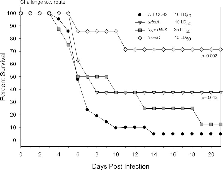
Animals infected with the ΔrbsA or the ΔvasK isogenic mutant by the s.c. route showed statistically significant attenuation of virulence, as 40% (P = 0.042) and 70% (P = 0.002), respectively, of the mice survived when challenged with the equivalent of 10 LD50 of WT CO92 (Fig. 4). The control animals infected with WT CO92 showed survival rates of less than 5% by day 14 p.i. in three combined independent experiments. When the Δypo0498 isogenic mutant was used to challenge mice by the s.c. route, an increase in mean time to death was noted at a much-higher 35 LD50, although the data did not reach statistical significance (Fig. 4).
FIG 4.
Survival of mice after challenge with the indicated isogenic mutants of Y. pestis CO92 in a bubonic plague model. After generation of the rbsA, vasK, and ypo0498 isogenic mutants, each strain was used to challenge 10 adult Swiss-Webster mice by the s.c. route at the indicated LD50. The data for challenge with WT CO92 represent pooled results from three independent experiments, with a total number of 22 mice. Survival data were analyzed for significance by Kaplan-Meier survival estimates with the Bonferroni post hoc test. The P values are for comparison of the results for each of the mutant strains to the pooled WT CO92 results.
Characterization of the ΔvasK mutant of Y. pestis CO92 in a mouse model of pneumonic plague.
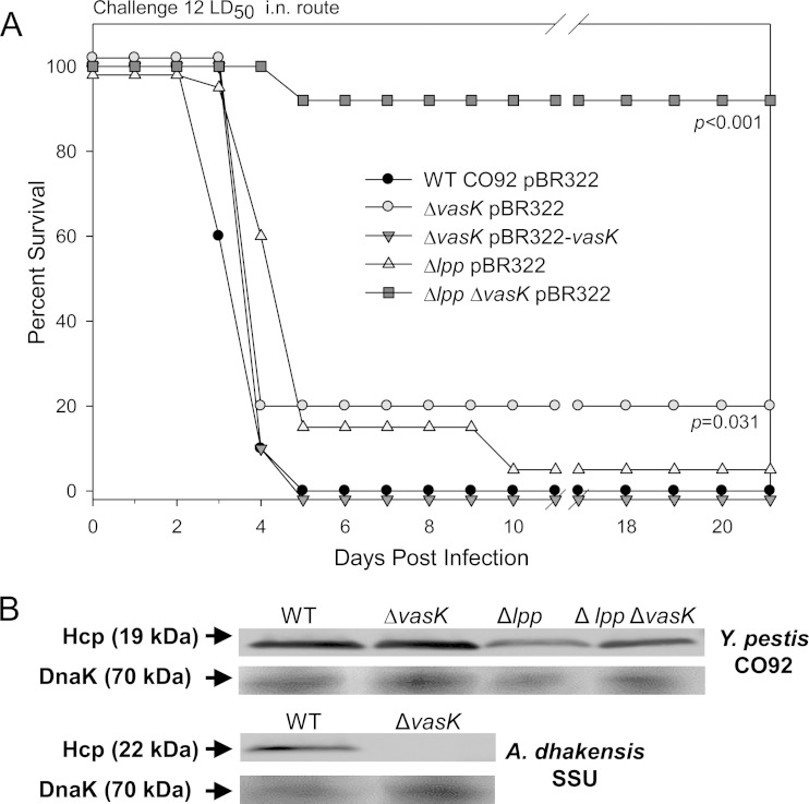
Following evaluation of the disease by the s.c. route (bubonic plague), the ΔvasK mutant was then assessed for attenuation in a mouse model of pneumonic plague. At a dose equivalent to 12 LD50 of WT CO92, the mice exhibited a 20% survival rate (P = 0.031) by day 21 p.i., with no survival of animals challenged with a similar dose of WT CO92 (Fig. 5A). Further confirming that deletion of the vasK gene resulted in this attenuated phenotype, mice challenged with the complemented strain [ΔvasK(pBR322-vasK)] (Table 1) recapitulated the WT phenotype, with no survival at an equivalent dose of the isogenic mutant. Although the attenuation of the ΔvasK mutant was not high in a stringent pneumonic plague mouse model, we obtained the first mutant which was attenuated in developing both bubonic and pneumonic plague.
FIG 5.
Survival of mice challenged with the ΔvasK isogenic mutants of Y. pestis CO92 in a pneumonic plague model, and secretion of Hcp through the T6SS. (A) Adult Swiss-Webster mice were challenged by the i.n. route with 12 LD50 of WT CO92(pBR322) (n = 10) or the ΔvasK(pBR322) (n = 10), ΔvasK(pBR322-vasK) (n = 10), Δlpp(BR322) (n = 20), or Δlpp ΔvasK(pBR322) (n = 10) mutant. Mice were followed for survival up to 21 days, and the data analyzed for significance by the Kaplan-Meier survival estimates with the Bonferroni post hoc test. The P values are for comparison of the results for each of the mutant strains to the results for WT CO92. (B) Western blot analysis was performed to detect Hcp in the supernatants of various bacterial cultures using a specific anti-Hcp antibody, and the levels of DnaK in bacterial pellets were examined as a loading control for the samples used during Western blot analysis. The molecular masses of Hcp and DnaK are indicated.
In addition to generating a ΔvasK single mutant, we also deleted the vasK gene from the Δlpp background strain of CO92. Braun lipoprotein (Lpp) has previously been shown in our laboratory to provide attenuation in mouse models of pneumonic and bubonic plague through decreased intracellular survival in macrophages (19, 20, 38). The rationale for deleting the ΔvasK gene from the Δlpp background strain of CO92 was to determine whether additive or synergistic attenuation could be achieved with the Δlpp ΔvasK double mutant in a mouse model of pneumonic plague.
Infection by the i.n. route with the Δlpp ΔvasK double mutant at a dose equivalent to 12 LD50 resulted in 90% survival (P < 0.001) of mice by day 21 p.i., compared to only a 5% survival rate of animals after challenge with the Δlpp single mutant at a comparable challenge dose (Fig. 5A). These data indicated synergistic attenuation of the Δlpp ΔvasK double mutant in a mouse model of pneumonic plague.
To determine whether the attenuating effect of the vasK gene deletion in WT CO92 or its Δlpp mutant in mice was related to inhibition of the secretion of a T6SS effector, Hcp, a Western blot analysis was performed on the culture supernatants of WT CO92 and its Δlpp single, ΔvasK single, and Δlpp ΔvasK double mutants. We showed previously that the secretion of Hcp was blocked when the vasK gene was deleted from diarrheal isolate SSU of A. dhakensis (43). As seen by the results in Fig. 5B, no differences in the secretion of Hcp were observed between the WT CO92 strain and the ΔvasK mutants, irrespective of whether the bacterial cultures were grown at 28 or 37°C. As expected, while the correct size Hcp was detected in the supernatant of WT A. dhakensis SSU, the protein band was absent from its corresponding ΔvasK mutant. Since the Hcp proteins of Y. pestis and A. dhakensis exhibit high homology (81%), the same antibody detected Hcp in both pathogens. A larger molecular size of Hcp was detected in A. dhakensis (22 kDa) than in Y. pestis (19 kDa), likely due to posttranslational modification, as was also observed in the Hcp from Vibrio cholerae (46).
The gene rbsA is required for full virulence of Y. pestis CO92 in a pneumonic plague mouse model and for the utilization of ribose.
In a pneumonic plague model, the ΔrbsA mutant was attenuated, with 30% of the mice (P = 0.0098) having survived the challenge, while the animals infected with the WT CO92 or those infected with the rbsA-complemented strain died by day 6 at a similar infectious dose of 11 LD50 (Fig. 6A).
Earlier studies suggested that domains with sequence homology to the RbsA protein function as a ribose transport system (44, 45). To explore the role of ribose utilization, the ΔrbsA mutant was grown in a modified minimal medium with a restricted carbon source. In a minimal medium supplemented with 0.4% ribose, the ΔrbsA mutant displayed a delayed growth pattern similar to the pattern seen when no ribose was added to the medium (Fig. 6B). However, the WT CO92 and the ΔrbsA(pBR322-rbsA) complemented strain exhibited normal and similar growth kinetics in a ribose-containing medium (Fig. 6B).
In a minimal medium, WT CO92 without any carbon source grew poorly after 16 h of incubation; however, the addition of 0.4% of either glucose or ribose resulted in luxuriant bacterial mass (P < 0.001) (Fig. 6C). As mentioned in regard to the data in Fig. 6B, the ΔrbsA mutant grew poorly in the minimal medium supplemented with ribose after 16 h of incubation and had a bacterial mass similar to that obtained when no carbon source was added to the medium (Fig. 6C). However, the ΔrbsA mutant exhibited growth similar to that noted for the WT CO92 when the medium was supplemented with glucose (Fig. 6C). The complementation of the ΔrbsA mutant strain restored the ability to utilize ribose and allowed the bacteria to grow to a density twice that of the noncomplemented strain (P < 0.001) (Fig. 6C) and similar to that of the WT CO92 strain when grown in the ribose- or glucose-supplemented medium.
To assess the potential of rbsA deletion as a component of the live attenuated vaccine and to further characterize its attenuating characteristics, we constructed double and triple isogenic mutants in which the rbsA gene was deleted from the Δlpp and Δlpp ΔmsbB background strains of CO92. The msbB gene encodes an acyltransferase that attaches lauric acid to the lipid A moiety to increase the biological potency of LPS (47). The Δlpp ΔmsbB double mutant exhibited increased attenuation compared to that of the respective single mutants alone (19). The resulting double (Δlpp ΔrbsA) and triple (Δlpp ΔmsbB ΔrbsA) isogenic mutants showed synergistic reductions in virulence (Fig. 7A).
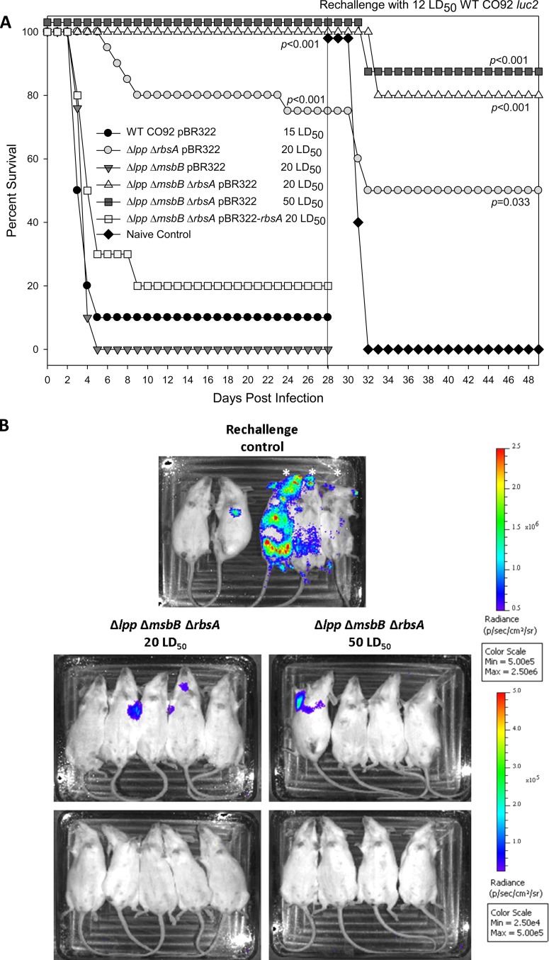
FIG 7.
Survival of mice after initial infection with the ΔrbsA isogenic mutants of Y. pestis CO92 in a pneumonic plague model, subsequent rechallenge with WT CO92 luc2, and evaluation of bacterial burden with the IVIS system. (A) Adult Swiss-Webster mice were challenged by the i.n. route with the WT CO92(pBR322) (n = 10), Δlpp ΔrbsA(pBR322) (n = 20), Δlpp ΔmsbB(pBR322) (n = 10), Δlpp ΔmsbB ΔrbsA(pBR322) (20 LD50, n = 10), Δlpp ΔmsbB ΔrbsA(pBR322) (50 LD50, n = 8), or Δlpp ΔmsbB ΔrbsA(pBR322-rbsA) (n = 10) strain at the indicated LD50 and observed for mortality over a period of 28 days. The mice that survived the initial challenge with a mutant strain and naive control animals (n = 5) were rechallenged or challenged with 12 LD50 of the WT CO92 luc2 (with luciferase gene) strain. Survival data were analyzed for significance by Kaplan-Meier survival estimates with the Bonferroni post hoc test. The P values are for comparison of the results for the indicated strains to the results for WT CO92 (challenge experiment) or naive control challenged with WT CO92 luc2 (rechallenge experiment). (B) On day 3 after rechallenge, selected groups of mice were imaged using the IVIS system to determine their relative bacterial burdens. In the WT CO92-challenged group, three mice, indicated by asterisks, were found dead before the imaging.
While none of the Δlpp ΔmsbB double mutant-infected mice survived past day 5 p.i. at 20 LD50, 90% of the mice challenged with 15 LD50 of the WT CO92 died (Fig. 7A). Animals infected with the Δlpp ΔrbsA double mutant showed 75% survival (P < 0.001) at a dose of 20 LD50. Challenge with 20 to 50 LD50 of the Δlpp ΔmsbB ΔrbsA triple mutant in mice by the i.n. route resulted in 100% survival for 28 days p.i. (P < 0.001) (Fig. 7A). As was observed with the ΔrbsA single mutant strain (Fig. 6A), when the Δlpp ΔmsbB ΔrbsA triple mutant was complemented with the rbsA gene and used to infect mice, 80% of the animals succumbed to infection, with no significant difference in the survival pattern compared to that of mice infected with the Δlpp ΔmsbB double mutant (Fig. 7A).
As part of the evaluation for its inclusion as a component in a live attenuated vaccine candidate, we were interested in testing the immune-protective potential of this strain. To accomplish this, we rechallenged the groups of mice that received 20 LD50 of the Δlpp ΔrbsA double mutant or the Δlpp ΔmsbB ΔrbsA triple mutant strain with a fully virulent bioluminescent Y. pestis CO92 strain on day 28 after the initial challenge. Likewise, the group that received 50 LD50 of the Δlpp ΔmsbB ΔrbsA triple mutant strain was subsequently challenged with the bioluminescent Y. pestis CO92 strain (Fig. 7B). This bioluminescent strain allowed us to evaluate the progression of the disease and survival data from the same groups of mice. By day 3 p.i., 60% of naive mice succumbed to the disease, while 1 of the remaining 2 showed heavy infection in the lungs (Fig. 7B) and died on the following day. The remaining naive mouse succumbed 2 days after the imaging.
Only 2 of the 10 mice (20%) from the group initially dosed with 20 LD50 of the Δlpp ΔmsbB ΔrbsA triple mutant strain showed a bacterial burden and later succumbed to infection due to challenge with WT CO92 (Fig. 7A and B). Only 1 of the 8 mice (13%) from the group initially dosed with 50 LD50 of the Δlpp ΔmsbB ΔrbsA triple mutant strain showed a bacterial burden and succumbed to infection during rechallenge with the WT (Fig. 7A and B).
Interestingly, animals initially challenged with the Δlpp ΔrbsA double mutant strain (75% protected) showed only a 50% survival rate after rechallenge (Fig. 7A), indicating that the Δlpp ΔmsbB ΔrbsA triple mutant strain was better attenuated than the Δlpp ΔrbsA double mutant and developed superior immunity in mice to subsequently protect animals from rechallenge with the WT CO92.
Third screen of selected signature-tagged transposon mutants of Y. pestis CO92 in a mouse model of pneumonic plague.
During the initial stage of pneumonic plague, Y. pestis suppresses the host immune system to downregulate the inflammatory response, thereby creating a highly permissive niche for the bacterium to multiply in an unrestricted manner (48, 49). Subsequently, this accumulation of proliferating bacteria leads to the induction of a massive inflammatory reaction, which causes lung edema and the death of the infected animals (48–50). Taking these findings together, it is likely that a Y. pestis mutant that is attenuated for dissemination to the peripheral organs after intranasal infection could still cause inflammatory changes in the lung tissue. Consequently, infected animals would succumb to infection due to pneumonia rather than the septicemic dissemination noted during bubonic plague. Therefore, we further evaluated the extent of attenuation in causing pneumonic plague of the remaining 18 signature-tagged mutants (ΔvasK and ΔrbsA isogenic mutants were already characterized) (Table 3) identified during the screening process by infecting animals by the i.n. route at a dose equivalent to 12 LD50 of WT CO92 (Table 3).
In general, a delayed pattern of death was noted for animals infected with any of the mutants on day 3 after infection (Table 3). Animals infected with 9 of the 18 mutants exhibited between 40 and 100% survival on day 9. Mutants 45-B9, 49-B1, 53-C3, and 53-G5 were unable to kill any mice, while 80% of mice survived challenge with mutants 52-B1 and 52-B5. These data implied that some mutants identified during the initial bubonic plague screen were attenuated in causing primary pneumonic plague as well, and these will be further characterized.
DISCUSSION
Knowledge of the virulence factors of Y. pestis is crucial to develop a new plague vaccine or design a better therapeutic intervention. As no FDA-approved plague vaccine is available for humans and antibiotics have a limited role when the disease progresses to a clinical stage, the search for novel virulence factors of the organism becomes a compulsive need in order to combat plague in the future. Here, we chose a high-throughput signature-tagged mutagenesis (STM) approach, because this technique offers the power to analyze multiple mutants for attenuation in virulence simultaneously, either in vitro or in vivo (29, 34).
In this study, more than 5,000 transposon mutants of Y. pestis CO92, an isolate obtained from a human case of pneumonic plague in the United States (51), were screened for impairment of dissemination to internal organs (e.g., spleen) in a mouse model of pneumonic plague (Fig. 1). Among these mutants, 118 were unable to reach detectable numbers in the spleen as identified by comparing the presence or absence of signature tags between the input and output pools. The detection rate of such mutants (∼2.4%) was close to that obtained in other studies using similar types of STM techniques (35, 52). We preferred to use a pneumonic plague mouse model for our initial screening of the mutants for the following reasons: (i) pneumonic plague is the deadliest form, with a high fatality rate compared to that of the bubonic form of plague, and (ii) the majority of the mice die due to pneumonic infection by day 3 p.i., with approximately 107 to 109 CFU of the plague bacterium in the peripheral organs (50). A high bacterial burden in the peripheral organs of mice is needed to obtain an adequate tag representation in the output pools during STM, and 104 CFU of bacteria is recommended as a threshold for obtaining authentic data (53). When animals are infected by the s.c. route to evoke bubonic plague, gauging consistent disease progression is somewhat challenging and the bacterial load in peripheral organs is relatively lower than that in the pneumonic plague mouse model (54, 55).
The complexity of the mutant pools during STM is a crucial parameter and has to be carefully considered for obtaining a high-quality screen in an animal model (34). Although an increased pool complexity would enable more mutants to be screened simultaneously, it might also enhance the probability that some virulent mutants would not be present in sufficient numbers in the organs of an infected animal, thus leading to false-positive data. In addition, the quantity of the labeled tag for each transposon is inversely proportional to the complexity of the tag pool during hybridization analysis (34). In our study, the number of signature tags was reduced from 56 to 53 due to cross-reaction noted for three tags during the prescreening step (35). The elimination of such tags is a prerequisite in performing STM-based screens (29). At a challenge dose of 5 LD50 (used in our study), the inocula for mutant pools with a complexity of 53 tags would provide ∼50 CFU of each mutant tested to ensure adequate bacterial numbers at the initial infection site, i.e., the lungs.
Unlike other STM studies, we neither opted for a second round of the animal infection-hybridization screening process nor used an in vitro assay to narrow down the number of mutants selected for further studies (35, 53). We rather chose to use the bubonic plague mouse model, tested each of the 118 mutants individually, and examined the animals for mortality. While 20/118 mutants exhibited an attenuated phenotype at 8 LD50, only 10 showed a promising level of attenuation at an infectious dose of 40 LD50 (Tables 3 and 4). The false-positive clones in our initial screen were most likely due to technical artifacts associated with the STM technique. Indeed, hybridization signals for 3 tags (2, 9, and 19) were consistently missing from the blots of our input pools (Fig. 2), suggesting that these three tags were insufficiently PCR amplified and labeled from the complex DNA pools. These data correlated with an earlier report that STM mutants were frequently found to contain unamplifiable tags or no tags at all (56). However, the genetic nature of the pathogen being studied may also play a role, as the same 56 signature tags were successfully used in studies of Salmonella Typhimurium and Aeromonas veronii (34, 35). Nevertheless, further reduction in the complexity of the pools should be strongly recommended to improve the specificity of the STM approach.
Our genomic characterization of the 10 mutants showed transposon insertion largely in uncharacterized genes (in 6 mutants) encoding hypothetical or putative proteins (Table 4). In the remaining four mutants (15-F2, 42-B8, 44-F11, and 52-B1), the interrupted genes encoded or were assigned some functions. In clone 15-F2, the interruption occurred in the well-known virulence gene pla, located on the pPCP1 plasmid of the plague bacterium. Pla plays an important role during bubonic plague, particularly in facilitating the systemic dissemination of Y. pestis (18, 42), so its inclusion among these mutations validates our screening process.
The STM technique has been used previously to identify virulence factors in several pathogenic bacteria, including Y. pestis (53, 57). These studies employed either Y. pestis strain CO99-3015, which lacks the pCD1 plasmid, in a cell culture-based in vitro screen or Y. pestis strain Kimberley53 in a bubonic plague mouse model (infection dose of 100 CFU) as the initial screen (53, 57). Strain Kimberley53 was obtained by infecting mice by the s.c. route with Y. pestis Kimberley (53, 58). More recently, a laboratory-reconstructed version of Y. pestis strain KIM10, KIM1001, was used for transposon mutagenesis with high-throughput sequencing (Tn-seq) to systematically probe the Y. pestis genome for elements contributing to fitness during infection. An intravenous route with an infection dose of 2.3 × 10⁷ CFU of KIM1001 was used in order to preserve the diversity of the mutant pools (30). Although all of the above-mentioned studies yielded a set of genes associated with the virulence of Y. pestis, several of them belonged to the category of uncharacterized genes. Importantly, none of these genes were shared with the interrupted genes identified in our mutant clones. This could be attributed to the different strains and infection routes used, as well as the stringency of the screening procedures and the threshold for accepting attenuated clones.
Two attenuated mutant clones (44-B5 and 44-F11) (Table 4) in which transposition occurred in the T6SS were identified during our screens. Although the role of T6SS has been demonstrated in some pathogenic bacteria (59, 60), its involvement in the pathogenesis of Y. pestis infections has not been elucidated. Based on in silico analysis, six T6SS clusters have been predicated in the Y. pestis genome (59). Mutant 44-B5 was interrupted in an uncharacterized gene, ypo0498, that is within the ypo0495-to-ypo0518 (ypo0495–ypo0518) locus, one of the six T6SS clusters found in Y. pestis CO92 (59, 61). Interestingly, the expression of the genes (ypo0499 to ypo0516) in this T6SS locus was found to be upregulated at 26°C compared to their expression at 37°C, and therefore, its role in the bacterial life cycle in fleas has been postulated (61). However, deleting a portion (ypo0499–ypo0516) of this T6SS locus from WT CO92 did not affect the ability of the bacterium to infect the oriental rat flea, Xenopsylla cheopis, or its associated disease dynamics in either the bubonic or pneumonic plague mouse model, although a decreased uptake by murine macrophage-like J774.A1 cells was noticed for the deletion mutant (59, 61).
Similarly, the expression of the ypo0498 gene was upregulated approximately 3.7-fold when Y. pestis CO92 was exposed to human plasma and was 14-fold higher at the mid-phase of exponential growth compared to its expression at the stationary phase of bacterial growth. Interestingly, switching the growth temperature from 28°C to 37°C downregulated the expression of the ypo0498 gene to approximately 9-fold (26). Furthermore, during infection in mice, the expression of the ypo0498 gene in the lung tissues was downregulated (2-fold) compared to its expression during bacterial growth in BHI broth at 37°C (27). Although we have shown a delayed time to death in mice infected with the Δypo0498 isogenic mutant in a bubonic plague model, this mutant is as virulent as the WT CO92 in a pneumonic plague mouse model (data not shown). Therefore, the role of YPO0498 and its associated T6SS locus in the pathogenesis of Y. pestis infections is still uncertain and needs further investigation.
In mutant 44-F11, the transposition occurred in the ypo3603 gene, which is homologous to the vasK gene and encodes a key component of the T6SS (43). The ypo3603 gene belongs to another T6SS locus (ypo3588–ypo3615) in Y. pestis CO92 (59). Our earlier study showed that VasK mediated the secretion of T6SS effectors (e.g., Hcp) in A. dhakensis strain SSU, as the deletion of this gene led to a failure of the mutant to secrete Hcp to the extracellular medium and the mutant was attenuated in a septicemic mouse model of infection (43). Although six possible T6SS gene clusters have been predicted, BLAST search of the hcp gene sequence against the other predicted protein-encoding genes on the Y. pestis CO92 genome revealed only one copy of the hcp gene. The amino acid sequence of this Y. pestis hcp gene shares 82% and 81% homology with Hcp of Vibrio cholerae and A. dhakensis strain SSU, respectively (http://blast.ncbi.nlm.nih.gov/Blast.cgi). Surprisingly, deletion of the ypo3603 gene (ΔvasK) from Y. pestis CO92 did not prevent secretion of Hcp (Fig. 5B), suggesting that the secretion of Hcp might operate through other T6SS channels in Y. pestis CO92; this needs further study. Nevertheless, our data demonstrated that the ΔvasK mutant of Y. pestis CO92 was attenuated in inducing both bubonic and pneumonic plague in mouse models. Importantly, the attenuated phenotype of the ΔvasK mutant could be fully complemented. To the best of our knowledge, this is the first report demonstrating a role of T6SS in the pathogenesis of Y. pestis infection.
In our screen so far, we did not identify any genes related to the T3SS, which is an important virulence mechanism for Y. pestis (16). We emphasize that our screening process for identifying Y. pestis mutants defective for dissemination to peripheral mouse organs in a pneumonic plague mouse model has not been completed as yet, and as of this study, only 50% of the total output pools have been successfully screened. We expect that further screening of the remaining output pools is likely to identify mutant clones related to the T3SS and its effectors, as well as other known virulence factors of Y. pestis.
In addition to the above-described T6SS-related mutants, clone 42-B8 was identified as having a transposon insertion in a gene encoding a putative sugar transport system ATP-binding protein, which is referred to as the rbsA gene. This gene is a part of the ribose transport (rbs) operon that encodes a ribose transport and modification system. The rbs operon consists of rbsDACBK, of which the rbsD and rbsK genes are involved in phosphorylation of the ribose sugar. Based on the genomic composition, the rbsACB genes are organized in a polycistronic transcript and form the ribose transportation channel (62, 63). RbsA carries an ATP binding domain and possesses a nucleotide-binding property, while RbsB is a ribose-binding protein in the periplasmic space and RbsC is a hydrophobic transmembrane protein (44, 45, 64).
In addition to the transport of ribose, RbsA was shown to mediate chemotaxis of ribose sugar for E. coli, and when this gene was mutated, the chemotactic activity and ribose transportation across the bacterial membrane were significantly affected (44). We have shown for the first time that deletion of the rbsA gene attenuated the Y. pestis bacterium in both bubonic and pneumonic plague mouse models and have clearly demonstrated a role of RbsA in the pathogenesis of Y. pestis infection. Although the underlying mechanism of the attenuation is currently not clear, considering the primary role of RbsA in sugar transportation, it is most likely that failure of the ΔrbsA mutant to utilize ribose would have a negative effect on bacterial fitness and survival inside the hostile environment of the host.
Interestingly, the rbs operon has been reported to be regulated by the AI-2 quorum-sensing system, and RbsB shares structural resemblance with the sensor protein LuxP (60, 65). Therefore, RbsA may regulate bacterial virulence through quorum sensing. In addition, RbsB has been reported to be a putative effector of T6SS in Rhizobium leguminosarum (60). In line with this finding, it is possible that the secretion of RbsB may be affected by the deletion of ypo3603, a homolog of vasK. There could possibly be an interplay between RbsA and VasK, which would constitute part of our future studies.
Similar to RbsA, a glucose importer (PtsG) from Y. pestis strain KIM1001 was recently reported to be required for in vivo growth in mouse spleen tissues, although the deletion of this gene did not change the pathodynamics of bubonic plague from that of the parental strain (30). Likewise, the chvE-gguAB operon in Agrobacterium tumefaciens encodes a glucose and galactose importer. Sugar binding to ChvE triggers a signaling response that results in virulence gene expression (66). These studies highlighted the importance of sugar transporters in bacterial pathogenesis.
We noted that the levels of attenuation of the isogenic mutants (Fig. 4) in a mouse model of bubonic plague were on the lower side compared to the attenuation of their respective transposon mutants (Tables 3 and 4). Likewise, the Δypo0498 mutant did not show any attenuation in a mouse pneumonic model (data not shown), while its corresponding STM mutant (44-B5) showed 60% survival of infected animals (Table 3). It is plausible that the presence of a kanamycin resistance cassette in transposon mutants could contribute to the organism becoming more sensitive to host antibacterial defense mechanisms (57). For example, the expression of neomycin phosphotransferase II (kanamycin resistance cassette) may interfere with bacterial signaling pathways that are critical for Y. pestis survival during host infections. When the antibiotic cassette was removed while creating isogenic mutants, the mutant strains reflected their actual level of attenuation. It is also possible that the transposon mutant clones corresponding to the isogenic mutants carried more than one transposon insertion.
Although the transposon DNA constructs used in this study did not carry any known prokaryotic transcription termination sites, the levels of mRNA transcription from the neighboring genes or from other genes of an operon (e.g., rbs operon) and the stability and secondary structure of their products could be altered when the transposon insertion occurs. However, these transcriptional alternations would be minimal in isogenic mutants, and thus, such mutants showed less attenuation than the corresponding transposon mutants.
Despite the reduced level of attenuation observed with each of the single isogenic mutants, they would be invaluable in the construction of a live attenuated plague vaccine strain carrying deletion mutations of multiple virulence genes. As evidence of this, when the rbsA gene was deleted from the Δlpp mutant of strain CO92, the level of attenuation increased synergistically in a mouse model of pneumonic plague (Fig. 6A). Similar synergistic attenuation for developing pneumonic plague was also noted for the Δlpp ΔvasK double mutant of CO92 (Fig. 5A). In agreement with these results, we have shown previously that deletion of msbB, which encodes an acetyltransferase to modify the lipid A portion of LPS, from the Δlpp background strain of CO92 further attenuated the Δlpp ΔmsbB double mutant in both models of plague (19). However, this synergistic augmentation of virulence attenuation was not observed in the Δlpp Δypo0498 double mutant of CO92 in a mouse model of pneumonic plague (data not shown).
Lipoproteins play various roles in different bacterial species, such as their involvement in host cell colonization and adhesion, bacterial cell division, protein folding, and signal transduction (67). Using Y. pestis strain KIM/D27 (with a deletion of the pigmentation [pgm] locus), we reported that deletion of the lpp gene led to increased production of cytokines, including gamma interferon (IFN-γ) and interleukin 2 (IL-2) from mouse splenic T cells and IL-12 from macrophages. Furthermore, this mutant caused fewer apoptotic changes and increased NF-κB signaling in both mouse splenocytes and macrophages (68). Similarly, we showed that the splenic T cells from mice infected with the Δlpp Δpla double mutant of CO92 showed increased tumor necrosis factor alpha (TNF-α) production when such immune cells were exposed ex vivo to heat-killed WT CO92 antigens (18). Furthermore, the Δlpp mutant of CO92 was defective in intracellular survival in RAW 264.7 murine macrophages, and it was attenuated in evoking both bubonic and pneumonic plague in a mouse model (19).
Therefore, it is likely that deletion of the rbsA and vasK genes from the Δlpp background strain of CO92 or the deletion of the rbsA gene from the Δlpp ΔmsbB background strain would augment the host immune system to increase cytokine production. Both IFN-γ and TNF-α contribute to host survival by inhibiting bacterial multiplication in vivo and increasing bacterial clearance by macrophages (69). However, further detailed studies are needed to fully understand this cytokine interplay in the synergistic attenuation of virulence of the double (Δlpp ΔvasK and Δlpp ΔrbsA) or triple (Δlpp ΔmsbB ΔrbsA) isogenic mutants in mouse models of plague infection.
In summary, we have identified 20 potential targets that could be associated with the full virulence of Y. pestis CO92 strain by using the STM approach. Among the respective mutants, 15 were attenuated in a mouse model of bubonic plague at a higher infectious dose of 40 LD50 and/or in a pneumonic mouse model with an infectious dose equivalent to 12 LD50 of WT CO92. For the first time, we have demonstrated a role of VasK and RbsA in the pathogenesis of Y. pestis infections. The Δlpp ΔvasK and Δlpp ΔrbsA double mutants that were generated, as well as the Δlpp ΔmsbB ΔrbsA triple mutant, showed promising potential for further development as live attenuated vaccines. Our future studies will continue to characterize the remaining genes that have been identified during this work, as well as their roles in causing plague.
ACKNOWLEDGMENTS
This research was supported by NIH/NIAID grant AI064389 awarded to A.K.C. We also acknowledge a UC7 grant (AI070083) that facilitated our research conducted in the Galveston National Laboratory, Galveston, TX. D.P. was supported by a James W. McLaughlin postdoctoral fellowship, UTMB. E.C.F. was supported in part by a T32 Biodefense Training grant (AI060549).
We thank Joerg Graf, Department of Molecular and Cell Biology, University of Connecticut, Storrs, CT, for providing the E. coli S17-1 strains carrying pUTminiTn5Km2STM plasmids with unique tags.
REFERENCES
- 1.Perry RD, Fetherston JD. 1997. Yersinia pestis—etiologic agent of plague. Clin Microbiol Rev 10:35–66. [DOI] [PMC free article] [PubMed] [Google Scholar]
- 2.Anonymous. 1999. Plague manual: epidemiology, distribution, surveillance and control. Wkly Epidemiol Rec 74:447 (In English, French). [PubMed] [Google Scholar]
- 3.Rollins SE, Rollins SM, Ryan ET. 2003. Yersinia pestis and the plague. Am J Clin Pathol 119(Suppl):S78–S85. [DOI] [PubMed] [Google Scholar]
- 4.Kumar RA, Nishanth T, Teja YR, Kumar DS. 2011. Biothreats: bacterial warfare agents. J Bioterrorism Biodefense 2:3. doi: 10.4172/2157-2526.1000112. [DOI] [Google Scholar]
- 5.Inglesby TV, Dennis DT, Henderson DA, Bartlett JG, Ascher MS, Eitzen E, Fine AD, Friedlander AM, Hauer J, Koerner JF, Layton M, McDade J, Osterholm MT, O'Toole T, Parker G, Perl TM, Russell PK, Schoch-Spana M, Tonat K. 2000. Plague as a biological weapon: medical and public health management. Working Group on Civilian Biodefense. JAMA 283:2281–2290. doi: 10.1001/jama.283.17.2281. [DOI] [PubMed] [Google Scholar]
- 6.Williams JE, Altieri PL, Berman S, Lowenthal JP, Cavanaugh DC. 1980. Potency of killed plague vaccines prepared from avirulent Yersinia pestis. Bull World Health Organ 58:753–756. [PMC free article] [PubMed] [Google Scholar]
- 7.Rosenzweig JA, Jejelowo O, Sha J, Erova TE, Brackman SM, Kirtley ML, van Lier CJ, Chopra AK. 2011. Progress on plague vaccine development. Appl Microbiol Biotechnol 91:265–286. doi: 10.1007/s00253-011-3380-6. [DOI] [PubMed] [Google Scholar]
- 8.Centers for Disease Control and Prevention. 2011. Fatal laboratory-acquired infection with an attenuated Yersinia pestis strain–Chicago, Illinois, 2009. MMWR Morb Mortal Wkly Rep 60:201–205. [PubMed] [Google Scholar]
- 9.Feodorova VA, Motin VL. 2012. Plague vaccines: current developments and future perspectives. Emerg Microbes Infect 1:e36. doi: 10.1038/emi.2012.34. [DOI] [PMC free article] [PubMed] [Google Scholar]
- 10.Sha J, Endsley JJ, Kirtley ML, Foltz SM, Huante MB, Erova TE, Kozlova EV, Popov VL, Yeager LA, Zudina IV, Motin VL, Peterson JW, DeBord KL, Chopra AK. 2011. Characterization of an F1 deletion mutant of Yersinia pestis CO92, pathogenic role of F1 antigen in bubonic and pneumonic plague, and evaluation of sensitivity and specificity of F1 antigen capture-based dipsticks. J Clin Microbiol 49:1708–1715. doi: 10.1128/JCM.00064-11. [DOI] [PMC free article] [PubMed] [Google Scholar]
- 11.Anisimov AP, Dentovskaya SV, Panfertsev EA, Svetoch TE, Kopylov P, Segelke BW, Zemla A, Telepnev MV, Motin VL. 2010. Amino acid and structural variability of Yersinia pestis LcrV protein. Infect Genet Evol 10:137–145. doi: 10.1016/j.meegid.2009.10.003. [DOI] [PMC free article] [PubMed] [Google Scholar]
- 12.Smiley ST. 2007. Cell-mediated defense against Yersinia pestis infection. Adv Exp Med Biol 603:376–386. doi: 10.1007/978-0-387-72124-8_35. [DOI] [PubMed] [Google Scholar]
- 13.Lin JS, Kummer LW, Szaba FM, Smiley ST. 2011. IL-17 contributes to cell-mediated defense against pulmonary Yersinia pestis infection. J Immunol 186:1675–1684. doi: 10.4049/jimmunol.1003303. [DOI] [PMC free article] [PubMed] [Google Scholar]
- 14.Deng W, Burland V, Plunkett G III, Boutin A, Mayhew GF, Liss P, Perna NT, Rose DJ, Mau B, Zhou S, Schwartz DC, Fetherston JD, Lindler LE, Brubaker RR, Plano GV, Straley SC, McDonough KA, Nilles ML, Matson JS, Blattner FR, Perry RD. 2002. Genome sequence of Yersinia pestis KIM. J Bacteriol 184:4601–4611. doi: 10.1128/JB.184.16.4601-4611.2002. [DOI] [PMC free article] [PubMed] [Google Scholar]
- 15.Parkhill J, Wren BW, Thomson NR, Titball RW, Holden MT, Prentice MB, Sebaihia M, James KD, Churcher C, Mungall KL, Baker S, Basham D, Bentley SD, Brooks K, Cerdeno-Tarraga AM, Chillingworth T, Cronin A, Davies RM, Davis P, Dougan G, Feltwell T, Hamlin N, Holroyd S, Jagels K, Karlyshev AV, Leather S, Moule S, Oyston PC, Quail M, Rutherford K, Simmonds M, Skelton J, Stevens K, Whitehead S, Barrell BG. 2001. Genome sequence of Yersinia pestis, the causative agent of plague. Nature 413:523–527. doi: 10.1038/35097083. [DOI] [PubMed] [Google Scholar]
- 16.Cornelis GR. 2002. Yersinia type III secretion: send in the effectors. J Cell Biol 158:401–408. doi: 10.1083/jcb.200205077. [DOI] [PMC free article] [PubMed] [Google Scholar]
- 17.Sing A, Rost D, Tvardovskaia N, Roggenkamp A, Wiedemann A, Kirschning CJ, Aepfelbacher M, Heesemann J. 2002. Yersinia V-antigen exploits toll-like receptor 2 and CD14 for interleukin 10-mediated immunosuppression. J Exp Med 196:1017–1024. doi: 10.1084/jem.20020908. [DOI] [PMC free article] [PubMed] [Google Scholar]
- 18.van Lier CJ, Sha J, Kirtley ML, Cao A, Tiner BL, Erova TE, Cong Y, Kozlova EV, Popov VL, Baze WB, Chopra AK. 2014. Deletion of Braun lipoprotein and plasminogen-activating protease-encoding genes attenuates Yersinia pestis in mouse models of bubonic and pneumonic plague. Infect Immun 82:2485–2503. doi: 10.1128/IAI.01595-13. [DOI] [PMC free article] [PubMed] [Google Scholar]
- 19.Sha J, Kirtley ML, van Lier CJ, Wang S, Erova TE, Kozlova EV, Cao A, Cong Y, Fitts EC, Rosenzweig JA, Chopra AK. 2013. Deletion of the Braun lipoprotein-encoding gene and altering the function of lipopolysaccharide attenuate the plague bacterium. Infect Immun 81:815–828. doi: 10.1128/IAI.01067-12. [DOI] [PMC free article] [PubMed] [Google Scholar]
- 20.Sha J, Agar SL, Baze WB, Olano JP, Fadl AA, Erova TE, Wang S, Foltz SM, Suarez G, Motin VL, Chauhan S, Klimpel GR, Peterson JW, Chopra AK. 2008. Braun lipoprotein (Lpp) contributes to virulence of yersiniae: potential role of Lpp in inducing bubonic and pneumonic plague. Infect Immun 76:1390–1409. doi: 10.1128/IAI.01529-07. [DOI] [PMC free article] [PubMed] [Google Scholar]
- 21.Bartra SS, Styer KL, O'Bryant DM, Nilles ML, Hinnebusch BJ, Aballay A, Plano GV. 2008. Resistance of Yersinia pestis to complement-dependent killing is mediated by the Ail outer membrane protein. Infect Immun 76:612–622. doi: 10.1128/IAI.01125-07. [DOI] [PMC free article] [PubMed] [Google Scholar]
- 22.Li B, Yang R. 2008. Interaction between Yersinia pestis and the host immune system. Infect Immun 76:1804–1811. doi: 10.1128/IAI.01517-07. [DOI] [PMC free article] [PubMed] [Google Scholar]
- 23.Du Y, Rosqvist R, Forsberg A. 2002. Role of fraction 1 antigen of Yersinia pestis in inhibition of phagocytosis. Infect Immun 70:1453–1460. doi: 10.1128/IAI.70.3.1453-1460.2002. [DOI] [PMC free article] [PubMed] [Google Scholar]
- 24.Sebbane F, Lemaitre N, Sturdevant DE, Rebeil R, Virtaneva K, Porcella SF, Hinnebusch BJ. 2006. Adaptive response of Yersinia pestis to extracellular effectors of innate immunity during bubonic plague. Proc Natl Acad Sci U S A 103:11766–11771. doi: 10.1073/pnas.0601182103. [DOI] [PMC free article] [PubMed] [Google Scholar]
- 25.Vadyvaloo V, Jarrett C, Sturdevant DE, Sebbane F, Hinnebusch BJ. 2010. Transit through the flea vector induces a pretransmission innate immunity resistance phenotype in Yersinia pestis. PLoS Pathog 6:e1000783. doi: 10.1371/journal.ppat.1000783. [DOI] [PMC free article] [PubMed] [Google Scholar]
- 26.Chauvaux S, Rosso ML, Frangeul L, Lacroix C, Labarre L, Schiavo A, Marceau M, Dillies MA, Foulon J, Coppee JY, Medigue C, Simonet M, Carniel E. 2007. Transcriptome analysis of Yersinia pestis in human plasma: an approach for discovering bacterial genes involved in septicaemic plague. Microbiology 153:3112–3124. doi: 10.1099/mic.0.2007/006213-0. [DOI] [PubMed] [Google Scholar]
- 27.Lathem WW, Crosby SD, Miller VL, Goldman WE. 2005. Progression of primary pneumonic plague: a mouse model of infection, pathology, and bacterial transcriptional activity. Proc Natl Acad Sci U S A 102:17786–17791. doi: 10.1073/pnas.0506840102. [DOI] [PMC free article] [PubMed] [Google Scholar]
- 28.Yang R, Du Z, Han Y, Zhou L, Song Y, Zhou D, Cui Y. 2012. Omics strategies for revealing Yersinia pestis virulence. Front Cell Infect Microbiol 2:157. doi: 10.3389/fcimb.2012.00157. [DOI] [PMC free article] [PubMed] [Google Scholar]
- 29.Mazurkiewicz P, Tang CM, Boone C, Holden DW. 2006. Signature-tagged mutagenesis: barcoding mutants for genome-wide screens. Nat Rev Genet 7:929–939. doi: 10.1038/nrg1984. [DOI] [PubMed] [Google Scholar]
- 30.Palace SG, Proulx MK, Lu S, Baker RE, Goguen JD. 2014. Genome-wide mutant fitness profiling identifies nutritional requirements for optimal growth of Yersinia pestis in deep tissue. mBio 5(4):e01385-14. doi: 10.1128/mBio.01385-14. [DOI] [PMC free article] [PubMed] [Google Scholar]
- 31.Darwin AJ, Miller VL. 1999. Identification of Yersinia enterocolitica genes affecting survival in an animal host using signature-tagged transposon mutagenesis. Mol Microbiol 32:51–62. doi: 10.1046/j.1365-2958.1999.01324.x. [DOI] [PubMed] [Google Scholar]
- 32.Karlyshev AV, Oyston PC, Williams K, Clark GC, Titball RW, Winzeler EA, Wren BW. 2001. Application of high-density array-based signature-tagged mutagenesis to discover novel Yersinia virulence-associated genes. Infect Immun 69:7810–7819. doi: 10.1128/IAI.69.12.7810-7819.2001. [DOI] [PMC free article] [PubMed] [Google Scholar]
- 33.Mecsas J, Bilis I, Falkow S. 2001. Identification of attenuated Yersinia pseudotuberculosis strains and characterization of an orogastric infection in BALB/c mice on day 5 postinfection by signature-tagged mutagenesis. Infect Immun 69:2779–2787. doi: 10.1128/IAI.67.5.2779-2787.2001. [DOI] [PMC free article] [PubMed] [Google Scholar]
- 34.Hensel M, Shea JE, Gleeson C, Jones MD, Dalton E, Holden DW. 1995. Simultaneous identification of bacterial virulence genes by negative selection. Science 269:400–403. doi: 10.1126/science.7618105. [DOI] [PubMed] [Google Scholar]
- 35.Silver AC, Rabinowitz NM, Kuffer S, Graf J. 2007. Identification of Aeromonas veronii genes required for colonization of the medicinal leech, Hirudo verbana. J Bacteriol 189:6763–6772. doi: 10.1128/JB.00685-07. [DOI] [PMC free article] [PubMed] [Google Scholar]
- 36.Silver AC, Kikuchi Y, Fadl AA, Sha J, Chopra AK, Graf J. 2007. Interaction between innate immune cells and a bacterial type III secretion system in mutualistic and pathogenic associations. Proc Natl Acad Sci U S A 104:9481–9486. doi: 10.1073/pnas.0700286104. [DOI] [PMC free article] [PubMed] [Google Scholar]
- 37.Datsenko KA, Wanner BL. 2000. One-step inactivation of chromosomal genes in Escherichia coli K-12 using PCR products. Proc Natl Acad Sci U S A 97:6640–6645. doi: 10.1073/pnas.120163297. [DOI] [PMC free article] [PubMed] [Google Scholar]
- 38.Agar SL, Sha J, Baze WB, Erova TE, Foltz SM, Suarez G, Wang S, Chopra AK. 2009. Deletion of Braun lipoprotein gene (lpp) and curing of plasmid pPCP1 dramatically alter the virulence of Yersinia pestis CO92 in a mouse model of pneumonic plague. Microbiology 155:3247–3259. doi: 10.1099/mic.0.029124-0. [DOI] [PMC free article] [PubMed] [Google Scholar]
- 39.Sha J, Rosenzweig JA, Kirtley ML, van Lier CJ, Fitts EC, Kozlova EV, Erova TE, Tiner BL, Chopra AK. 2013. A non-invasive in vivo imaging system to study dissemination of bioluminescent Yersinia pestis CO92 in a mouse model of pneumonic plague. Microb Pathog 55:39–50. doi: 10.1016/j.micpath.2012.09.011. [DOI] [PMC free article] [PubMed] [Google Scholar]
- 40.Grim CJ, Kozlova EV, Ponnusamy D, Fitts EC, Sha J, Kirtley ML, van Lier CJ, Tiner BL, Erova TE, Joseph SJ, Read TD, Shak JR, Joseph SW, Singletary E, Felland T, Baze WB, Horneman AJ, Chopra AK. 2014. Functional genomic characterization of virulence factors from necrotizing fasciitis-causing strains of Aeromonas hydrophila. Appl Environ Microbiol 80:4162–4183. doi: 10.1128/AEM.00486-14. [DOI] [PMC free article] [PubMed] [Google Scholar]
- 41.Suarez G, Sierra JC, Kirtley ML, Chopra AK. 2010. Role of Hcp, a type 6 secretion system effector, of Aeromonas hydrophila in modulating activation of host immune cells. Microbiology 156(Pt 12):3678–3688. doi: 10.1099/mic.0.041277-0. [DOI] [PMC free article] [PubMed] [Google Scholar]
- 42.Lathem WW, Price PA, Miller VL, Goldman WE. 2007. A plasminogen-activating protease specifically controls the development of primary pneumonic plague. Science 315:509–513. doi: 10.1126/science.1137195. [DOI] [PubMed] [Google Scholar]
- 43.Suarez G, Sierra JC, Sha J, Wang S, Erova TE, Fadl AA, Foltz SM, Horneman AJ, Chopra AK. 2008. Molecular characterization of a functional type VI secretion system from a clinical isolate of Aeromonas hydrophila. Microb Pathog 44:344–361. doi: 10.1016/j.micpath.2007.10.005. [DOI] [PMC free article] [PubMed] [Google Scholar]
- 44.Barroga CF, Zhang H, Wajih N, Bouyer JH, Hermodson MA. 1996. The proteins encoded by the rbs operon of Escherichia coli. I. Overproduction, purification, characterization, and functional analysis of RbsA. Protein Sci 5:1093–1099. doi: 10.1002/pro.5560050611. [DOI] [PMC free article] [PubMed] [Google Scholar]
- 45.Zaitseva J, Zhang H, Binnie RA, Hermodson M. 1996. The proteins encoded by the rbs operon of Escherichia coli. II. Use of chimeric protein constructs to isolate and characterize RbsC. Protein Sci 5:1100–1107. doi: 10.1002/pro.5560050612. [DOI] [PMC free article] [PubMed] [Google Scholar]
- 46.Williams SG, Varcoe LT, Attridge SR, Manning PA. 1996. Vibrio cholerae Hcp, a secreted protein coregulated with HlyA. Infect Immun 64:283–289. [DOI] [PMC free article] [PubMed] [Google Scholar]
- 47.Karow M, Georgopoulos C. 1992. Isolation and characterization of the Escherichia coli msbB gene, a multicopy suppressor of null mutations in the high-temperature requirement gene htrB. J Bacteriol 174:702–710. [DOI] [PMC free article] [PubMed] [Google Scholar]
- 48.Bubeck SS, Cantwell AM, Dube PH. 2007. Delayed inflammatory response to primary pneumonic plague occurs in both outbred and inbred mice. Infect Immun 75:697–705. doi: 10.1128/IAI.00403-06. [DOI] [PMC free article] [PubMed] [Google Scholar]
- 49.Price PA, Jin J, Goldman WE. 2012. Pulmonary infection by Yersinia pestis rapidly establishes a permissive environment for microbial proliferation. Proc Natl Acad Sci U S A 109:3083–3088. doi: 10.1073/pnas.1112729109. [DOI] [PMC free article] [PubMed] [Google Scholar]
- 50.Agar SL, Sha J, Foltz SM, Erova TE, Walberg KG, Parham TE, Baze WB, Suarez G, Peterson JW, Chopra AK. 2008. Characterization of a mouse model of plague after aerosolization of Yersinia pestis CO92. Microbiology 154:1939–1948. doi: 10.1099/mic.0.2008/017335-0. [DOI] [PubMed] [Google Scholar]
- 51.Williamson EDP, Waters PJ, Simpson EL, Dyer AJ, Hartings D, Twenhafel JN. 2011. Recombinant (F1+V) vaccine protects cynomolgus macaques against pneumonic plague. Vaccine 29:4771–4777. doi: 10.1016/j.vaccine.2011.04.084. [DOI] [PubMed] [Google Scholar]
- 52.Mei JM, Nourbakhsh F, Ford CW, Holden DW. 1997. Identification of Staphylococcus aureus virulence genes in a murine model of bacteraemia using signature-tagged mutagenesis. Mol Microbiol 26:399–407. doi: 10.1046/j.1365-2958.1997.5911966.x. [DOI] [PubMed] [Google Scholar]
- 53.Flashner Y, Mamroud E, Tidhar A, Ber R, Aftalion M, Gur D, Lazar S, Zvi A, Bino T, Ariel N, Velan B, Shafferman A, Cohen S. 2004. Generation of Yersinia pestis attenuated strains by signature-tagged mutagenesis in search of novel vaccine candidates. Infect Immun 72:908–915. doi: 10.1128/IAI.72.2.908-915.2004. [DOI] [PMC free article] [PubMed] [Google Scholar]
- 54.Bonacorsi SP, Scavizzi MR, Guiyoule A, Amouroux JH, Carniel E. 1994. Assessment of a fluoroquinolone, three beta-lactams, two aminoglycosides, and a cycline in treatment of murine Yersinia pestis infection. Antimicrob Agents Chemother 38:481–486. doi: 10.1128/AAC.38.3.481. [DOI] [PMC free article] [PubMed] [Google Scholar]
- 55.Walker DL, Foster LE, Chen TH, Larson A, Meyer KF. 1953. Studies on immunization against plague. V. Multiplication and persistence of virulent and avirulent Pasteurella pestis in mice and guinea pigs. J Immunol 70:245–252. [PubMed] [Google Scholar]
- 56.Zhao H, Li X, Johnson DE, Mobley HL. 1999. Identification of protease and rpoN-associated genes of uropathogenic Proteus mirabilis by negative selection in a mouse model of ascending urinary tract infection. Microbiology 145:185–195. doi: 10.1099/13500872-145-1-185. [DOI] [PubMed] [Google Scholar]
- 57.Leigh SA, Forman S, Perry RD, Straley SC. 2005. Unexpected results from the application of signature-tagged mutagenesis to identify Yersinia pestis genes required for adherence and invasion. Microb Pathog 38:259–266. doi: 10.1016/j.micpath.2005.02.004. [DOI] [PubMed] [Google Scholar]
- 58.Ben-Gurion R, Hertman I. 1958. Bacteriocin-like material produced by Pasteurella pestis. J Gen Microbiol 19:289–297. doi: 10.1099/00221287-19-2-289. [DOI] [PubMed] [Google Scholar]
- 59.Boyer F, Fichant G, Berthod J, Vandenbrouck Y, Attree I. 2009. Dissecting the bacterial type VI secretion system by a genome wide in silico analysis: what can be learned from available microbial genomic resources? BMC Genomics 10:104. doi: 10.1186/1471-2164-10-104. [DOI] [PMC free article] [PubMed] [Google Scholar]
- 60.Pukatzki S, McAuley SB, Miyata ST. 2009. The type VI secretion system: translocation of effectors and effector-domains. Curr Opin Microbiol 12:11–17. doi: 10.1016/j.mib.2008.11.010. [DOI] [PubMed] [Google Scholar]
- 61.Robinson JB, Telepnev MV, Zudina IV, Bouyer D, Montenieri JA, Bearden SW, Gage KL, Agar SL, Foltz SM, Chauhan S, Chopra AK, Motin VL. 2009. Evaluation of a Yersinia pestis mutant impaired in a thermoregulated type VI-like secretion system in flea, macrophage and murine models. Microb Pathog 47:243–251. doi: 10.1016/j.micpath.2009.08.005. [DOI] [PMC free article] [PubMed] [Google Scholar]
- 62.Horazdovsky BF, Hogg RW. 1987. High-affinity L-arabinose transport operon. Gene product expression and mRNAs. J Mol Biol 197:27–35. doi: 10.1016/0022-2836(87)90606-1. [DOI] [PubMed] [Google Scholar]
- 63.Park Y, Cho YJ, Ahn T, Park C. 1999. Molecular interactions in ribose transport: the binding protein module symmetrically associates with the homodimeric membrane transporter. EMBO J 18:4149–4156. doi: 10.1093/emboj/18.15.4149. [DOI] [PMC free article] [PubMed] [Google Scholar]
- 64.Park Y, Park C. 1999. Topology of RbsC, a membrane component of the ribose transporter, belonging to the AraH superfamily. J Bacteriol 181:1039–1042. [DOI] [PMC free article] [PubMed] [Google Scholar]
- 65.Bladergroen MR, Badelt K, Spaink HP. 2003. Infection-blocking genes of a symbiotic Rhizobium leguminosarum strain that are involved in temperature-dependent protein secretion. Mol Plant Microbe Interact 16:53–64. doi: 10.1094/MPMI.2003.16.1.53. [DOI] [PubMed] [Google Scholar]
- 66.Davidson AL, Dassa E, Orelle C, Chen J. 2008. Structure, function, and evolution of bacterial ATP-binding cassette systems. Microbiol Mol Biol Rev 72:317–364. doi: 10.1128/MMBR.00031-07. [DOI] [PMC free article] [PubMed] [Google Scholar]
- 67.Kovacs-Simon A, Titball RW, Michell SL. 2011. Lipoproteins of bacterial pathogens. Infect Immun 79:548–561. doi: 10.1128/IAI.00682-10. [DOI] [PMC free article] [PubMed] [Google Scholar]
- 68.Liu T, Agar SL, Sha J, Chopra AK. 2010. Deletion of Braun lipoprotein gene (lpp) attenuates Yersinia pestis KIM/D27 strain: role of Lpp in modulating host immune response, NF-kappaB activation and cell death. Microb Pathog 48:42–52. doi: 10.1016/j.micpath.2009.09.002. [DOI] [PubMed] [Google Scholar]
- 69.Nakajima R, Brubaker RR. 1993. Association between virulence of Yersinia pestis and suppression of gamma interferon and tumor necrosis factor alpha. Infect Immun 61:23–31. [DOI] [PMC free article] [PubMed] [Google Scholar]
- 70.Galindo CL, Sha J, Moen ST, Agar SL, Kirtley ML, Foltz SM, McIver LJ, Kozlova EV, Garner HR, Chopra AK. 2010. Comparative global gene expression profiles of wild-type Yersinia pestis CO92 and its Braun lipoprotein mutant at flea and human body temperatures. Comp Funct Genomics 2010:342168. doi: 10.1155/2010/342168. [DOI] [PMC free article] [PubMed] [Google Scholar]


