Abstract
We investigated the influence of thermal initiation pathway on the irradiance threshold for laser induced breakdown in transparent, absorbing and scattering phantoms. We observed a transition from laser-induced optical breakdown to laser-induced thermal breakdown as the absorption coefficient of the medium is increased. We found that the irradiance threshold after correction for the path length dependent absorption and scattering losses in the medium is lower due to the thermal pathway for the generation of seed electrons compared to the laser-induced optical breakdown. Furthermore, irradiance threshold gradually decreases with the increase in the absorption properties of the medium. Creating breakdown with lower irradiance threshold that is specific at the target chromophore can provide intrinsic target selectivity and improve safety and efficacy of skin treatment methods that use laser induced breakdown.
OCIS codes: (190.0190) Nonlinear optics, (170.0170) Medical optics and biotechnology, (190.4180) Multiphoton processes
1.Introduction
The spectrum of laser based skin rejuvenation methods based on selective photothermolysis ranges from ablative to non-ablative, and more recently to fractional resurfacing. These methods differ in technology, efficacy of treatment, patient discomfort, side effects and social downtime [1]. To overcome the limitations of these existing technologies, Philips Research has developed a novel minimally invasive laser technology for skin rejuvenation using laser induced optical breakdown [2,3]. The optical breakdown caused by tightly focused near-infrared laser pulses in a grid of intradermal lesions leads to skin rejuvenation without affecting the epidermis [2]. This skin rejuvenation technique using laser induced optical breakdown has been indicated a strong potential for wrinkle and fine-line reduction [3].
Laser induced optical breakdown (LIOB) is a non-linear absorption process leading to plasma formation at locations where the irradiance threshold for breakdown is surpassed [4–6]. The initiation of breakdown can occur either through multiphoton absorption as in the case of LIOB or through the thermal initiation pathway as in the case of laser induced thermal break down or LITB [7,8]. Once the starting seed electrons (or “lucky electrons”) have been generated either by multiphoton absorption or by thermionic emission, the plasma grows through the mechanism of “electron avalanche” and leads to a subsequent breakdown event in the sample. Laser induced optical breakdown (LIOB) via multiphoton absorption occurs when the irradiance threshold of ~1013 W/cm2 is surpassed in the focal region. In order to initiate the multiphoton absorption for creating a LIOB event in skin, six to twelve photons (λ = 1064 nm) with same polarization state and with total energy (Nhυ) exceeding the ionization potential of water (Δ~6.5-12.5 eV) are required [4]. Therefore skin needs to be exposed to high intensities and the pulse parameters required for creating optical breakdown become very critical. Laser induced thermal breakdown through thermionic emission occurs via heating of the target chromophore in the focal region. The seed electrons are then generated from the absorbing chromophore (e.g., melanin, blood) in the skin. The chromophore absorbs energy from the laser beam and quickly converts it into heat, increasing the probability of thermally liberating an electron. Breakdown through the thermionic emission pathway starts with the linear absorption of energy, which is later followed by non-linear absorption via avalanche ionization.
The irradiance threshold to create breakdown is a function of both medium characteristics, such as ionization energy and impurity level, and laser beam characteristics, such as wavelength, pulse width and spot size. Recently, we reported for the first time the effects of polarization and apodization on laser induced optical breakdown threshold using linearly and radially polarized light [9]. The irradiance threshold for optical breakdown was experimentally measured for different conditions of polarization and apodization in transparent and scattering phantoms. We found that optical breakdown can be created with lower irradiance threshold by exploiting the properties of radially polarized light.
In this manuscript, we experimentally investigated the influence of the thermal initiation pathway on the irradiance threshold required for creating laser induced breakdown. Laser induced optical and thermal breakdown irradiance thresholds are measured in aqueous transparent, absorbing and scattering phantoms using ns and ps pulses. We demonstrate that the thermal initiation pathway used for generating seed electrons in thermal breakdown results in a lower irradiance threshold compared to multiphoton initiation pathway used for optical breakdown. We experimentally demonstrate the transition from multiphoton initiation to thermal excitation and establish the optimal range of parameters required for creating breakdown at lower irradiance threshold using ns and ps pulses.
2. Materials and methods
The experimental setup used for the breakdown threshold measurements comprises a pulsed laser source, beam shaping optics and mirrors to guide the laser beam via an articulated arm. The ns laser source is a flash lamp pumped SLM TEM00 Nd:YAG laser which delivers 10ns laser pulses of 1064 nm with maximum pulse energy of less than 10mJ and ps laser source is a SLM TEM00 Nd: YAG laser which delivers 200 ps laser pulses of 1064 nm with maximum pulse energy of less than 1 mJ. Both lasers were coupled to the same optical path using a flipping mirror and were focused into the cuvette using an aspheric focusing lens (NA = 0.67, f = 2.84 mm, AR: 1050-1600 nm).The pulse energy was controlled using a polarization beam splitter and a half lambda wave-plate.
Distilled water (1064 nm: μa = 0.2 cm−1) was used as transparent medium and water with varying concentrations of Indian ink was used to make calibrated absorption phantoms with absorption coefficients ranging from 0 to 365 cm−1. Water suspensions of Polystyrene microspheres (Polysciences, Inc) with diameters of ϕ0.75 μm (anisotropy factor, g = 0.85) were used to make calibrated scattering phantoms with reduced scattering coefficient (µs’) of 2.8 cm−1.
Laser induced breakdown in the samples was created at three different focusing depths, i.e. 130 μm, 150 μm and 170 μm relative to the sample surface. The pulse energy was initially set below threshold and increased until a visible flash and an audible sound were detected inside the cuvette at the desired focusing depth, which is the standard detection method used for flash endpoint measurements indicating that laser induced breakdown has occurred. Measurements were repeated three times for each focusing depth and for each concentration. After measuring the pulse energy to induce breakdown in the medium, we calculated, for each focusing depth, the irradiance threshold in the focal region correcting the measured pulse energy using the refractive index and the absorption/scattering losses by the laser pulse-width and the focal area.
3. Results
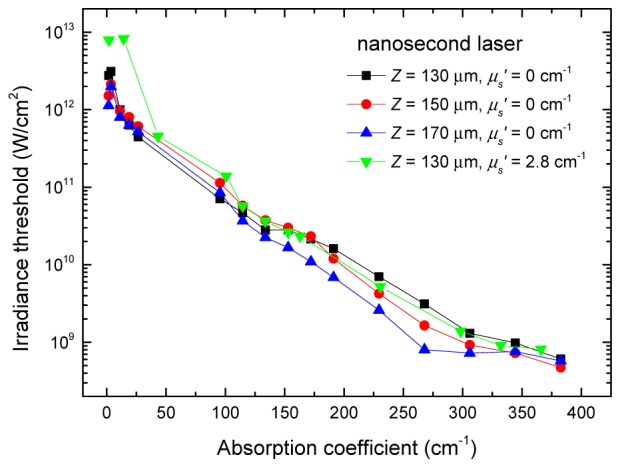
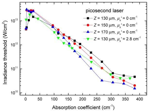
The irradiance threshold measured for ns and ps pulses in distilled water are shown in Figs. 1 and 2 respectively. These results show that as the absorption coefficient of the medium is increased, the irradiance threshold for laser induced breakdown decreases. Depending on the absorption properties of the medium three different breakdown regimes can be observed: (i) in the regime of lowest absorption (μa< 10 cm−1) laser induced optical breakdown occurs through multiphoton-initiation and irradiance threshold for optical breakdown is less dependent on the absorption properties of the medium compared to the high-absorption regime occurring through thermal breakdown; (ii) in the regime of medium to high absorption (10 cm−1<μa <250 cm−1), the thermal breakdown occurs through thermionic initiation and the irradiance threshold decreases with the increase in the absorption properties of the medium; (iii) in the highly absorbing regime (μa>250 cm−1) ‘surface breakdown’ occurs at the interface and the irradiance threshold becomes less dependent on the absorption properties of the samples.
Fig. 1.

Irradiance threshold for laser induced breakdown as a function of the absorption coefficient for the nanosecond laser pulses.
Fig. 2.

Irradiance threshold for laser induced breakdown as a function of the absorption coefficient for the picosecond laser pulses.
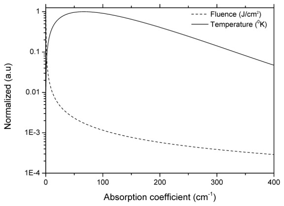
The initiation of ionization requires at least one seed electron in the focal volume. In the regime of lowest absorption (μa< 10 cm−1), free carrier electron density is negligible [10]. In this regime of lowest absorption multiphoton absorption must produce the seed electrons [11]. Here irradiance threshold is found to be insensitive to the linear absorption coefficient and can be very sensitive to the laser beam spatial and temporal profiles and to polarization as we reported before [9]. On the other hand, in the regime of medium to high absorption (10 cm−1 <μa< 250 cm−1) the exponential decrease of lowering of threshold is found and this is due to the generation of seed electrons through thermal initiation pathway. Here the initial free electron density is generated by thermal excitation of molecules which follows Maxwell-Boltzmann probability distribution. The likelihood of finding one thermally ionized electron in the focal volume follows Maxwell Boltzmann distribution (Exp(-ΔE/(kBT)) and depends on the temperature and ionization energy. Assuming for sake of simplicity that each water molecule could potentially be ionized once and using the total number of molecules in the focal volume as the initial density of states, the temperature where at least 0.5 electrons (breakdown threshold definition of 50%) is freed in the focal volume can be derived. For short pulses for which thermal diffusion from the focal volume can be ignored, temperature in the focus can be calculated (T = T0 + µaIx0exp(-µad)/(ρCp)), where Ix0 is the intensity of the un-attenuated focus and d is the focusing depth in the linearly absorbing medium. Typical temperatures for these effects to occur are in the order of 2000-4000K, depending on the assumption of ionization energy and focal spot volume. Focal temperature calculated for different values of absorption coefficient is shown in Fig. 3 . Once an electron has been freed, the breakdown avalanche still needs time to complete, however, these processes occur at a much faster rate in the nanosecond and picosecond time frame. The threshold intensity plots as shown demonstrate that irradiance threshold of nanosecond and picosecond pulses are merely scaled by the actual pulse durations used for these experiments, i.e. the breakdown fluence is constant, suggesting that indeed the breakdown temperature is a constant that is dictating the threshold value for these cases.
Fig. 3.

Fluence required for reaching the breakdown threshold temperature and the temperature in the focus calculated as a function of the absorption coefficient of the medium.
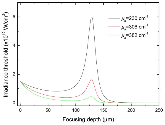
The irradiance threshold for strongly absorbing samples becomes less dependent on the absorption properties of the samples. This is probably because once critical temperature required for generating seed electrons has been reached, increasing the probability of thermal excitation does not necessarily lead to further lowering of threshold. For the highest values of the absorption coefficient and at higher focusing depths, the breakdown is not occurring in the focus but rather a ‘surface breakdown’ is occurring at the interface (glass-medium)of the sample. To further demonstrate this hypothesis, we calculated the irradiance by normalizing the pulse energy measured by the area as a function of focusing depth inside the sample (Fig. 4 ) assuming Gaussian beam propagation. For higher values of the absorption coefficient, the irradiance at the interface is higher than the irradiance in the focus, implying that the breakdown is occurring at the interface, due to a process called “surface breakdown”. In this highly absorbing regime two phenomena can be observed: (i) decreasing of the irradiance in the focus and (ii) the appearance of an irradiance peak at the interface, which is first lower (μa = 267 cm−1), then comparable (μa = 306 cm−1) and finally higher (μa = 382 cm−1) than the irradiance peak in the focus. The irradiance threshold in this regime of surface breakdown is two orders of magnitude less than the threshold for optical breakdown, suggesting that surface breakdown occurs through thermal breakdown. When the absorption coefficient reaches a critical value wherein the surface irradiance exceeds that of the focal region irradiance, then no matter how much higher absorption coefficient is, the surface irradiance will remain the same value, and thus the threshold to create breakdown in the surface. In this study, we demonstrated the gradual transition of breakdown initiation mechanism from multiphoton absorption where irradiance threshold is insensitive to material absorption properties to thermal excitation for which linear absorption controls the laser energy deposition.
Fig. 4.

Irradiance for the picosecond laser pulses calculated at the focus (130 µm) and at the interface for different absorption properties of the medium.
4. Discussion
In this paper we have presented experimentally the influence of the thermal initiation pathway on the irradiance threshold for breakdown in transparent, absorbing and scattering phantoms. The influence of medium (ionization energy, impurity level and absorption) as well as beam (wavelength, pulse width, spot size, and polarization) characteristics on irradiance threshold has been the subject of several theoretical as well as experimental studies [11–16]. These investigations were primarily motivated to produce the same surgical effect in ophthalmic laser surgery applications at lower pulse energies and with less collateral tissue damages [17–20] and therefore have so far concentrated predominantly on transparent and low absorbing tissues. The mechanisms of laser tissue interactions have not been thoroughly studied in the transition regime from multiphoton to thermal initiation pathways in low to very high absorbing samples. In [21] the influence of linear absorption on optical breakdown in transparent media was for the first time considered. The numerical model of optical breakdown was extended to include the thermal electron emission. To date, the dominance of the thermal breakdown process has been demonstrated only for intermediate and long pulsewidths. However, to the best of our knowledge, laser induced thermal breakdown as an independent process was not analyzed in any of the previously published models. An empirical justification for this approach is that the optical breakdown thresholds in water and in transparent tissues such as cornea were found to be very similar for laser wavelengths exhibiting small linear absorption in those tissues [22]. However, for laser wavelengths that are highly absorbed in tissues, the dynamics of laser induced breakdown is completely different. The interaction of linear and nonlinear absorption is also relevant for near and mid IR wavelengths because they are well absorbed in water in the skin.
In the case of transparent and weakly absorbing tissues such as such as the cornea, plasma is the only means to initiate ablation and therefore multiphoton initiated optical breakdown is the optimal choice. Optical breakdown does not differentiate between the target chromophore and the surrounding tissue as the process is independent of the absorption properties of the target. This makes the treatment non-selective for the target chromophore and the surrounding tissue. In contrast during the treatment of pigmented spots, optical breakdown would not differentiate between the target chromophore and the surrounding tissue as the process is less dependent on the absorption properties of the target. This will make the treatment to be non-selective for the target chromophore and the surrounding tissue. This implies that treatment location has to be precisely controlled by the clinician or by an automated feedback system in order to limit the treatment zone to the pigmented zones.
Results obtained here confirm two of the most significant benefits of employing the thermal initiation pathway in skin rejuvenation applications: (i) the selectivity of the thermal initiation pathway for the target chromophore and the surrounding tissue, as the process is dependent on the absorption properties of the target; (ii) the less demanding laser pulse parameters as a result of the lower irradiance threshold, and; (iii) reduction in the required peak power and the associated collateral damage to the superficial layers of skin arising from the peak intensity of laser pulses. If the wavelength of the source is chosen so that the absorption coefficient of the target chromophore at the selected wavelength is in the range of 10 - 250 cm−1, breakdown can be created at significantly lower intensities. On the other hand, this may lead to spatial variation in the ablation efficiency depending on the inherent variation in tissue optical properties.
The absorption coefficient of melanin and oxy hemoglobin are 85 cm−1and 10 cm−1 at 1064 nm. For melanin, this would lead to an irradiance threshold of ~5x10¹¹ W/cm2, which is approximately two orders of magnitude lower than the irradiance threshold for optical breakdown. For oxy-hemoglobin, lowering of threshold will occur only at wavelengths shorten than 1064 nm. For skin rejuvenation applications targeting water, the preferred wavelengths are 1.4 μm (deep treatment) 1.9 μm (superficial treatment).When a chromophore is present everywhere, there exists an optimal range of absorption coefficient (10 cm−1< μa < 250 cm−1), where the irradiance threshold is 10-100 times lower compared to optical breakdown threshold and breakdown occurs in the focal point without any surface breakdown. This occurs when the chromophore is present everywhere within the sample (eg: water) whereas in the case of skin chromophore such as melanin and blood, breakdown can occur at the focal position without surface breakdown.
The irradiance threshold also depends on the M2 value of the laser and optical aberrations in the optical path. Irradiance threshold calculations based on the assumption of diffraction-limited focusing can lead to erroneous results [23]. In our experiments, for calculating the irradiance threshold, we have experimentally measured the spot size using knife edge method and laser pulse energy was kept low enough to avoid plasma formation at the knife edge. The M2 values of the ps and ns laser are 1.6 and 1.03 respectively. The lasers were coupled along the same optical path and are focused by an aspheric lens (Geltech Molded Glass Aspheric Collimator Lens). The aspheric lens has a RMS WFE (axial @ 632.8nm) < diffraction limit. This implies that, in our experiments, the absolute values of irradiance threshold calculated may be also influenced by the changes in the refractive index of the medium as the absorption coefficient is increased.
5. Conclusions
In this manuscript, we report for the first time the effects of thermal initiation pathway on irradiance threshold for laser induced breakdown in transparent, absorbing and scattering phantoms. The initiation mechanism of breakdown shows transition from multiphoton absorption where irradiance threshold is insensitive to material absorption properties to thermal excitation for which linear absorption controls the laser energy deposition. We found that thermal breakdown can be created with lower irradiance threshold in an optimal range of absorption coefficient in the range of 10 cm−1 < μa< 250 cm−1compared to laser-induced optical breakdown. Moreover, the benefits of using thermal breakdown over optical breakdown in lowering the irradiance threshold increase as the absorbing coefficient of the medium are increased. Lower irradiance threshold may allow deeper layers of tissue to be reached, resulting in higher efficacy of treatment. Furthermore since a lower power is delivered over the same target area, the risks of collateral damage can also be reduced. These results have important implications on laser-based biomedical applications and in particular for the novel minimally invasive skin rejuvenation technique recently demonstrated by Philips Research using laser induced breakdown [2,3].
References and links
- 1.Papadavid E., Katsambas A., “Lasers for facial rejuvenation: a review,” Int. J. Dermatol. 42(6), 480–487 (2003). 10.1046/j.1365-4362.2003.01784.x [DOI] [PubMed] [Google Scholar]
- 2.Habbema L., Verhagen R., Van Hal R., Liu Y., Varghese B., “Minimally invasive non-thermal laser technology using laser-induced optical breakdown for skin rejuvenation,” J. Biophotonics 5(2), 194–199 (2012). 10.1002/jbio.201100083 [DOI] [PMC free article] [PubMed] [Google Scholar]
- 3.Habbema L., Verhagen R., Van Hal R., Liu Y., Varghese B., “Efficacy of minimally invasive nonthermal laser-induced optical breakdown technology for skin rejuvenation,” Lasers Med. Sci. 28(3), 935–940 (2013). 10.1007/s10103-012-1179-z [DOI] [PMC free article] [PubMed] [Google Scholar]
- 4.Hammer D. X., Thomas R. J., Noojin G. D., Rockwell B. A., Kennedy P. P., Roach W. P., “Experimental investigation of ultrashort pulse laser-induced breakdown thresholds in aqueous media,” IEEE J. Quantum Electron. 32(4), 670–678 (1996). 10.1109/3.488842 [DOI] [Google Scholar]
- 5.Kennedy P. K., Hammer D. X., Rockwell B. A., “Laser-induced breakdown in aqueous media,” Prog. Quantum Electron. 21(3), 155–248 (1997). 10.1016/S0079-6727(97)00002-5 [DOI] [Google Scholar]
- 6.Noack J., Vogel A., “Laser-induced plasma formation in water at nanosecond to femtosecond time scales: calculation of thresholds, absorption coefficients, and energy density,” IEEE J. Quantum Electron. 35(8), 1156–1167 (1999). 10.1109/3.777215 [DOI] [Google Scholar]
- 7.Niemz M. H., Laser Iissue Interactions: Fundamentals and Applications (Springer; 1996) Chap. 3. [Google Scholar]
- 8.Shen Y. R., The Principles of Nonlinear Optics (John-Wiley & Sons, Canada: 1993). [Google Scholar]
- 9.Varghese B., Turco S., Bonito V., Verhagen R., “Effects of polarization and apodization on laser induced optical breakdown threshold,” Opt. Express 21(15), 18304–18310 (2013). 10.1364/OE.21.018304 [DOI] [PubMed] [Google Scholar]
- 10.Williams F., Varma S. P., Hillenius S., “Liquid water as a lone-pair amorphous semiconductor,” J. Chern. Phys. 64(4), 1549–1554 (1976). 10.1063/1.432377 [DOI] [Google Scholar]
- 11.Kennedy P. K., “A first-order model for computation of laser-induced breakdown thresholds in ocular and aqueous media. I. Theory,” IEEE J. Quantum Electron. 31(12), 2241–2249 (1995). 10.1109/3.477753 [DOI] [Google Scholar]
- 12.Hammer D. X., Thomas R. J., Noojin G. D., Rockwell B. A., Kennedy P. P., Roach W. P., “Experimental investigation of ultrashort pulse laser-induced breakdown thresholds in aqueous media,” IEEE J. Quantum Electron. 32(4), 670–678 (1996). 10.1109/3.488842 [DOI] [Google Scholar]
- 13.Kennedy P. K., Boppart S. A., Hammer D. X., Rockwell B. A., Noojin G. D., Roach W. P., “A first-order model for computation of laser-induced breakdown thresholds in ocular and aqueous media. II. Comparison to experiment,” IEEE J. Quantum Electron. 31(12), 2250–2257 (1995). 10.1109/3.477754 [DOI] [Google Scholar]
- 14.Noack J., Vogel A., “Laser-induced plasma formation in water at nanosecond to femtosecond time scales: calculation of thresholds, absorption coefficients, and energy density,” IEEE J. Quantum Electron. 35(8), 1156–1167 (1999). 10.1109/3.777215 [DOI] [Google Scholar]
- 15.Vogel A., “Nonlinear absorption: intraocular microsurgery and laser lithotripsy,” Phys. Med. Biol. 42(5), 895–912 (1997). 10.1088/0031-9155/42/5/011 [DOI] [PubMed] [Google Scholar]
- 16.Oraevsky A., Da Silva L. B., Rubenchik A. M., Feit M. D., Glinsky M. E., Perry M. D., Mammini B. M., Small W., Stuart B. C., “Plasma mediated ablation of biological tissues with nanosecond-to-femtosecond laser pulses: relative role of linear and nonlinear absorption,” IEEE J. Sel. Top. Quantum Electron. 2(4), 801–809 (1996). 10.1109/2944.577302 [DOI] [Google Scholar]
- 17.Zysset J. B., Fujimoto J. G., Deutsch T. F., “Time-resolved measurementsof picosecond optical breakdown,” Appl. Phys. B 48(2), 139–147 (1989). 10.1007/BF00692139 [DOI] [Google Scholar]
- 18.Vogel A., Busch S., Jungnickel K., Birngruber R., “Mechanisms of intraocular photodisruption with picosecond and nanosecond laser pulses,” Lasers Surg. Med. 15(1), 32–43 (1994). 10.1002/lsm.1900150106 [DOI] [PubMed] [Google Scholar]
- 19.Gitomer S. J., Jones R. D., “Laser-produced plasmas in medicine,” IEEE Trans. Plasma Sci. 19(6), 1209–1219 (1991). 10.1109/27.125042 [DOI] [Google Scholar]
- 20.van Gemert M. C., Welch A. J., “Clinical use of laser-tissue interactions,” IEEE Eng. Med. Biol. Mag. 8(4), 10–13 (1989). 10.1109/51.45950 [DOI] [PubMed] [Google Scholar]
- 21.Venugopalan V., Nishioka N. S., Mikić B. B., “The thermodynamic response of soft biological tissues to pulsed ultraviolet laser irradiation,” Biophys. J. 69(4), 1259–1271 (1995). 10.1016/S0006-3495(95)80024-X [DOI] [PMC free article] [PubMed] [Google Scholar]
- 22.Vogel A., “Nonlinear absorption: intraocular microsurgery and laser lithotripsy,” Phys. Med. Biol. 42(5), 895–912 (1997). 10.1088/0031-9155/42/5/011 [DOI] [PubMed] [Google Scholar]
- 23.Vogel A., Nahen K., Theisen D., Birngruber R., Thomas R. J., Rockwell B. A., “Influence of optical aberrations on laser-induced plasma formation in water and their consequences for intraocular photodisruption,” Appl. Opt. 38(16), 3636–3643 (1999). 10.1364/AO.38.003636 [DOI] [PubMed] [Google Scholar]


