Abstract
Hydatid cyst disease may develop in any organ of the body, most frequently in the liver and lung, but occasionally can affect other organs such as the thyroid gland. Although the prevalence of thyroidal cyst disease varies by region, literature data suggest that it ranges between 0% and 3.4%. The aim of this report was to share 2 cases with thyroid hydatid cyst. Two female patients aged 26 and 57 years were admitted to our outpatient clinic with different complaints. While the first case presented with front of the neck swelling and pain, the second case presented with hoarseness, sore throat, and neck swelling. Both patients were living in a rural area in the southeastern region of Turkey and had had a long history of animal contact. Both patients had undergone previous surgeries for hydatid cyst disease. Both patients presented with a clinical picture consistent with typical multinodular goiter, and both underwent total thyroidectomy after detailed examinations and tests. The exact diagnosis was made after histopathologic examination in both patients. They both had a negative indirect hemagglutination test studied from blood samples. They both have had no recurrences during a 4-year follow-up. In conclusion, although thyroid gland is rarely affected, hydatid cyst disease should not be overlooked in differential diagnosis of cystic lesions of thyroid gland in patients who live in regions where hydatid cyst disease is endemic and who had hydatid cysts in other regions of their body.
Key words: Hydatid cyst, Unusual location, Thyroid, Differential diagnosis
Hydatid disease, also known as echinococcal disease, is a zoonotic disease caused by the parasite species Echinococcus belonging to the Taeniidae family of the cestode class. Four different Echinococcus species have been defined that cause human infection.1–3 The most common species causing human infection are E. granulosus, causing cystic echinococcosis and E. multilocularis causing alveolar echinococcosis.4 Cystic echinococcosis (hydatid cyst disease) is responsible for 95% of all echinococcal diseases in humans. Hydatid cysts may reside almost all organs of human body, although liver (50–77%), lungs (15–47%), spleen (0.5–8%), and kidneys (2–4%) are the most frequently involved organs.1–3 Less commonly, brain, musculoskeletal system, heart, retroperitoneal organs, pancreas, and thyroid gland can also be affected by the disease process. Thyroid gland, however, is rarely involved even in the regions where hydatid cyst disease is endemic.2,3 Hydatid cyst disease can develop in the thyroid gland either primarily (involving the thyroid gland only) or secondarily (with multiple organ involvement).3 This condition is related to the blood circulation of thyroid gland and the entry routes of the larvae into the systemic circulation. E. granulosus larvae arriving at thyroid gland grow slowly within the gland to acquire properties mimicking cystic nodular lesions of the thyroid gland. In some cases, a history of extrathyroidal hydatid cyst disease and observing cyst elements in radiologic tests help making preoperative diagnosis while, in a significant majority of cases, the disease can only be diagnosed by postoperative histopathologic examination. In this study, we aimed to present 2 cases with secondary thyroid hydatid cyst.
Case Reports
Case 1
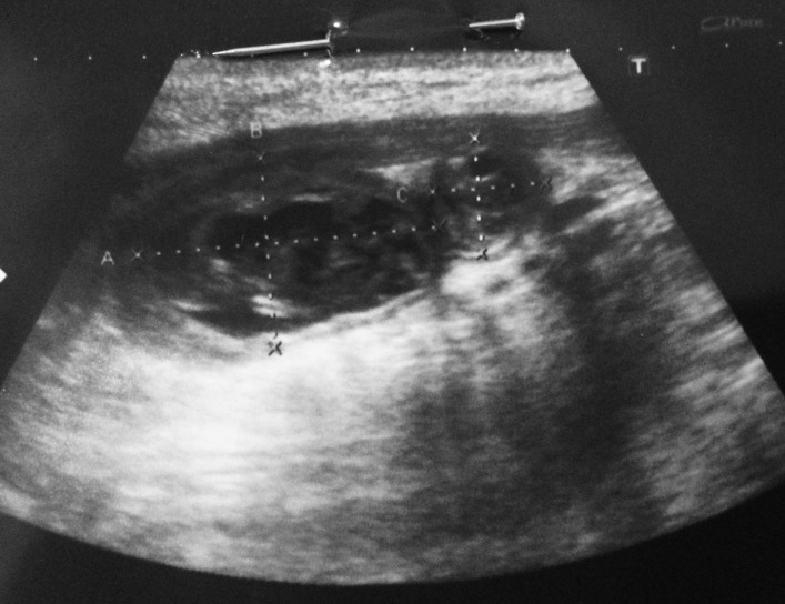
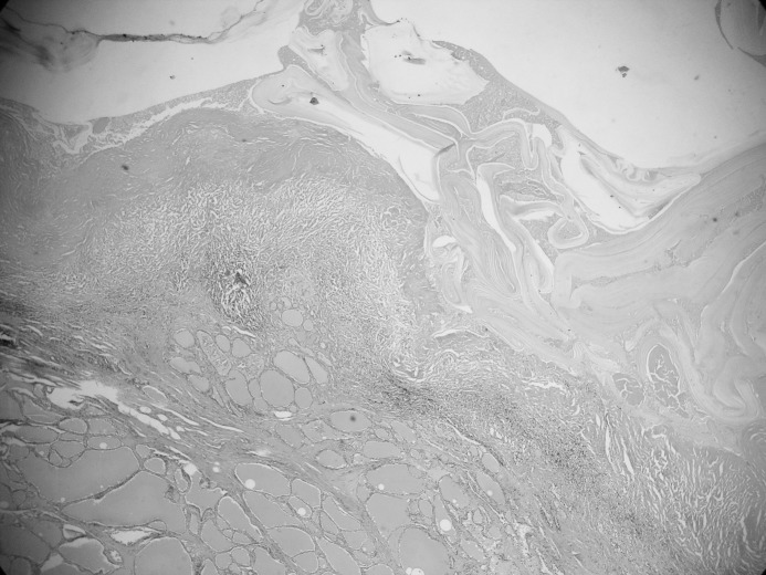
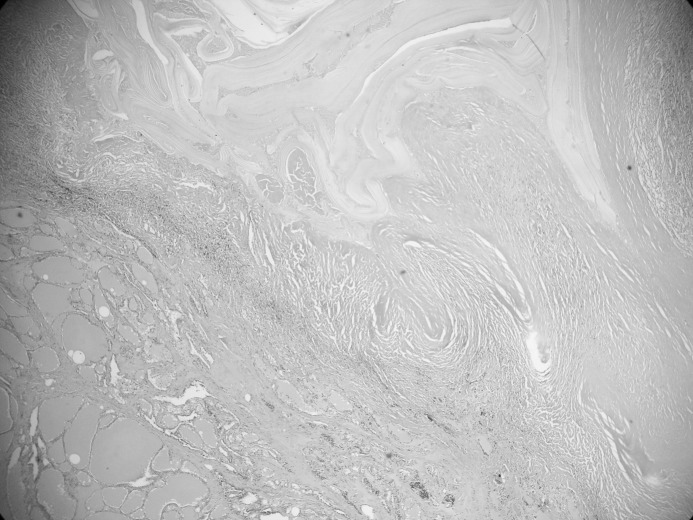
A 26-year-old female patient presented to our outpatient clinic with front of the neck swelling and pain. She stated that the swelling had been there for a long time, but it increased in size recently. She was living in a rural area in the southeastern region of Turkey and she also had a history of animal contact. She told the examiners that she had had an operation for a hydatid cyst in the lung 10 years ago. On systemic examination, no pathologic finding was detected. Neck examination revealed a grade II–III multinodular goiter. Thyroid function tests, hemogram, and liver and renal function tests were all normal. Review of her past medical history revealed that she underwent a thyroid ultrasonography in our hospital in 2007, which revealed a solid nodule of 11 × 6 mm in the neighborhood of isthmus in the right lobe, and a separate well-bordered, lobulated cystic lesion of 40 × 10 mm located in the isthmus just adjacent to the nodule described above. A separate thyroid ultrasonography performed in 2010 in a different outside clinic showed 2 neighboring nodular, hypoheterogenous lesions of 31 × 15 mm and 11 × 10 mm that had a well-defined border and a thick wall and contained cystic degenerated areas within (Fig. 1). Additionally, there were also bilateral multiple cervical lymphadenopathies, with the largest sized at 17 × 6 mm. A fine-needle aspiration biopsy at that time was reported to be nondiagnostic at that time. Based on the findings of physical examination and radiologic tests and upon patient request, the patient was taken into operation and total thyroidectomy was performed. The pathology report was returned as diffuse hyperplasia, multinodular goiter, and presence of hydatid cyst in isthmus (Figs. 2, 3). Despite extensive workup against cyst presence in liver, lungs, and other regions with both ultrasonography and tomography, no other foci could be localized. Furthermore, a serum IHA test at postoperative first month was also negative. Thus, no antihelmintic therapy was administered during the postoperative period.
Fig. 1.
Thyroid ultrasonography showed two neighboring nodular, hypoheterogenous lesions of 31 × 15 mm and 11 × 10 mm that had a well-defined border and a thick wall and contained cystic degenerated areas within.
Fig. 2.
The cyst wall (upper) consists of a laminated, faintly staining chitinous membrane. The adjacent thyroid tissue shows pressure atrophy (HE × 40).
Fig. 3.
The cyst wall is surrounded by a fibrous capsule (so-called pericyst layer; HE × 40).
Case 2
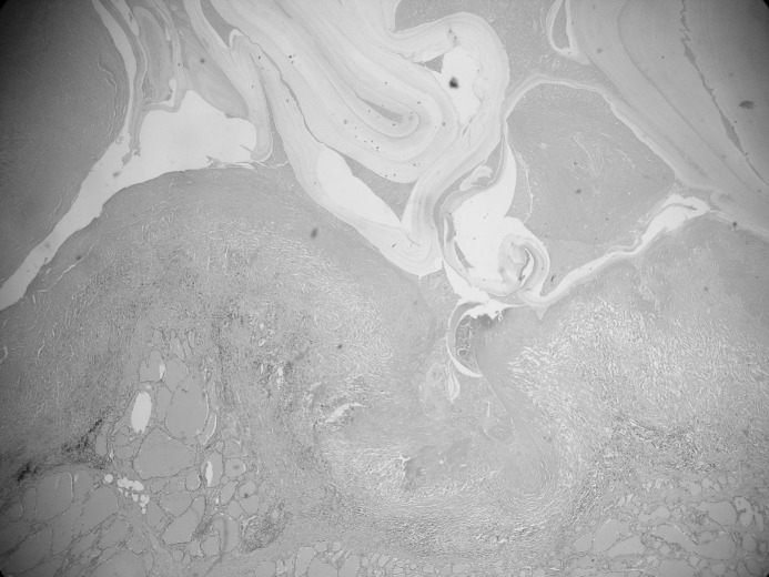
A 57-year-old female patient presented to our outpatient clinic with hoarseness, sore throat, and neck swelling. She stated that the swelling started 10 years ago and increased in size recently. She was living in a rural area in the southeastern region of Turkey, and she also admitted history of animal contact. She had no systemic disease except for hypertension regulated with drugs. She also revealed that she had undergone an operation for hydatid cyst several years ago but did not know what had been done in the operation. On neck palpation, she had no nodular lesion in the left thyroid region but there was a nodular lesion of approximately 5 cm in the right thyroid region in addition to a hyperplastic thyroid gland. Thyroid function tests, hemogram, liver and renal function tests were all normal. A preoperative IHA test was reported negative. Preoperative abdominal CT examination revealed a lesion consistent with a hydatid cyst in the caudate lobe of liver. Thyroid ultrasonography showed a 45 × 29 mm cystic nodule with a peripheral halo, a smooth contour, and internal echoes; it was filling the right lobe completely and making it hyperplastic. Left lobe, on the other hand, was free of any nodular lesion. Considering the history of having a hydatid cyst and the ultrasonographic features of the cyst, the patient was administered preoperative albendazole treatment for 2 weeks, followed by a total thyroidectomy. Her pathology results returned as diffuse hyperplasia, multinodular goiter, and a 5-cm hydatid cyst in the right lobe (Fig. 4). The patient developed signs and symptoms of hypocalcaemia at the postoperative period and she first received calcium treatment followed by calcitriol (Rocaltrol, 0.50 mcg) upon lack of symptom resolution. The parathormone levels ranged between 11 and 11.75 (15–65 pg/mL) and calcium levels between 6.4 and 8.2 mg/dL (8.6–10.2 mg/dL) despite treatment during her 4-year follow-up.
Fig. 4.
The cyst wall contains laminated membrane, an avascular refractile membrane; and an outer dense fibrovascular tissue (HE × 40).
Discussion
Hydatid cyst disease (cystic echinococcosis) is a major public health issue seen in many regions of the world where the ovine and bovine breeding is prevalent as the primary source of income.1,2 As a result of its geographic location, Turkey is found in the center of the Middle Eastern and Mediterranean countries where cystic echinococcosis is endemic, and thus hydatic cyst disease is among the most common endemic diseases in Turkey.1,2 Humans who have no role in the biologic life cycle of the parasite are infected after accidental ingestion of echinococcus eggs in dog feces.2,4,5 Larvae that emerge after rupture of the eggs in the gastrointestinal system of the host first penetrate intestinal wall and then pass into the portal system through which they finally arrive at hepatic sinusoids.4 Larvae of small sizes pass the liver's filtration system and travel to the lungs.2 Most of the larvae that can make it into the lungs are trapped here by a second capillary filtration system.2 Larvae that are capable of avoiding the filtration system of the lung may reach other organs/regions of the body including spleen, kidneys, heart, bone, muscle tissue, pancreas, retroperitoneum, breast, and thyroid gland.1,9 Larvae may sometimes pass into lymphatic channels from intestinal mucosa and into systemic circulation from there through lymphatic vessel without stopping in filtering systems in the liver and lungs. They can also rarely pass to adjacent tissues and organs.
Thyroid gland is only rarely involved in hydatid cyst disease even in countries like Turkey where the disease has a high prevalence.7,8 Although the prevalence of thyroidal cyst disease varies by region, literature data suggest that it ranges between 0% and 3.4%.2 To our knowledge, the first case of thyroid hydatid cyst was published in the literature by Reddy et al in 1946.13 A literature search in PubMed, Medline, and Google Scholar databases by using the key words “thyroid,” “echinococcosis,” “hydatid cyst,” and “hydatid disease” in different combinations retrieved more than 85 articles published in various languages. Pathology reports of 3053 thyroidectomy operations performed in our clinic between June 2006 and March 2014 were retrospectively assessed and only 2 (0.065%) patients were found to have hydatid cysts.
Hydatid cyst disease of the thyroid gland is divided into primary and secondary forms.2,3 The primary form refers to thyroid hydatid cyst disease diagnosed by clinical and radiologic means, with no evidence of extrathyroidal hydatid disease.2 The secondary form is associated with additional hydatid cyst disease of the liver, lung, or other organs.2 Hydatid cyst of the thyroid is generally the primary focus of the infestation; only a few patients are reported to have had secondary hydatid cysts in the liver, lung, or other organ besides the thyroid.3,8,9 Both cases presented in this paper probably had thyroid hydatic cyst disease that developed secondary to hydatid cyst disease in the lung or liver.
Although thyroid hydatid cyst disease can be seen in every age group and both genders, it is usually observed in young adults. The disease is usually confined to a single lobe and tends to be solitary. However, more than 1 cyst can be found in the same lobe or in both lobes in some cases. As with other organs, hydatid cysts located in thyroid gland show a slow growth pattern, and therefore, remain clinically silent for long years.3,6,8 When sufficiently grown, hydatid cysts may adhere to surrounding tissues and organs like trachea, esophagus, carotid sheath, inferior laryngeal nerve, and strap muscles.3,6 Depending on the adhesions and organ compression, dyspnea, inability to swallow, and hoarseness can be produced.3,5,6,8 The most common complications developing secondary to hydatid cyst disease located to thyroid gland include anaphylaxis after spontaneous or iatrogenic rupture, pyogenic abscess formation, and cystotracheal fistulae secondary to erosion of tracheal wall by cyst wall.3,6
Living in an endemic area and a previous hydatid cyst surgery are important clues for preoperative diagnosis of thyroid hydatid cysts. The chance of a correct preoperative diagnosis may increase when these findings are supported by radiologic and serologic tests. The most commonly used radiologic tests for diagnosis are ultrasonography (USG), computed tomography (CT), magnetic resonance imaging (MRI), scintigraphy, and fine-needle aspiration cytology (FNAC).2,4–6,12 These tests also help for differential diagnosis. This is because hydatid cysts of thyroid origin are commonly confused with benign or malignant tumors, abscesses, cysts of other tissues, and other space-occupying thyroid lesions.6 USG is very effective in showing the daughter vesicles, which is central to a preoperative diagnosis of hydatid disease.6 CT and MRI are usually used for advanced diagnosis when the relationship between cystic lesions and the adjacent structures cannot be clearly established by USG.6,7 On scintigraphy, hydatid cysts usually appear as cold nodules and this condition may frequently be confused with thyroid cancers.6,8,11 The most commonly employed serologic tests are indirect hemagglutination, latex agglutination, enzyme-linked immunosorbent assay, and immune electrophoresis.12 However, serologic tests have limited sensitivity and specificity in primary thyroid hydatid cyst disease, limiting their use in this indication.6,9 FNAC is a universally accepted diagnostic tool for determining the approach to thyroid nodules.6,8,10 Especially when performed in experienced hands, it has a quite higher sensitivity and specificity. However, FNAC should be avoided when differential diagnosis include thyroid hydatid cyst.10 This is because FNAC may cause both the development of anaphylactic reactions and the dissemination of cystic elements.6,7 Nevertheless, no complication has been reported in some patients in whom hydatid cyst has not been considered on clinical grounds but incidentally discovered after FNAC.14 Thus, use of FNAC in diagnosis of hydatid cyst disease remains unclear.
As with other regions of hydatid cyst location, the thyroid hydatid cysts are primarily treated with surgical excision.4,8 Removing the cyst(s) radically with utmost care not to rupture the main cystic lesion is the most recommended surgical approach.2,6 However, subtotal thyroidectomy can be recommended in cysts localized to a certain region of a thyroid lobe or in small cysts.6,9 To avert anaphylactic reactions and dissemination of cyst, it is also recommended to protect the surgical field with a hypertonic saline-soaked sponge, to manipulate the cyst as little as possible, and to administer a several-day course of medical therapy in preoperatively diagnosed cysts.6,11 Anthelmintic drugs such as mebendazole, albendazole, and praziquantel can be used not to achieve complete cure, but to kill the living parasites and to prevent possible contamination of cyst content during surgical operation.8
In conclusion, despite being rare, hydatid disease of thyroid gland should not be overlooked in differential diagnosis of cystic lesions of thyroid gland in patients having hydatid cysts in other organs of the body and/or coming from an endemic region.
Acknowledgments
Sami Akbulut and Firat Demircan designed the report; Nilgun Sogutcu provided histopathologic information; and Sami Akbulut organized the report and wrote the paper. The authors declare that they have no conflict of interest.
References
- 1.Akbulut S, Sogutcu N, Eris C. Hydatid disease of the spleen: single-center experience and a brief literature review. J Gastrointest Surg. 2013;17(10):1784–1795. doi: 10.1007/s11605-013-2303-5. [DOI] [PubMed] [Google Scholar]
- 2.Yilmaz M, Akbulut S, Sogutlu G, Arabaci E, Kayaalp C. Hydatid cyst of the thyroid gland: report of three cases. Surg Today. 2013;43(8):937–941. doi: 10.1007/s00595-012-0269-7. [DOI] [PubMed] [Google Scholar]
- 3.Dey AK, Mandal PK, Dutta A, Pramanik S, Maji S, Chakraborti N. A case of hydatid cyst of thyroid gland. Asian J Med Science. 2014;5:143–145. [Google Scholar]
- 4.Oksuz S, Pektas E, Yavuz M, Aksungur N, Cayir Y, Akcay MN. An unusual cause of hoarseness: hydatid cyst of the thyroid. Trop Biomed. 2013;30(4):642–644. [PubMed] [Google Scholar]
- 5.Geramizadeh B. Unusual locations of the hydatid cyst: a review from Iran. Iran J Med Sci. 2013;38(1):2–14. [PMC free article] [PubMed] [Google Scholar]
- 6.Azendour I, Boulaich M. Primary hydatid cyst of the thyroid gland. Int J Otolaryngol. 2011;2011:713089. doi: 10.1155/2011/713089. [DOI] [PMC free article] [PubMed] [Google Scholar]
- 7.Erbil Y, Barbaros U, Baspinar I. Hydatid cyst of the thyroid gland: two case eports. Infect Dis Clin Pract. 2005;13:318–320. [Google Scholar]
- 8.Gokce C, Patiroglu T, Aksehirli S, Durak AC, Kelestimur F. Hydatic cyst in the thyroid gland diagnosed by fine-needle aspiration biopsy. Thyroid. 2003;13(10):987–989. doi: 10.1089/105072503322511409. [DOI] [PubMed] [Google Scholar]
- 9.Ozaydin I, Ozaydin C, Oksuz S, Yildirim M. Primary echinococcus cyst of the thyroid: a case report. Acta Med Iran. 2011;49(4):262–264. [PubMed] [Google Scholar]
- 10.Zulfikaroglu B, Ozalp N, Keskek M, Koc M. Primary echinococcal cyst of the thyroid: report of a case. Surg Today. 2008;38(9):833–835. doi: 10.1007/s00595-007-3796-x. [DOI] [PubMed] [Google Scholar]
- 11.Moghimi M, Kamrava SK, Asghari AM. Heshmatzade Behzadi A, Jalessi M, Naraghi MM, Ehteshamia Afshar E. Primary echinococcal cyst in the thyroid gland: a case report from Iran. J Infect Dev Ctries. 2009;3(9):732–734. doi: 10.3855/jidc.172. [DOI] [PubMed] [Google Scholar]
- 12.Avcu S, Unal O, Kotan C, Oztürk M, Ozen O. Submandibular and thyroid gland involvement of hydatid cysts: a very rare association with percutaneous treatment. Diagn Interv Radiol. 2010;16(3):251–254. doi: 10.4261/1305-3825.DIR.1967-08.2. [DOI] [PubMed] [Google Scholar]
- 13.Reddy DG, Thangavelu M. Hydatid cyst-thyroid. Indian J Surg. 1946;849 [PubMed] [Google Scholar]
- 14.Sodhani P, Gupta S, Jain S. Unusual presentation of disseminated echinococcosis with thyroid involvement: diagnosis by fine needle aspiration cytology in a clinically unsuspected case. Acta Cytol. 2002;46(1):75–76. [PubMed] [Google Scholar]


