Abstract

Introduction: A growing interest in apoptosis, programmed cell death, in the last years is observed and leads to better understanding of molecular mechanisms during cell–cell signaling, cell-environment interaction and screening of drugs. This in turn results in emerging of new assays and development of more accurate kits for fast and early detection of apoptosis. However, their sensitivity and reliability have often been scrutinized. Here we introduce a rapid and improved method of DNA ladder apoptosis assay for evaluating apoptosis in mammalian cells.
Methods: NIH-3T3 cell line was used in this study. After treatment of cells with apoptotic agent, 500 μM H2O2 at 48 hours, DNA was extracted. Then an update protocol of DNA ladder assay was applied for detection of apoptosis. Flow cytometry and DAPI staining were performed to verify apoptosis.
Results: Primary and late apoptosis in the H2O2-treated cells was determined by flow cytometry analysis. DAPI Staining used to show DNA damage and DNA ladder assay using 1.5% gel electrophoresis showed fragmentation in the DNA of treated cells.
Conclusion: In this research we aimed to improve DNA ladder assay to the high quality detection of apoptosis in mammalian cells. In our strategy, employing a practical DNA extraction protocol, DNA ladder assay could be applied as an easy/fast method for apoptosis detection. This improved method is able to detect apoptosis in a cost effective/timely manner without need for commercial kits and special equipment.
Keywords: DNA extraction, DNA ladder assay, Apoptosis, Signaling pathway, Cancer
Introduction
Apoptosis is a distinct form of a programmed cell death (PCD) or cell suicide, first described by Kerr et al. in the 1970s.1,2 It is a normal physiological phenomenon that plays pivotal role during embryonic development, retention of tissue homeostasis and pathology.3 Apoptosis is triggered by extrinsic and intrinsic stimuli as radiation, oxidative stress and genotoxic chemicals. It is defined by characteristic changes in the nucleus morphology including chromatin condensation and fragmentation, overall cell shrinkage/rounding and formation of apoptotic cell bodies. Among the many markers of apoptosis, DNA fragmentation is considered a hallmark.3-5
Defect in apoptotic pathways plays an important role in the development and progression of cancer.3 Modulation of the apoptotic signaling pathways towards normality by means of drugs or treatment strategies have the potential to eliminate cancer cells.3,6 There are different methods to assess the DNA fragmentation due to apoptosis including many commercial kits, morphological identification, DNA ladder observation, flow cytometry and fluorescent assays.7,8 The DNA ladder assay described here is a simple, sensitive, cost-effective and rapid method for estimating apoptosis in single cells. The aim of present study was to develop a fast method for evaluating apoptotic DNA fragments from mammalian cells by providing an easy-to-use DNA extraction protocol. These modified and updated protocol lessens multi-step manipulations including multiple stages of incubation, elution, drying and etc that may cause reduction in DNA yield and quality of results.
Methods and material
Materials
The NIH-3T3 cell line was obtained from the national cell bank of Iran (Tehran, Iran). Cell culture flasks and plates were purchased from JET BioFIL (Elgin IL, USA). Cell culture dishes were obtained from TPP (Trasadingen, Switzerland). RPMI1640 medium and Trypsin – EDTA (1X) were obtained from Sigma-Aldrich (Poole, UK). Normal melting agarose and fetal bovine serum (FBS) were bought from Invitrogen-Gibco (Paisley, UK). The other chemical materials such as paraformaldehyde, H2O2, 2% lysis buffer (Tris-HCl 100 mM, EDTA 20 mM, NaCl 1.4M, C-TAB (N-Cetyl-N,N,N-trimethylammonium bromide) were bought from Merck (Kenilworth, NJ, USA). Annexin V-FITC apoptosis detection kit was obtained from EMD Chemicals (Gibbstown, NJ, USA).
Cell culture
The NIH-3T3 cell line were cultured in RPMI1640 medium supplemented with 10% FBS and antibiotics (penicillin 0.1 μg/μL and streptomycin 0.1 μg/μL) in a humidified incubator with an atmosphere of 95% air and 5% CO2 at 37°C. Induction of apoptosis occurred by 500 µM H2O2 for 48 hours.
DNA extraction protocol
Because of floating apoptosis cells, the media of cultures was collected and centrifuged at 5000 rpm for 5 minutes, the supernatant was discarded and added 500 μL lysis buffer, after then, 500 μL of lysis buffer was added to vacant plates, incubated in room temperature for 10 minutes and harvested lysate cells from plates/flasks into the tubes harboring pellet of centrifuged cells then incubated in 65°C for 5 minutes. After cooling in room temperature for 5 minutes, 700 μL chloroform-isoamyl alcohol was added, and then centrifuged at 12000 rpm for 5 minutes. The aquatic (upper) phase was transfered into new eppendrof tubes. An equal volume of cold isopropanol was added into tubes, and mixed gently by inversion. The tubes were then centrifuged at 12000 rpm for 5 minutes. The supernatant was discarded, and the pellet was air-dried for 30 minutes. Then, the dried DNA was dissolved in 50 μL distilled water. The extracted DNA was quantified by a spectrophotometer, NanoDrop 1000 (NanoDrop, Wilmington, USA). The DNA samples were electrophoresed on a 1.5% agarose gel containing 1μL/100mL SYBR-Safe DNA gel stain (Invitrogen, Cat No. S33102). The gel was examined and photographed by an ultraviolet gel documentation system (iNTAS, Goettingen, Germany).
Flow cytometry
Trypsinized cells with 1 mL trypsin-EDTA at 37°C for 5 minutes until cells were detached completely. Then, 10% FBS–RPMI1640 medium was added to terminate the trypsinization, which were then gently pipetted to avoid the formation of cell clumps. The cells were transferred into an eppendrof tube and centrifuged at 1000 rpm for 5 minutes. The supernatant was discarded, then the cells were washed with PBS (1X). Afterward, the cells were treated with H2O2 (500 μM), and then the cells were collected and resuspended in 100 μL of annexin V binding buffer. Afterward, 5 μL annexin V-FITC was added (Cat. No. 88-8005). After 15 minutes incubation at room temperature in the dark, the cells were centrifuged at 1000 rpm for 5 minutes. Then, the cell pellet was resuspended in 200 mL annexin V binding buffer, and the cells were counter-stained with 5 μL propidium iodide (PI) before analysis. The cells were analyzed using FACS-Calibur flow cytometry (Becton Dickinson, San Jose, USA) with emission filters of 515-545 nm for FITC (green) and 600 nm for PI (red). A total event of 10000 cells per sample were acquired, and the data were analyzed with Cell Quest software (Becton Dickinson, San Jose, USA).
DAPI staining
NIH-3T3 cells were seeded in 6-well plates containing coverslips and treated 500 µM H2O2 for 48 hours, then fixed with 4% paraformaldehyde for 10 minutes. The cells were washed with PBS (3×), then permeabilized with 0.1% Triton X-100 for 10 minutes, washed again with PBS (3×), then stained with DAPI (4,6-diamidino-2-phenylindole) for 5 minutes. The cells were evaluated by a fluorescent microscope, Olympus IX81 invert fluorescence microscope equipped with Olympus DP70 camera (Olympus Corp., Tokyo, Japan).9
Technical hints
(i) To avoid loss of majority of apoptotic cells for DNA ladder assay, floating cells should harvested since apoptosis cause detachment of the cells.
(ii) Before performing flow cytometry assay, the media of treated cell cultures should be collected and centrifuged for harvesting the detached cells.
Results
DNA fragmentation assay
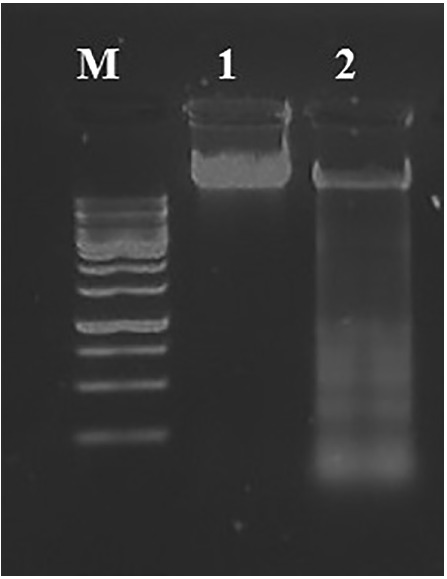
Apoptosis can be visualized as a ladder pattern of 180-200 bp due to DNA cleavage by the activation of a nuclear endonuclease by standard agarose gel electrophoresis.10,11 Thus, we showed the formation of the DNA ladder in gel electrophoresis by induction of apoptosis in NIH-3T3 cell line ( Fig. 1).
Fig. 1 .

Quick detection of apoptotic DNA ladder in NIH-3T3 cell line. Lane M: Standard molecular size marker (1 Kb). Lane 1: untreated cells, Lane 2: H2O2- treated cells.
Flow cytometry assay
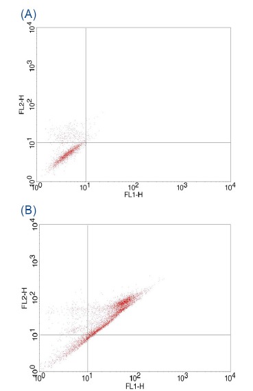
Flow cytometry was applied to distinguish apoptotic and necrotic cell death. Apoptosis includes translocation of membrane phosphatidylserine (PS) from the inner side of the plasma membrane to the surface. Annexin V, a Ca+2-dependent phospholipid-binding protein, has high affinity for PS, cells were stained with Annexin V-FITC/PI.12,13 Our results demonstrated that 58.14% of H2O2- treated cells showed apoptosis in this cell line ( Fig. 2).
Fig. 2 .

FITC-labeled Annexin V flowcytometric detection of apoptosis in NIH-3T3 cells. (A) Untreated control cells. (B) H2O2- treated cells.
DAPI staining
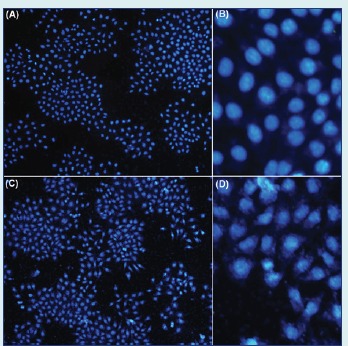
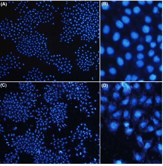
Induction of apoptosis on NIH-3T3 cells was also investigated by microscopic analysis of DAPI stained cells. Fig. 3 shows the untreated control and the H2O2 treated cells. The H2O2 treated cells demonstrated significant fragmentation in the chromatin within the nucleus cells but their morphology did not changed in untreated normal cells.
Fig. 3 .

Fluorescent microscopy of NIH-3T3 cells stained with DAPI. A and B) Untreated control cells. C and D) H2O2 - treated cells.
Discussion
Flow cytometry, DAPI staining and DNA ladder assays for detection of apoptosis are now in widespread use due to importance of apoptosis in molecular biology and drug screening in cancer studies.14 Despite their worldwide use, we demonstrate that DNA ladder assay protocol described in this report, has some advantages over other assays specially DNA ladder kits and previous reports: (i) as mentioned before DNA ladder assay demonstrate the hallmark of apoptosis – mono and oligonucleosomal DNA fragments; (ii) no pre-labeling of the cells is required, not limited to cells that proliferate in vitro; (iii) finally this method is rapid, easy-to-perform because of excluding laborious and multi-step manipulations during DNA extraction, cost effective due to less chemical materials used in this protocol, non-radioactive and yet reliable , and suitable for routine detection of apoptosis in mammalian cells.
DNA ladder assay is an easily available method and seems to be very useful for quick screening of apoptotic changes in cell populations. This method allows working with cell lysates and dose not require any special laboratory equipment.11 It is a simple assay to perform, however being time consuming procedure, prompted us to enhance its sensitivity and reliability through some modification in DNA extraction protocol. The present study results showed that our modified and updated protocol, can be considered as a useful method for evaluating DNA damage and fragmentation without need for laborious laboratory procedures or expensive kits/equipments.
Conclusion
Although flow cytometry is a quantitative technique for determining apoptosis, and DAPI staining shows the morphological changes in the chromatin but they need sophisticated instrumentation and both are time consuming. We believe that the reported improved method of DNA ladder assay will be very useful for numerous laboratories that routinely study cell death or carry out routine experimental/clinical screening of drugs and chemotherapeutics.
Acknowledgments
This work was fully supported by the Research Center for Pharmaceutical Nanotechnology (RCPN) at Tabriz University of Medical Sciences.
Ethical issues
No ethical issues to be declared.
Competing interests
The authors declare no competing interests.
Research Highlights
What is current knowledge?
√ Current DNA extraction protocols appear to be tedious, costly and time-consuming.
What is new here?
√ The Current method is an efficient and high yielded cost-effective DNA extraction protocol.
√ There is no need for commercial kits or advanced instruments.
References
- 1. Rastogi RP, Sinha RP. Apoptosis: molecular mechanisms and pathogenicity; 2010.
- 2.Rennert OM. Apoptosis: Reprogramming and the fate of mature cells. ISRN Cell Biology. 2012;2012:685852. [Google Scholar]
- 3.Wong R. Apoptosis in cancer: from pathogenesis to treatment. J Exp Clin Cancer Res. 2011;30:87. doi: 10.1186/1756-9966-30-87. [DOI] [PMC free article] [PubMed] [Google Scholar]
- 4.Vernon WI, Printen JA. Accelerated Apoptotic DNA Laddering Protocol. Biotechniques. 2002;34:207–15. doi: 10.2144/02334bm03. [DOI] [PubMed] [Google Scholar]
- 5.Zhang G, Gurtu V, Kain SR, Yan G. Early detection of apoptosis using a fluorescent conjugate of annexin V. Biotechniques. 1997;23:525–31. doi: 10.2144/97233pf01. [DOI] [PubMed] [Google Scholar]
- 6.de Bruin EC, Medema JP. Apoptosis and non-apoptotic deaths in cancer development and treatment response. Cancer Treatment Reviews. 2008;34:737–49. doi: 10.1016/j.ctrv.2008.07.001. [DOI] [PubMed] [Google Scholar]
- 7.Suman S, Pandey A, Chandna S. An improved non-enzymatic “DNA ladder assay” for more sensitive and early detection of apoptosis. Cytotechnology. 2012;64:9–14. doi: 10.1007/s10616-011-9395-0. [DOI] [PMC free article] [PubMed] [Google Scholar]
- 8.Koopman G, Reutelingsperger C, Kuijten G, Keehnen R, Pals S, Van Oers M. Annexin V for flow cytometric detection of phosphatidylserine expression on B cells undergoing apoptosis. Blood. 1994;84:1415–20. [PubMed] [Google Scholar]
- 9.Chazotte B. Labeling nuclear DNA using DAPI. Cold Spring Harb Protoc. 2011;2011:5556. doi: 10.1101/pdb.prot5556. [DOI] [PubMed] [Google Scholar]
- 10.Chen FW. Quantitation of DNA fragmentation in apoptosis. Nucleic Acids Research. 1996;24:992–3. doi: 10.1093/nar/24.5.992. [DOI] [PMC free article] [PubMed] [Google Scholar]
- 11.Matalová E, Španová A. Detection of apoptotic DNA ladder in pig leukocytes and its precision using LM-PCR (ligation mediated polymerase chain reaction) Acta Veterinaria Brno. 2002;71:163. [Google Scholar]
- 12. Telford WG, Komoriya A, Packard BZ. Multiparametric analysis of apoptosis by flow and image cytometry. Flow Cytometry Protocols. Springer; 2004. p. 141-59. [DOI] [PubMed]
- 13. Diaz D, Prieto A, Reyes E, Barcenilla H, Monserrat J, Alvarez-Mon M. Flow cytometry enumeration of apoptotic cancer cells by apoptotic rate. Apoptosis and Cancer. Springer; 2008. p. 23-33. [DOI] [PubMed]


