Abstract
Introduction: Pathological features of disease appear to be quite different. Despite this diversity, the common feature of various disorders underlies physicochemical and biochemical factors such as surface tension. Human biological fluids comprise various proteins and phospholipids which are capable of adsorption at fluid interfaces and play a vital role in the physiological function of human organs. Surface tension of body fluids correlates directly to the development of pathological states.
Methods: In this review, the variety of human diseases mediated by the surface tension changes of biological phenomena and the failure of biological fluids to remain in their native state are discussed.
Results: Dynamic surface tension measurements of human biological fluids depend on various parameters such as sex, age and changes during pregnancy or certain disease. It is expected that studies of surface tension behavior of human biological fluids will provide additional information and might become useful in medical practice. Theoretical background on surface tension measurement and surface tension values of reference fluids obtained from healthy and sick patients are depicted.
Conclusion: It is well accepted that no single biomarker will be effective in clinical diagnosis. The surface tension measurement combined with routine lab tests may be a novel non-invasive method which can not only facilitate the discovery of diagnostic models for various diseases and its severity, but also be a useful tool for monitoring treatment efficacy. We therefore expect that studies of surface tension behavior of human biological fluids will provide additional useful information in medical practice.
Keywords: Interfacial tension, Biological fluid, Pathological disease, Medical diagnosis
Introduction
Surface tension is a phenomenon that we see in our everyday life. Many biological performances and natural processes involve an understanding of wetting and interfacial tension where most biochemical reactions occur not in solution but at the surface and interface. Human biological fluids such as serum, urine, gastric juice, amniotic fluid, digestive, urinary and reproductive tracts, endocrine glands, middle ear, cerebrospinal and alveolar lining fluid contain numerous surfactants, proteins, and lipids. These low and high-molecular weight surfactants are the common materials in various tissues of the body which control surface tension of human interfaces. The physicochemical processes that take place in these interfaces are of fundamental importance for various tissues and the vital function of body organs.1-4
Pathological features of diseases vary in the nature and the magnitude. Despite this diversity, the common feature of various disorders underlies the physicochemical and biochemical factors such as surface tension. Changes in the surface tension behavior of human biological fluid are characteristic for some diseases. Studying these interfaces and the changes that occur will provide valuable information relating to various diseases and help to monitor the treatment efficacy. In medicine, surface tension measurement is above all used in connection with various pathological states of lung surfactants such as adult respiratory distress syndrome, bronchial asthma, and pneumonia. In addition to pneumology, there are other studies evaluating the surface tension of plasma, urine and other biological fluids, however beside this work there has been very little work in relating rheology and these forces to pathological disease states.5,6
The impact of surface tension on pharmaceutical formulation and technology has been studied by our group.7-13 Surface tensiometry of biological fluids with an impact on medicine is still in its infancy. A reason for this discrepancy could be that the surface rheology is not introduced to medicine and theoretical studies and characteristics of biological fluids are not well established.3,6 This article will review the significant discoveries and milestone to the current state of our knowledge in the area of biological surfaces and provide a context for the discussion of surface tension as a tool in medical diagnostics, which combined with routine lab tests may be used as an indicator for differential diagnosis.
Theoretical background on surface tension and its measurement in biological samples
The molecules of a fluid experience attractive forces exerted on it by all its neighboring molecules. In the bulk phase, molecules are attracted equally in all directions leading to a net force of zero; however molecules at or near the surface experience attractive force which tends to pull them to the interior of the fluid. Surface tension, γ is energy of the surface at any air/fluid interface defined as force per unit length or energy per unit area. The surface free energy of a fluid is measured by its surface tension and the surface free energy of a solid can be revealed by contact angle measurements.14
Surface tension is dependent on many variables such as temperature, measurement time, materials of the apparatus and the viscosity of the fluid. When a new surface is being formed, surface active chemicals diffuse to the surface and align. During this process, the surface tension is changing rapidly and continuously. Dynamic surface tension measurements allow us to track these changes. When the process reaches equilibrium, static surface tension is obtained by measuring the maximum force at a fluid/gas interface on a sample.
Surface tension, as an intricate property of a fluid, is associated markedly with many variables such as temperature, composition of the solution, presence of impurities in the fluid, measurement time, materials of the apparatus, and viscosity of the liquid.15,16
Human biological fluids contain numerous low-and high-molecular weight surfactants, proteins and lipids that adsorb at fluid interface. The composition of these fluids varies with age, sex, health condition and therapeutic treatment. The processes which take place at these interfaces could reflect the age and sex of a person. The low content of enzyme, lipoprotein and carbohydrate components in female serum is responsible for the high equilibrium surface tension of biological fluids when compared to that of the male. A person’s age reflects the dynamic surface tension of his biologic fluids. As the age increases, the surface tension of serum increases while the surface tension of urine decreases. That is, due to the compositional changes in biological fluids during pregnancy, surface tension of serum decreases.2,6 In biological fluids interfacial forces are involved in maintenance of cell morphology and cell-to-cell cohesion.16,17
Surface tension in human pathophysiology
Cell sorting behavior and morphology
Biological tissues are viscoelastic materials and the cells in a tissue behave very much like molecules in a fluid. This property enables them to change their position and move against each other. The morphology and shape of the organism are driven by the events that occur at the cellular level. The shape of a cell is the result of a balance of intracellular and extrinsic forces exerted on it. This behaviour is defined through surface tension which tends to minimize the exposed area of the cell aggregate and maximize the cohesive forces. The intracellular forces on the membrane are a result of the cytoskeleton reorganization. Energy at the cellular level is usually measured through physical properties such as cell adhesion, viscosity, and cortical tension.9,18 Cell adhesion can be expressed in the following manner:
| (1) |
Where ∆F adh is the free energy of adhesion, γcs, γcl and γsl are the cell-substrate interfacial tensions, cell-fluid interfacial tension and substrate-fluid interfacial tension, respectively.
Intracellular adhesion and repulsion between various cell types cause mobility within cells. Cellular forces are essential for the process of cell coupling, cell morphology and tissue rearrangement.19-21 The resulting balance of intracellular and extracellular forces defines the geometrical shape of the cell and lead to rounding of the tissue which helps to minimize its surface tension and to maximize the intracellular adhesion. Fig. 1 is the schematic representation of interaction between cortical tension and cellular adhesion during the formation of cell–cell contact. Adhesion contact on neighbouring cells ensures stable organization of tissues. Three kinds of forces, cortical tension, cellular adhesion and cellular viscoelastic properties are mechanically coupled to each other. Interaction between these mechanical properties governs cell shape and cell arrangement in tissues. These forces regulate cellular behaviours such as tissue development, embryogenesis, protein expression, cell proliferation and differentiation and will help to study the cell behaviour in normal and pathological conditions and invasiveness of malignant tumors.18-22
Fig. 1 .
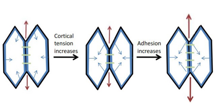
Schematic image of the interaction between cortical tension and cellular adhesion during the formation of cell–cell contacts. Directions of the forces are depicted with arrows, the red line represents cortical cytoskeleton, black lines are plasma membrane and the purple rods depict adhesion sites, adapted from reference 24.
Research has revealed that forces generated by surface tension play a leading role in the position rearrangement of cells and act in minimizing the exposed area of the aggregate. Cell-cell adhesion is maintained through specific adhesion molecules such as cadherins which are expressed on the cell surface. Due to the different adhesion molecules on various cell types, intensity of adhesion among cells happen to markedly varry.23,24 This phenomenon is responsible for the cell migration and is explained by differential adhesion hypothesis. This hypothesis explains adhesion-driven morphogenic processes in the early embryonic development. Expression of different cadherins changes the cell surface contact and controls the cell shape. Regulation of these adhesion molecules in different cell groups determines cell shape and helps to gain information about cellular aggregates in order to decide on the next migration step. As shown in Fig. 2, it can be deduced that when cells with different adhesive forces are put together, the less adhesive cells migrate gradually and surround the more adhesive cells.17,25,26 Taken all, cell adhesion studies will help to understand how organisms are built and how controlling these forces can help to prevent cancer and other human diseases. Both cell-cell cohesion and cell-matrix interactions are responsible for cell configuration and malignant invasion.27,28
Fig. 2 .
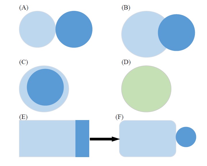
Configuration of two different cell populations with varying adhesion forces, the dark sphere represents cells with high selfadhesion whereas the white sphere represents cells with lower self-adhesion. (A) no cross-adhesion, (B) relatively weak crossadhesion, (C) intermediate cross-adhesion and (D preferential cross-adhesion between the cells and (E) differential adhesion and morphological changes during somatogenesis. Figures were adapted to reference 17.
Tissue grafting
Surface-modified biomaterials are synthetic materials exhibiting specific physiological behavior. They are currently being employed to create cardiovascular, ophthalmic (contact lens) and dental orthopedic materials. Cell adhesion plays a crucial role in medicinal implants. Some factors that influence the cell behavior in contact with foreign material include surface charge, hydrophobicity and surface energy. Extracellular matrix proteins such as fibronectin, collagen and laminin provide an attachment framework for the cell adhesion in vitro. When such materials are exposed to biological environments, protein adsorption and cell adhesion take place and interfacial properties of the material and cells determine the degree of biocompatibility of the device.29-31
Oncology
Tumor morphology and metastasis
Tumors can arise from cells of all types of tissues. Neoplasms are associated with compositional changes of blood, hormones and immune imbalance. The compositional changes that occur during tumor development depend on the localization, size and histological structure of the tumor, treatment pattern, duration of the disease and patient’s sex and age.32
Tumor cell biology and cell thermodynamic function could represent substantial information on cell fate, apoptosis and proliferation. Bending energy, the amount of energy needed to transform a cell to its lowest energy state; i.e., circle, is inversely correlated with surface tension.33 Changes in cell shape and bending energy can trigger modification in gene expression and enzymatic function and result in cancerous cell proliferation, tissue invasion and metastasis spreading.33 In malignant tumors there is a decreased number of spot desmosomes which correspond to a decrease in cell-cell-adhesion resulting in lack of adhesiveness of the cancerous cells. Surface tension of tumor cell can be measured from its surface map. The surface of a tumor can be described by Gaussian curvature with corresponds to excess growth and deficiency of growth in cells. Regions of the tumor with positive Gaussian curvature denote excess cellular growth which is an early indication of the tumour invasion. In regions with low surface tension values, the internal pressure force could overcome it, which could lead to the detachment of carcinoma and possible metastasis and the onset of invasion. In carcinomas, there is a minimum critical value for the surface tension, below this point invasion and/or metastasis of the tumor occurs.17,32 This correlation is shown as:
γ≥ αH*, no invasion/metastasis
γ< αH*, invasion/metastasis
Where γ is surface tension, H is the mean curvature of the solid tumor, and α is dependent on the number of spot desomoses. Tumor classification to benign or malignant types, grading and staging of tumors can be studied using surface tension measurements. A mathematical model for the growth of a solid tumor was introduced using nonlinear elasticity theory. This model describes the basic relationship between stress and cell shape. These correlations also help to determine if tumor invasion or metastasis has taken place. The theoretical suggestion may be useful at cellular level in medical profession and can help make objective decisions on the treatment plan.32
Morphologies of benign and malignant tumor can be used in diagnosis of tumor stages. The interface of malignant tumor is blurred and irregular where as the surface of benign tumor is smooth and clear. During tumor transition, interfacial tension between neoplastic and normal cells decrease and cause the two cell populations to mix.34
Tensiometric parameters of biological fluids in patients with various malignancies
There is a direct correlation between the extent of tumor process and the concentration of γ-globulin, immune complexes, C-reactive protein, ferritin, haptoglobin and α-antitrypsin in blood of patients suffering from cancer of various organs.3,35-37 The concentration of albumin, pre-albumin and α2NS-glycoprotein inversely correlate with the extent of tumor invasion. These factors change the dynamic surface tension parameter and will help to evaluate cancerous phenotype, cell metastasis and therapeutic efficacy. A decrease in blood glucose level is observed in patients with extended tumors, where interfacial tensiometric parameters correlate negatively with the blood glycemic level.3
For a cell to be able to detach from a primary tumour and migrate to a distant location, cancerous cells often undergo some morpho-physiological changes such as pH dysregulation, diminished intercellular adhesion and enhanced motility. These changes enable subsequent metastasis through local invasion and spreading to other tissues which may induce chemoresistance and immunoresistance in cancerous cells.38 Changes in dynamic surface tension parameter in serum obtained from patients with various tumor locations indicate that each type of tumor has its own specific feature which may be considered as informative data from a practical point of view. Most changes in the blood composition were observed for carcinoma of stomach, lung and liver. A decrease in equilibrium surface tension of serum for patients with stomach or lung carcinoma may indicate involvement of the liver and metastatic spreading of the tissue into this organ. However for mammary gland tumors, no changes were observed in the average surface tension values of serum.3,38
In a study E-cadherin was down-regulated in the cancer cells with liver metastasis. This indicates that reduced expression of E-cadherin is closely related to liver metastasis. Interfacial tension serves as a marker of pathological stage of a tumor disease and changes that occur during carcinogenesis have potential application in diagnosis of certain tumors and monitoring of their treatment plan.3,38-40
Tumor of the female reproductive organs
For tumors of the female reproductive organs such as malignant neoplasm of corpus uteri and cervix, dynamic surface tension of the serum is less than normal value. These changes correlate to the increased concentration of γ-globulin, circulating immune complexes and β2-microglobulin. Malignant cervical tissues contain low triglyceride as compared to the normal tissues. Proteins are down regulated in cervical cancer tissues whereas the total lipid and cholesterol content of the cervical tumor tissue is higher than that of a normal tissue. This alteration in lipid composition and cell fluidity could make cells less susceptible to antitumor treatment.41,42
The total phospholipid content of the cancerous cervical tissue appears to be 2.5 folds higher than that of the relevant normal tissue; however significant reduction is seen after radiotherapy. Following successful radiotherapy, the amount of serum proteins and concentration of prostaglandins E2 and F2α decrease and surface pressure parameter of cervical tissue normalize. It is interesting to note that these variations in serum are accompanied by a decrease in the dynamic surface tension of urine.23,41,42 In the course of radiotherapy, interfacial tension measurements may also serve as a predictor of radio-resistance in cervical cancer. Influence of various stages of radiation therapy on the surface tension of cervical tissue is shown in Table 1. After the first dose of radiation, tissues with a minimum surface tension greater that 50 mN/m are considered partially radio-responsive. Tissues with a minimum surface tension less than 47 mN/m are considered completely radio-responsive. Radiation-induced changes in the surface tension values are due to the changes in cholesterol and phospholipid contents. After radiation therapy, tensiometric parameters of the radio-responsive tissue shift towards that of the normal cervical tissue as shown in Table 1. This makes tensiometric measurements a simple and effective tool to study radio-responsiveness in clinical therapy.43
Table 1 . The effect of radiotherapy on the surface tension of cancerous cervical tissue .
| Cervical cancer |
Minimum surface tension mN/m
a) Pre-radiation b) Post-radiation |
Maximum surface tension mN/m
a)Pre-radiation b) Post-radiation |
| Radio-responsive |
a) 26.6±3.6 b) 41.1 ±2.8 |
a) ~45 b) ~31 |
| Partially radio-responsive |
a) 24.9 ±1.2 b) 25.1 ± 3.1 |
a) ~47 b) ~47 |
*data adapted to reference 43.
Brain tumor
Brain tumors are the most fatal types of cancers because of their aggressive and invasive characters. In the course of brain tumor, due to the high expression level of metalloproteinases (MMP), cells are more migratory and less adhesive. Cells with high MMPs expression exhibit faster migratory pattern whereas brain cells with slower invasion usually have denser matrix. MMP activity seems to be more dominant in glioblastomas. Further, higher level of N-cadherin was observed with increasing tumor grade in astrocytic tumors. Surface tension of serum depends on the type and location of brain tumor and inversely correlates with the duration of the disease. For example, serum of patients with cerebellum, ventricle IV and posterior cranial fossa contain lower amounts of γ-globulin and β2-microglobulin but higher concentrations of amino acids.3,28
Respiratory diseases
Pulmonary surfactant and breathing cycle
Airway is the main port of entry for pathogens and allergens. For this reason both upper airways (trachea and brohchi) and lower airways (alveoli) present an epithelium lining cell that secret mucus and pulmonary surfactant, respectively. Respiratory mucus represents a complex mixture of water, protein and glycoproteins, lipids and salts, which provides viscoelastic properties as a physical barrier for the entry of pathogens and allergens during breathing cycles. Pulmonary surfactant (PS) has been delineated as a surface-active material that coats the alveolar space of human lung to reduce the surface tension and prevent collapse of the alveoli during breathing; it also plays a significant part in the lung’s defense against bacterial or chemical invasion. PS is composed of lipids (~90 weight percentage (wt%)) and surfactant associated proteins (~10 wt%). The most abundant lipid with surface tension lowering properties is dipalmitoylphosphatidylcholine (DPPC), ~60-70% wt%. DPPC is necessary to achieve surface tension of near zero mN/m during the film compression and expiration. Surfactant specific proteins (SP) include SP-A, SP-B, SP-C and SP-D. SP-A and SP-D are hydrophilic proteins with no surface properties. SP-B and SP-C have low molecular weight, hydrophobic molecules which lower surface tension and prevent the alveolar collapse at the end of expiration. Air pollutants can significantly interfere with the ability of lung surfactants to decrease the surface tension during breathing cycles.44,45
Lung surfactants possess a key role in normal pulmonary function. Dynamic surface tension of lung surfactants allows the alveoli to remain inflated. An effective lung surfactant must have three properties, as follows: i) The rate of its surface adsorption should generate surface tension of 25 mNm-1 to match that of the air-fluid interface in the lung, during the first breaths and inspiration throughout life. ii) It should be able to prevent alveolar collapse during surface compression/expansion cycle. Surfactant should reduce surface tension to nearly 0 mNm-1 to avoid alveoli collapse. iii) During surface expansion, surfactants should reduce surface tension and decrease the pressure difference needed to maintain ventilatory cycles throughout life ( Fig. 3). Alteration in the surfactant system during pulmonary diseases can be detected with the surface tension analysis. Stress and damage can cause leakage of the blood proteins (such as albumin and fibrinogen) into the lung and interfere with its ability to lower the surface tension and cause difficulty in breathing.46-50
Fig. 3 .
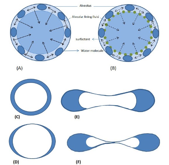
Schematic illustration of surface tension properties at the alveolous, (A) without surfactants, solid arrows represent strong attractive forces and high interfacial tension leading to alveolar collapse, (B) with surfactant coating, broken arrows represent reduced tension leading to lung stability, (C) cross-section of alveolous, surface tension-induce fluid pressure and (D) the external pressure causes the initially axisymmetric alveolous wall, (E) to buckle non-axisymmetrically to a low surface area and energy state, (F) figures were adapted to references 46,47.
Respiratory distress syndrome and hyaline membrane disease
Hyaline membrane disease (HMD), more commonly known as the neonatal respiratory distress syndrome (RDS), is one of the most common causes of morbidity and mortality in pre-term babies. The disease is caused by lung immaturity and surfactant deficiency in the alveolar space. RDS is more frequent in male infants and infants of diabetic mothers. RSD rate decreases with advances in gestational age. Functional residual capacity in babies with RDS is insufficient and leads to collapse and re-expansion of the lung causing reduction in ventilation and lung failure. The supernatants from the airway samples of infants with RDS had a mean minimal surface tension of 35.0 ± 1.4 dynes/cm. These infants have little or no phospholipid in their surfactant content which causes a high surface tension value. In contrast, the mean minimal surface tension of infants without RDS was 6.3 ± 1.1 dynes/cm. Luckily this disease can be treated by applying naturally modified or synthetic surfactants to replace surfactant content of the lung and lower the surface tension properties of the lung and help for an easier breathing cycle.51,52
Chronic bronchitis
Chronic bronchitis is caused by the excess mucus production or hypertrophy of the mucous gland which causes thickening of the bronchial mucosa. An increase in the mucus secretion dilutes the surfactant and increases the surface tension of surfactant lining of the lung airways and causes narrowing and obstruction of the airway.
Drugs are used to ameliorate the mucus production and restore the normal condition of the bronchial mucosa and the pulmonary airways.53
Cystic fibrosis
Cystic fibrosis (CF) is a genetic disease caused by mutation of the transmembrane conductance regulator protein. Little is known about the exact mechanisms by which the pathology arises and progresses. In these patients decrease in phosphatidylcholine fraction and an increase in phosphatidylinositol fraction causes abnormally viscous bronchial secretion. This deficiency in the essential fatty acids attributes to higher minimum surface tension compared to patients with chronic bronchitis. Minimum surface tension in the bronchial lipid fraction samples from CF (27.6 ± 5.9 mN/m) was significantly higher than that of the healthy control samples (22.6 ±1.0 mN/m).54,55
Obstructive sleep apnea
Obstructive sleep apnea is caused by the negative pressure in upper airway and the imbalance of forces to maintain an open airway. Surface tension forces between mucosal surfaces play an important role in the pathogenesis of the disease. The obstruction of upper airway elicit the activation of autonomic nervous system, hypoxemia, and arousal from sleep. Further, continuous positive airway pressure may be an important form of treatment to help eradicate episodes of airway collapse during sleep. Topical application of synthetic surfactant to the upper airway at night may reduce the severity of the disease, decrease surface tension and facilitate opening of the upper airways. For example, phosphocholinamine, derived from lecithin, has some chemical similarity to human surfactants. It has long duration of action and when administered to the nose before sleep it can significantly reduce the overnight snoring.56,57
Atelectatic human lung
Atelectasis refers to the collapse of part or the entire lung which may be due to alveolar collapse or fluid consolidation. Development of the atelectasis is associated with impairment of oxygenation, increased pulmonary vascular resistance and lung injury. Table 2 demonstrates that the surface activity of the non-atelectatic portion of the lung is similar to that of the control lung and markedly higher than that of the atelectatic section of the same patient. It has been shown that the minimum surface tension attained on compression of surface film in atelectatic lung did not reach the low surface tension attained by the controls.58
Table 2 . Maximum and minimum surface tension on expansion and compression of surface film in atelectatic human lung .
| Human volunteers | Surface tension (dyne/cm) | |||
| Atelectatic portion | Non-atelectatic portion | |||
| Maximum | Minimum | Maximum | Minimum | |
| 38.5 | 22.0 | 38.2 | 4.2 | |
| 43.1 | 18.1 | 43.2 | 5.4 | |
| 41.8 | 14.2 | 33.6 | 5.4 | |
| 39.7 | 22.7 | 41.0 | 7.9 | |
| 43.1 | 13.6 | 41.9 | 7.9 | |
| 39.7 | 21.5 | 32.9 | 4.5 | |
| 44.2 | 13.6 | 51.0 | 4.5 | |
| 44.2 | 18.1 | 39.7 | 9.1 | |
| 47.6 | 23.8 | 37.4 | 6.8 | |
*Data adapted to reference (58.
Asthma and allergy
Asthma is defined as the obstruction of pulmonary airway caused by the contraction of smooth muscle together with the edema and hypersecretion. There are indications that the surfactant deficiency and interfacial forces can play a role in asthma, however exact mechanisms have not been elucidated. One of the major mechanisms of asthma is eosinophilic inflammation. These inflammatory cells secrete various cytotoxic proteins which are potent surfactant inhibitors. One study indicates that eosinophil cationic protein (an inflammatory mediator) concentration correlates well with the surfactant dysfunction. This can significantly increase surface tension (γmin> 20 mN/m) which in turn could lead to airway obstruction.59-61
Renal and genitourinary diseases
Urine constituents comprise of over 150 chemicals including; electrolytes, nitrogenous compounds, vitamins, hormones, organic acids, and miscellaneous organic compounds. None of these normal constituents have any considerable effect(s) on the urine surface tension. Urea, uric acid, creatinine, and acetone do not alter the surface tension. The urinary constituent that influences the surface tension includes bile salt and sodium chloride. Urine exhibits inverse relationship between the surface tension and the bile salt concentration. In other words surface tension of urine decreases with age and certain conditions such as acidity and jaundice.6 The surface tension characteristics of urine depend on the person’s sex, age and diet (Table 3). These differences may be due to various levels of eicosanoids, palmitic and hyaluronic acids, urea, creatinine and uric acid contents. It is found that increased levels of calcium in the urine can potentially increase bacterial adhesion to uroepithelial cells.6,62-64
Table 3 . Dynamic surface tension characteristics in urine of healthy volunteersa .
| Sex | Age | Surface tension parameters b | ||
| γ 1 (mNm -1 ) | γ 2 (mNm -1 ) | γ 3 (mNm -1 ) | ||
| Males | >20 | 72-73 | 69-63 | 61-63 |
| 20-35 | 70-72 | 68-70 | 59-62 | |
| 36-50 | 70-72 | 68-70 | 57-60 | |
| ≤50 | 70-73 | 67-71 | 56-61 | |
| Females | >20 | 72-73 | 69-71 | 65-68 |
| 20-35 | 70-72 | 68-70 | 64-66 | |
| 36-50 | 70-72 | 68-70 | 61-65 | |
| ≤50 | 70-73 | 67-71 | 60-65 | |
a Data adapted to reference 6.
b γ1 surface tension for t=0.01 s; γ2 surface tension for t=1 s; γ3 surface tension t→∞.
Urinary stone disease
Urinary stone disease is a chronic disorder in humans and the most common types of kidney stones are those of calcium oxalate. Stone formation involves different phases; nucleation is the first stage of stone formation which is influenced by supersaturation. This causes crystallization of the urinary stone, where molecules start forming clusters. After nucleation, the crystal growth is the next step where the total free energy of the cluster is increased by surface energy. This causes crystal aggregation and attachment to the renal epithelial cells. Non-adherent surface of tubules, bladder and the urethra provide a natural defense mechanism against crystal retention. When the physical mechanisms that actively inhibit crystal retention and adhesion are compromised, urolithiasis and stone disease may occur.65
Proteinuria
Changes in the lipid and protein content of urine are symptoms for kidney diseases.6 The traces of protein-like substances in urine samples are rarely detected by routine laboratory tests and tensiometric methods are more sensitive for such measurements.66 It is demonstrated that surface tension of urine increases in proteinuria. Interfacial tension measurements can help to predict nephropathy and to estimate the effects of therapy, which can also serve as a surrogate parameter to complement biochemical, spectroscopic and other quantitative and qualitative analyses.2,67
Dialysis
Continuous ambulatory peritoneal dialysis (CAPD) is a method used for the treatment of patients with chronic renal failure. There are various complications such as peritonitis, infection and loss of ultrafiltration which can lead to failure and hospitalization of patients. Surface active materials such as phospholipids (mainly phosphatidylcholine and palmitic acid) have been detected in CAPD dialysis effluent of patients via chromatographic methods. It has been found that phospholipid concentration correlates with the time the patient has been on CAPD modality. The decrease in phospholipid content with time on dialysis and a subsequent decrease in surface activity cause loss of ultrafiltration. Infection is another complication of CAPD that causes the failure of therapy. Infection could be prevented by lubricating the peritoneal surface using surface active materials. A relationship is found between the dialysis time and length and the number of infection episodes. Data indicates that long-standing and infected patients have lower phospholipid content in their peritoneal dialysis effluent.68
Glomerulonephritis
Misbalance of protein, lipid, carbohydrate and electrolyte exchange are typical in glomerulonephritis and Pyelonephritis. There are various types of glomerulonephritis which can be verified with surface tension measurements. In general the pathogenesis of all types of glomerulonephritis are similar, however the parameters of protein composition within specific groups are rather different. Morphological changes that occur in kidney tissue influence the tubular reabsorption and the glomerular filtration. These changes can easily be characterized with tensiometric measurements. There is an increase in γ1 and γ2 of patients suffering from acute glomerulonephritis (AGN) and hemorrhagic vasculitis (Genoch glomerulonephritis (GGN)). In Lupus glomerulonephritis all these characteristics are increased, whereas in chronic glomerulonephritis (CGN) λ increases. From Table 4 it is clear that the increase in urine γ1 is characteristic for all groups of patients where as γ1 and γ2 increase for GGN only. A decrease in the surface tension of urine was observed in patients with GGN. AGN and CGN are characterized by very low concentrations of serum proteins, albumin, C-reactive protein, fibrinogen and other proteins as compared to Lupus glomerulonephritis (LGN) and glomerulonephritis accompanying hemorrhagic vasculitis (GHV). Treatment of glomerulonephritis includes application of glucocorticoids, ultrafiltration and hemodialysis and kidney transplantation. Glucocorticoids have different effects on the dynamic surface tension characteristics of various types of glomerulonephritis. In patients with chronic disease all surface tension parameters are increased. In patients with Genoch, γ2 and γ3 is increased and in patients with acute and lupus no changes in the initial dynamic interfacial characteristics were observed. Ultrafiltration influences the interfacial tension of plasma without affecting its viscosity, osmolarity and electrolyte concentration.6 After kidney transplantation or hemodialysis surface active materials removal, blood constituents change and significant increase has been observed in surface tension values. Thus interfacial tension can be used as a promising tool to estimate the efficiency of treatment and diagnose transplant rejection.6
Table 4 . Surface tension of blood and urine for patients with various forms of glomerulonephritisa .
| Types of glomerolonephritis | Serum | Urine | ||||||
| γ 1 | γ 2 | γ 3 | λ | γ 1 | γ 2 | γ 3 | λ | |
| Acute | + | + | + | |||||
| Chronic | + | + | - | - | - | |||
| Lupus | + | + | + | + | + | |||
| Genoch | + | + | + | + | + | |||
a Data adapted to reference 6.
+ Statistically significant increase of parameters compared to normal.
- Statistically significant decrease of parameters compared to normal.
Pyelonephritis
Primary and secondary pyelonephritis are types of kidney diseases with sex-related tensiometric parameters. In female patients, the values of λ decreases significantly irrespective of the type of pyelonephritis, while in cases of primary pyelonephritis in male λ values are higher. In secondary pyelonephritis the electrolyte misbalance and hyperglycemia will decrease serum γ1 and γ2.6
Tensiometric studies of biological fluids can help to differentiate the glomerulonephritis and the amyloidosis without implementation of invasive methods such as kidney biopsy. Multiple-myeloma causes a decrease in blood γ2 and γ3, whereas the values of γ1 in urine are inversely related to the total proteinuria. After treatment with plasmapheresis, the surface tension normalizes and reaches values corresponding to that of healthy humans.6
Eye, ear, nose and throat diseases
Human tear film and dry eye syndrome
In the normal healthy eye, the lipid layer has a thickness of less than 0.1 micron. The pilosebaceous meibomian glands located in the upper and lower eyelids produce most of the outermost lipid layer. This lipid layer is the most superficial layer of the tear film and contains both polar and non polar lipids mainly waxy and cholesterol esters. An important effect of the tear mucus glycoprotein is its ability to lower the surface tension of the tear and permit wetting of an otherwise non-wettable surface of the corneal. This allows easier distribution of the tear fluid. Alteration of the polarity and molecular weight of tear film may result in disease such as blepharitic.69,70
The low surface tension of human tear, 43.6±2.7 mN/m-1, is due to the presence of certain tear lipids of meibomian origin or lipid binding proteins such as lipocalins which are able to reduce the surface tension of tear film. The surface tension is deemed to be a direct measure of the wettability of tear film. In the presence of the superficial lipid layer, surface tension significantly decreases to enhance the spreading of the tear fluid over the cornea. Excessive loss or maldistribution of the tear film, ineffective lid blinking or insufficient tear supply increase the surface tension value and cause dry eye syndrome.69,70
Tear film breakup time is defined as the time needed for a tear film to break up following a blink, this normally takes 15-20 seconds. A break up time of less than 10 seconds is an indicator of the abnormal tear film. This may be due to contamination of the mucin layer which decreases the surface tension of the tear film.65 Tear dysfunction may occur when tear mucins are deficient in number [avitaminosis A, conjunctival destruction], excess in number [due to simulation of foreign object; allergy and giant papillary conjunctivitis] or biochemically altered [keratoconjunctivitis]. Nearly all artificially tear preparations contain one or more chemical surfactants that can enhance the wettability of cornea. Adhesion and biocompatibility of the contact lens with the tear film requires an intimate understanding of the lens surface chemistry and the wetting agents including those that are normally found in the eye.71
Eustachian tube and otitis media
Surfactant of the Eustachian tube is a weak surface tension lowering agent which facilitates the discharge of the effusion in the ear. Eustachian tube surfactants are quiet similar to the lung surfactants. However there are some differences in the ratio of the phospholipids and Eustachian tube lavage has 2:1 ratio, phosphatidylcholine to sphingomyelin content, versus a 67:1 ratio in pulmonary lavage. Surface tension in the healthy Eustachian tube is 58 mN/m. This is because surface tension lowering surfactant lipids are present in the Eustachian tube at low concentration.4 These surface active agents lower the opening pressure and facilitate the fluid effusion in the middle ear of healthy individuals and prevent the accumulation of fluid and inflammation within the middle ear and prevent the otitis media.72
Caries and teeth decay
Saliva is composed of 99% water and 1% organic and inorganic compounds. The surface tension of saliva has been studied and results show that there is no significant differences between the surface tension of samples taken from various locations of the mouth, however there are age and sex-related differences in some tensiometric characteristics of the saliva. The equilibrium surface tension is the highest (48.0±2.3 mN/m) at age 5-9 and decreases with age and reaches 42.3±3.5 mN/m at age 40-55. However, at ages above 55 years this value increases again to 45.7±2.9 mN/m. Sex related difference in the tensiometric parameter is thought to be due to the biochemical composition of saliva and its high protein content in women. The most significant difference in the tensiometric parameter of saliva sample is seen among children with and without caries and tooth decay. Thus tensiometric measurements can be used as new diagnostic criteria in dentistry for the estimation of tooth enamel decay. Table 5 depicts surface tension parameters of oral fluid in caries-free and caries-active group children of age 4-6 years at time range between 0.001-100 seconds. It can be seen that equilibrium surface tension, γ∞, is significantly different in two groups.73,74
Table 5 . Tensiometric parameters of oral saliva in caries-free and cries active children of age 4-6 yearsa .
| Tensiometric parameters (mNm -1 ) | Examined group | |
| Carries-free | Caries-active | |
| γ0.001 | 72.6±0.3b | 73.9±0.2 |
| γ1 | 69.9±0.4 | 70.1±0.6 |
| γ100 | 61.9±0.5c | 62.5±0.4 |
| γ∞ | 46.0±0.7b | 43.1±1.0 |
Interfacial tension is also an important factor in adhesion and formation of dental plaque on the tooth surface. The only preventive regimen proven successful is the use of anti-tartar ingredient in toothpaste or dentifrices.75 Dental fissures are good sites for microbial plaque. Fluoride application has proven to be effective against dental caries. Viscosity and surface tension influence the penetration of fluoride gels into fissures. By optimizing these values, penetration and therefore treatment of dental caries can be achieved.76 Gargle mouthwashes and toothpastes contain surfactants such as sodium dodecyl sulfate to loosen the surface deposits and to emulsify the debris.77
Neurology diseases
Cerebrospinal fluid
The chemical composition of cerebrospinal fluid (CSF)is regulated via peripheral blood circulation, and thus its content is similar to that of the blood serum. CSF consists of 90% water, 10% solid residues and traces of inorganic and organic compounds mainly surfactants. The protein content of CSF is 200-400 times lower than serum. CSF helps maintain an osmotic pressure and protects the brain from the physical injuries. Any pathological processes in the central nervous system are accompanied by some changes in CSF in terms of the composition and surfactant properties (Table 6).3,78 CSF surface tension of patients with CSF infection, hemorrhage, congenital and tumor hydrocephalus, and CNS malformations has been studied. It is found that the surface tension of CSF is dependent on its protein concentration.5,79
Table 6 . Surface tension of serum and cerebrospinal fluid during various types of nervous system diseasea .
| Disease of the nervous system | Surface tension Serum | Urine | ||||||
| γ 1 | γ 2 | γ 3 | λ | γ 1 | γ 2 | γ 3 | λ | |
| Infection | - | - | + | + | + | |||
| Vascular | + | + | + | + | ||||
| Spondylogenic | - | + | + | + | + | |||
| Neoplasm | - | - | + | |||||
| Trauma | - | - | + | + | + | |||
a Data adapted from reference 3.
+ Statistically significant increase of parameters compared to normal.
- Statistically significant decrease of parameters compared to normal.
Neurosyphilis
Neurosyphilis (NS) is caused by a syphilitic infection and involves the nervous system at multiple sites followed by changes in the CSF. The disease is asymptomatic and does not exhibit any clear clinical indications which may complicate its diagnosis using traditional serological studies of the blood and the CSF. Patients not treated for the persistent CSF abnormalities are at risk of developing clinically apparent disease; therefore development of a diagnostic method is of high interest. Dynamic surface tension studies of CSF can help to diagnose neurosyphilis and other pathologies as well as concomitant disease of syphilitic patients. Elasticity parameter of more than 40 mN/m indicates the absence of other nervous system pathologies for the syphilitic patient. However, elasticity values of less than 32 mN/m correspond to the presence of other neurological disease such as encephalopathy accompanied by syphilis.78
In the cases of malignancies, trauma and infection, there is a decrease in the γ2 and γ3. Form Table 6, it can be seen that infection causes a decrease in the surface tension parameters and an increase in the plasma viscosity. The equilibrium surface tension is inversely dependent on the total concentration of protein in the CSF of patient with spondylogenic disease.3
Gastrointestinal diseases
Human digestion system
Gastric mucosal cells secrete unsaturated phosphatidylcholines to protect the mucosa against acid-back diffusion. Surface tensions of gastrointestinal fluid of stomach, duodenum and jejunum are 35-45, 32.3 and 28 mN. m-1, respectively.80,81
Gastrointestinal microflora consists of a complex of micro organism species that live in the digestive tract and help maintain ecological balance in the body. Microflora of healthy individuals is different from that of the diseased and changes through the consumption of antibiotics and probiotics. Microflora of the gastrointestinal tract provides adhesive characteristics which are considered useful as a tool to prevent pathogenic adhesion of bacteria.82
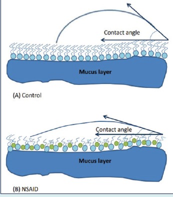
Gastric mucus glycoproteins form an adherent gel of 100-200 μm thick between the tissue and the bulk lumen tissue. Furthermore, the mucus cells secrete a hydrophobic surfactant-like phospholipid monolayer that adsorbs directly onto the mucus gel layer. Contact angle studies of this layer directly correlate with the barrier property of the gastrointestinal tissue (shown in Fig. 4). This membrane is able to stretch and cover the gastrointestinal tract during digestion. These hydrophobic forces increase the surface tension at the aqueous-lipid interface and help to pull the lipid molecules together, hence the membrane can stretch during digestion and prevent deformation. This weak barrier is disrupted when the luminal pH is lower than 2. Amphiphilic molecules such as non steroidal anti inflammatory drugs (NSAIDs) interact and thereby change membrane surface tension. This influences the bending rigidity of the membrane, changes its hydrophobicity fluidity and decreases the mucosal barrier property of the membrane.83
Fig. 4 .
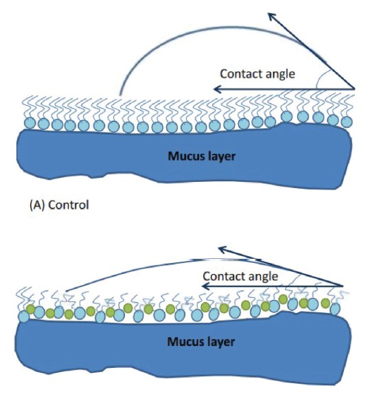
Schematic figure of interfaces between the gastric mucus gel layer and the gastric juice measured by contact angle measurements. (A) contact angle reading between 60-80°, (B) contact angle reading of &40° after exposure to NSAID and reduction of hydrophobicity. Figures were adapted to reference 83.
Biles in the stomach have been found to be responsible for the gastritis and gastric ulcer. Gastric biopsy and surface tension measurements from patients have shown that surface tension lowering agents are presumed to be from bile.84 The fundamental stages in understanding digestion involve knowledge of acid pH, body temperature, salt and bile acid concentrations, and the interfacial β-lactoglobulin protein network. These factors will influence the action of digestive enzymes and phospholipids and ultimately change the digestion process. The study of human digestion system involves an understanding of the interfacial mechanism and the activity of lipase and lipid adsorption. Interfacial tension studies can be used to monitor enzymatic action of lipase and hence control digestion of food. Atomic force microscopy may be used to study the effect of bile salts on the interfacial forces of the intestinal system.85,86
Inflammatory bowel syndrome
The gastrointestinal tract contains <200 ml of gas. Nitrogen, carbon dioxide, methane and hydrogen gases are formed from the colonic bacterial fermentation of the ingested food. Gas-related symptoms depend partly on interfacial tension of the tract and how fast the gastrointestinal system moves. Various studies have been undertaken to investigate the pathogenesis of bloating. Symptoms include flatulence, bloating, gas production and abnormal muscle activity of abdomen.87 Evaluation of such patients should be based on the underlying condition and treatment should be based upon the pathophysiologic findings. Agents which reduce the surface tension are often used for the gas symptoms. Other therapies include dietary modification, gut transit alteration and enzymatic preparation.87
Hematologic diseases
Erythrocyte shape and malaria parasitic invasion
The normal human red blood cell (RBC or erythrocyte) with a diameter of 8 μm is shaped like a flattened biconcave disc often referred to as discocyte. The outer leaflet is composed of phospholipids and cholesterol such as phosphatidylcholine, sphingomyelin, and glycophospholipids, whereas phosphatidylserine and phosphatidylethanolamine are located in the inner leaflet. Due to elastic properties and charge difference between the two leaflets, the resting shape of the red blood cells is formed. Environmental factors such as pH and chemical agents like amphiphiles can increase the area difference between the two leaflets and deform its shape by causing curvature on its lipid bilayer. The resting shape is in equilibrium with other shapes such as echinocytes (crenated shapes) and stomatocytes (cup-like shapes). An increase in the pH of the medium or addition of amphiphiles can lead to an increase in the surface pressure. The repulsion between the two surface charges and the tension difference between the inner and outer membrane surface leads to cell-shape transformation in the sequence of stomatocyte → discocyte → echinocyte ( Fig. 5).88,89
Fig. 5 .
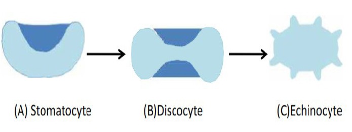
Scheme of erythrocyte shapes: increase in ionic strength of the medium causes transformation of the red blood cells in the sequence stomatocyte → discocyte → echinocyte. Figures were adapted to reference 88.
Atherosclerosis and coronary artery disease
Surfactant system is probably the most genetically consistent system amongst all vertebrate species. The differences in interfacial tension of plasma and serum amongst various species including human, canine, porcine and equine are relatively small.2,90 Albumin with a concentration of 35-50 g/L is the main active compound of serum. Surface tension is sensitive to pH values as well as presence of inorganic and organic electrolytes, carbohydrates and other surfactants. Dynamic surface tension measurements of serum can be used as an indicator of some pathologic disturbances.3
There are sex related differences in the surface tension of human serum (Table 7). High values of equilibrium surface tension in female are due to the low physiological content of albumin, fatty acids and phospholipids, carbohydrate compound, lipoproteins, fibronectin and enzymes.3,6 Due to the metabolism of proteins and lipids and alterations in the surfactant concentration of biological fluids, the surface tension of serum increases with the age.3,6
Table 7 . Tensiograms of serum obtained from healthy people of different agea .
| Sex | Age | Surface tension parameters b | ||
| γ 1 (mNm -1 ) | γ 2 (mNm -1 ) | γ 3 (mNm -1 ) | ||
| Males | >20 | 64-68 | 64-68 | 56-59 |
| 20-35 | 68-72 | 65-69 | 57-60 | |
| 36-50 | 68-72 | 66-69 | 57-63 | |
| ≤50 | 68-74 | 66-71 | 57-66 | |
| Females | >20 | 66-70 | 65-69 | 58-61 |
| 20-35 | 68-74 | 66-70 | 59-62 | |
| 36-50 | 70-74 | 67-70 | 59-65 | |
| ≤50 | 70-76 | 67-72 | 59-68 | |
a Data taken from reference 6.
b γ1 surface tension for t=0.01 s; γ2 surface tension for t=1 s; γ3 surface tension t→∞.
Endothelial surface is covered by surface acting substances that reduce surface tension. Change in endothelial surface tension and hydrophobicity will influence bubble formation, activate platelet and coagulation cascade and increase the risk of atherosclerosis, hypertension and coronary artery disease.91 Nitric oxide is essential to preserve the capsular integrity and prevent the synthesis of adhesion molecules. It can also change the surfactant composition at the endothelial surface, decrease hydrophobicity and increase bubble detachment and dissolution to help prevent the coronary artery disease.91
Musculoskeletal and connective tissue disease
Synovial fluid and joint disease
Synovial fluid is a dialysate of plasma that contains lubricin, hyaluronic acid and surface-active phospholipids. It lubricates the articular surface to help provide mechanical function of the joint and supports pressures up to 200 atm.92,93 Synovial fluid decreases the friction forces and prevents the cartilage-cartilage adhesion. Surface-active phospholipids impart the thin outermost lining of the articular surface and help to reduce friction. In pathological states, the superficial layer disappears and leads to deterioration and aggregation of the joint function.82,83 Analysis of synovial fluid may be a diagnostic screening tool for the joint disease including osteoarthritis as well as septic arthritis and inflammatory arthropathies. Osteoarthritis is a degenerative disease associated with change in the phospholipid content of the synovial fluid. Injection of exogenous surface-active phospholipid effectively reduces the joint stiffness in osteoarthritis. 92,93
Rheumatic disease
Connective tissue contains various types of collagens, proteoglycans and glycoproteins. During the development of rheumatoid arthritis proteolysis and formation of biologically-active substances causes degradation of the connective tissue. Rheological properties of the blood change during the course of disease and augmentation of immunoglobulin and circulating immune complexes increase blood viscosity. These changes alter the surface tension of blood and urine (Table 8). Synovial fluid of patients with rheumatoid disease contains large amounts of immunoglobulin complexes, fibrinogen and fibrin, therefore synovial fluid has lower surface tension value than serum. Glucocorticoids are the first line therapy in the management of rheumatic disease. They normalize the dynamic tensiographic parameters and increase the surface tension values of serum.3
Table 8 . Surface tension changes during various rheumatic diseasesa .
| Diseases | Surface tension Serum | Urine | ||||||
| γ1 | γ2 | γ3 | λ | γ1 | γ2 | γ3 | λ | |
| Rheumatism | + | + | - | |||||
| Systemic lupus erythematosus | + | + | - | - | ||||
| Scleroderma systematica | + | + | + | + | + | - | ||
| Haemorrhagic vasculitis | + | + | + | + | ||||
| Rheumatoid arthritis | + | + | + | - | ||||
| Bechterew’s disease | - | + | ||||||
| Reiter’s disease | + | - | + | + | ||||
| Psoriatic arthropathy | + | + | - | |||||
| Gout | - | + | + | + | ||||
| Osteoarthrosis | + | + | + | - | ||||
a Data taken from reference 3.
+ Statistically significant increase of parameters compared with normal values.
- Statistically significant decrease of parameter compares with normal values.
Infectious diseases
The initial step in bacterial infection is its adhesion to host enteric, oral or respiratory organs. Bacterial adhesion to host organs leads to colonization of the bacteria, receptor-specific bacterial interaction, cellular invasion and subsequent development of the disease state.84 Bacterial adhesion can be expressed through ligand-receptor interactions. Adherent state is a key step in bacterial survival and pathogenesis, therefore an important approach is to interfere with the ability of the bacteria to adhere to host cells and thus help prevention of disease. Measurement of interfacial forces will help in the prophylaxis and treatment of various infections such as, enteric disease, urinary tract infection as well as endocardiatis.94
Infection of the human cervix
Human cervical mucus contains mucin and soluble proteins. Mucin is secreted from the epithelium cells of the cervix and plays an important role in the physicochemical properties of cervical mucus including viscosity and surface tension. Mucin acts as a lubricant to keep pathogens out. A relationship has been found between the surface tension of the mucous and the permeation rate of microbicides. Cervical compounds can be used in the treatment of dry vagina or as microbicidal agents against Sexually Transmitted Disease (STD) pathogens. The degree of irritation of these compounds depends upon their surface tension. Compounds with low surface tension have lower binding of the membrane to the mucosa proteins and thus will cause less cervical irritation.95
Tuberculosis
Tuberculosis (TB) infection is usually initiated by the entry of the mycobacterium TB to the respiratory system. Mycolic acid and cord factor are the most abundant surface lipids of mycobacterial cell with moderate surface active effect. Presence of such surface active lipids at the alveolar interface will cause reorientation of the surfactant molecules, poor surface activity of the DPPC and symptoms similar to adult respiratory distress syndrome. In such cases minimum surface tension is not reached, the alveoli will collapse and the patient will require increased breathing efforts to re-expand and stabilize the atelectatic alveoli.
TB contains multiple lipids that can inhibit the function of endogenous surfactants in respiration. Inhibitions of mycolic acid synthesis by antitubercular drug, isoniazid, will destroy the mycobacterial cell wall, and improve the breathing process.96
Gastrointestinal tract
Infection
The internal lining of the gastrointestinal tract blocks the passage of toxic substances to the systemic circulation. Any substance that compromises the integrity of the GI epithelium could be involved in the induction of disease. Mucus is a viscoelastic gel coating of gastrointestinal track which provides the hydrophobic property of the stomach and prevents adherence and invasion of bacteria. Contact angle measurement of the lining of the gastrointestinal track demonstrates that hydrophobicity is highest in the antrum of the stomach and decreases through rectum and ileal mucosa. Microbial attachment and adherence to human biological surfaces can determine the rate and extent of infection. The rate of bacterial attachment is greater in hydrophobic surfaces. Pathogen adhesion and specific stereochemical interactions between ligand and gastrointestinal tract receptor is important in pathogenesis and infection. Therefore, development of therapeutic agents for microbial pathogens involves knowledge of these surfaces and the adherence forces between them. Helicobacter pylori produce protease and lipases which reduce hydrophobicity of the mucus layer and cause gastric infection. The reduction in hydrophobicity is greater in patients infected with H. pylori as well as duodenal ulcer than it in patients with H. pylori and gastritis.97
Concluding remarks
Surfaces are critically important for nearly all aspects of biological phenomena. The concept of surface has been applied in many biological fields such as disease diagnosis and treatment, biomaterial implant, blood compatibility and biosensors. Various diseases influence the composition and interfacial tension of body fluids. To further confirm these changes, the surface tensiogram can be used as an indicator of some pathological disturbances.
It is well accepted that no single biomarker will be effective in clinical diagnosis. A better understanding of pathological disease is possible if various techniques are used. The surface tensiometry is a sensitive method which can reveal subtle changes in the phospholipid and surfactant content of biological fluids. Surface tension methods are easy to use and require a finite amount of sample. A decrease in the surface tension of blood and urine appears to be characteristics for some diseases, while other diseases result from an increase of these parameters.
Well established dynamic surface tension measurements will widen our understanding on the surface rheology in biological fluids. In this review article, the opportunity of the interfacial tensiometric measurements as a diagnostic tool in medical application and monitoring device in the treatment efficacy was discussed. We will continue the research on the surface tension of biological fluids with a greater focus on urine and blood samples. We envision that studies on the surface tension behavior of human biological fluids will provide additional useful information in medical practice.
Ethical issues
There is none to be declared.
Competing interests
There is none to be declared.
Research Highlights
What is current knowledge?
√ Experimental methods dealing with surface tension of biological systems.
√ Correlation of biochemical parameters to surface tension of biological fluids.
√ Correlation of tensiometric parameters with various pathologies.
What is new here?
√ Review of available knowledge on the surface tension of biological fluids.
√ Providing a theoretical framework for modeling and understanding of surface tension as a tool in medical diagnostic.
√ Presenting a clear and straightforward account of the biointerface studies to understand physiological functions of human biology.
√ Bridging the gap between laboratory research and clinical medicine.
References
- 1.Castner DG, Ratner BD. Biomedical surface science: Foundations to frontiers. Surf Sci. 2002;500:28–60. doi: 10.1016/S0039-6028(01)01587-4. [DOI] [Google Scholar]
- 2.Trukhin DV, Sinyachenko OV, Kazakov VN, Lylyk SV, Belokon AM, Pison U. Dynamic surface tension and surface rheology of biological Liquids, Colloid Surf B. 2001;21:231–238. doi: 10.1016/S0039-6028(01)01587-4. [DOI] [PubMed] [Google Scholar]
- 3.Kazakov VN, Vozianov AF, Sinyachenko OV, Trukhin DV, Kovalchuk VI, Pison U. Studies on the application of dynamic surface tensiometry of serum and cerebrospinal liquid for diagnostics and monitoring of treatment in patients who have rheumatic, neurological or oncological diseases. Adv Colloid Int Sci. 2000;86:1–38. doi: 10.1016/S0001-8686(00)00031-2. [DOI] [PubMed] [Google Scholar]
- 4.McGuire JF. Surfactant in the middle ear and eustachian tube: A review. Int J Ped Otorhinolaryngol. 2002;66:1–15. doi: 10.1016/S0165-5876(02)00203-3. [DOI] [PubMed] [Google Scholar]
- 5.Depalma RG. Surface forces in biological material; measurement of surface tension by drop volume. J Surg Res. 1967;7:317–322. doi: 10.1016/0022-4804(67)90072-8. [DOI] [PubMed] [Google Scholar]
- 6.Kazakov VN, Sinyachenko OV, Trukhin DV, Pison U. Dynamic interfacial tensiometry of biologic liquids -does it have an impact on medicine. Colloid Surf A. 1998;143:441–459. doi: 10.1016/S0927-7757(98)00612-8. [DOI] [Google Scholar]
- 7.Khoubnasabjafari M, Jouyban V, Fathi Azarbayjani A, Jouyban A. Application of Abraham solvation parameters for surface tension prediction of mono-solvents and solvent mixtures at various temperatures. J Mol Liq. 2013;178:44–56. doi: 10.1016/j.molliq.2012.11.010. [DOI] [Google Scholar]
- 8.Fathi Azarbayjani A, Khu JV, Chan YW, Chan SY. Development and characterization of skin permeation retardants and enhancers: A comparative study of levothyroxine-loaded PNIPAM, PLA, PLGA and EC microparticles. Biopharm Drug Disposition. 2011;32:380–388. doi: 10.116/j.powtec.2012.04.060. [DOI] [PubMed] [Google Scholar]
- 9.Fathi-Azarbayjani A, Jouyban A, Chan SY. Impact of surface tension in pharmaceutical sciences. J Pharm Pharm Sci. 2009;12:218–228. doi: 10.18433/j32p40. [DOI] [PubMed] [Google Scholar]
- 10.Fathi Azarbayjani A, Lin HS, Yap CW, Chan YW, Chan SY. Surface tension and wettability in transdemal delivery: a study on the in-vitro permeation of haloperidol with cyclodextrin across human epidermis. J Pharm Pharmacol. 2010;62:770–778. doi: 10.1211/jpp.62.06.0014. [DOI] [PubMed] [Google Scholar]
- 11. Jouyban A, Fathi Azarbayjani A. Experimental and computational methods pertaining to surface tension of pharmaceutical. In: “Toxicity and Drug Testing”. 2012.
- 12.Fathi Azarbayjani A, Tan EH, Chan YW, Chan SY. Transdermal delivery of haloperidol by proniosomal formulations with non-ionic surfactants. Biol Pharm Bull. 2009;32:1453–1458. doi: 10.1248/bpb.32.1453. [DOI] [PubMed] [Google Scholar]
- 13.Jouyban A, Fathi Azarbayjani A, Acree WE. Surface tension calculation of mixed solvents with respect to solvent composition and temperature by using Jouyban-Acree model. Chem Pharm Bull. 2004;52:1219–1222. doi: 10.1248/cpb.52.1219. [DOI] [PubMed] [Google Scholar]
- 14.Buckton G. The assessment and pharmaceutical importance, of the solid/liquid and the solid/vapour interface: A review with respect to powders. Int J Pharm. 1988;44:1–8. [Google Scholar]
- 15. Drelich J, Fang C, White CL. In: Hubbard AT, editor.Measurement of interfacial tension in fluid-fluid systems in Encyclopedia of Surface and Colloid Science. New York: Marcel Dekker; 2002. p. 3152.
- 16.Grima R, Schnell S. Can tissue surface tension drive somite formation? Dev Biol. 2007;307:248–257. doi: 10.1016/i.ydbio.2007.04.032. [DOI] [PMC free article] [PubMed] [Google Scholar]
- 17. Thiessen DB, Man KF. Surface tension measurements. CRC Press; 1999.
- 18.Wozniak MA, Modzelewska K, Kwong L, Keely PJ. Focal adhesion regulation of cell behavior. Colloids Surf B. 2004;1692:103–119. doi: 10.1016/j.bbamcr.2004.04.007. [DOI] [PubMed] [Google Scholar]
- 19.Steinberg MS. Reconstruction of tissues by dissociated cells. Science. 1963;141:401–408. doi: 10.1126/science.141.3579.401. [DOI] [PubMed] [Google Scholar]
- 20.Davis GS, Phillips HM, Steinberg MS. Germ-layer surface tensions and ‘‘tissue affinities’’ in rana pipiens gastrulae: quantitative measurements. Dev Biol. 1997;192:630–644. doi: 10.1006/dbio.1997.8741. [DOI] [PubMed] [Google Scholar]
- 21.Kalantarian A, Ninomiya H, Saad SMI, David R, Winklbauer R, Neumann AW. Axisymmetric drop shape analysis for estimating the surface tension of cell aggregates by centrifugation. Biophys J. 2009;96:1606–1616. doi: 10.1016/j.bpj.2008.10.064. [DOI] [PMC free article] [PubMed] [Google Scholar]
- 22.Paluch E, Heisenberg CP. Biology and physics of cell shape changes review in development. Curr Biol. 2009;19:R790–799. doi: 10.1016/j.cub.2009.07.029. [DOI] [PubMed] [Google Scholar]
- 23.Walker JL, Fournier AK, Assoian RK. Regulation of growth factor signaling and cell cycle progression by cell adhesion and adhesion-dependent changes in cellular tension, Cytokine & Growth Factor Rev. 2005;16:395–405. doi: 10.1016/j.cytogfr.2005.03.003. [DOI] [PubMed] [Google Scholar]
- 24.Mombach JC, Robert D, Graner F, Gillet G, Thomas GL, Idiart M. et al. Rounding of aggregates of biological cells: Experiments and simulations. Physica A. 2005;352:525–534. doi: 10.1016/j.physa.2005.02.008. [DOI] [Google Scholar]
- 25.Balaban NQ, Schwarz US, Riveline D. Force and focal adhesion assembly: a close relationship studied using elastic micropatterned substrates. Nat Cell Biol. 2001;13:466–472. doi: 10.1038/35074532. [DOI] [PubMed] [Google Scholar]
- 26.Voß-Böhme A, Deutsch A. The cellular basis of cell sorting kinetics. J Theor Biol. 2010;263:419–436. doi: 10.1016/j.jtbi.2009.12.011. [DOI] [PubMed] [Google Scholar]
- 27.Lecuit T. Adhesion remodeling underlying tissue morphogenesis. Trends Cell Biol. 2005;15:34–42. doi: 10.1016/j.tcb.2004.11.007. [DOI] [PubMed] [Google Scholar]
- 28.Hegedüs A, Marga F, Jakab K, Sharpe-Timms KL, Forgacs G. The interplay of cell-cell and cell-matrix interactions in the invasive properties of brain tumors. Biophys J. 2006;91:2708–2716. doi: 10.1529/biophysj.105.077834. [DOI] [PMC free article] [PubMed] [Google Scholar]
- 29.Moussy F, Lin FY, Lahooti S, Policova Z, Zingg W, Neumann AW. A micropipette aspiration technique to investigate the adhesion of endothelial cells. Colloids Surf B. 1994;2:493–503. doi: 10.1016/0927-7765(94)80057-X. [DOI] [Google Scholar]
- 30.de Queiroz AA, Barrak ÉR, de Castro SC. Thermodynamic analysis of the surface of biomaterials. J Mol Struct (Theochem) 1997;394:271–279. doi: 10.1016/S0166-1280(96)04843-9. [DOI] [Google Scholar]
- 31.Harnett EM, Alderman J, Wood T. The surface energy of various biomaterials coated with adhesion molecules used in cell culture. Colloids Surf B. 2007;55:90–97. doi: 10.1016/j.colsurfb.2006.11.021. [DOI] [PubMed] [Google Scholar]
- 32.Chaplain MA, Sleeman BD. A Mathematical model for the growth and classification of a solid tumor: A new approach via nonlinear elasticity theory using strain-energy functions. Math Biosci. 1992;111:169–215. doi: 10.1016/0025-5564(92)90070-D. [DOI] [PubMed] [Google Scholar]
- 33.D’Anselmi F, Valerio M, Cucina A, Galli L, Proietti S, Dinicola S. et al. Metabolism and cell shape in cancer:A fractal analysis. Int J Biochem Cell Biol. 2011;43:1052–1059. doi: 10.1016/j.biocel.2010.05.002. [DOI] [PubMed] [Google Scholar]
- 34.Knewitz MA, Mombach JC. Computer simulation of the influence of cellular adhesion on the morphology of the interface between tissues of proliferating and quiescent cells. Comput Biol Med. 2006;36:59–69. doi: 10.1016/j.compbiomed.2004.08.002. [DOI] [PubMed] [Google Scholar]
- 35.Guy K, Di Mario U, Irvine WJ. Circulating immune complexes and autoantibodies in lung cancer. Br J Cancer. 1981;43:276. doi: 10.1038/bjc.1981.45. [DOI] [PMC free article] [PubMed] [Google Scholar]
- 36.Mattison P, Cove DH, Walsh L. Serum proteins as tumor markers for breast cancer. Br J Cancer. 1981;43:542–545. doi: 10.1038/bjc.1981.78. [DOI] [PMC free article] [PubMed] [Google Scholar]
- 37. Kazakov VN, Sinyachenko OV, Fainerman VB, Pison U, Miller R. Interfacial tensiometry in oncology. In: Dynamic surface tension in medicine. Chapter 8. 2007; 8:324-358.
- 38.Hasler WL. Irritable bowel syndrome and bloating. Best Pract Res Clin Gastroenterol. 2007;21:689–707. doi: 10.1016/j.bpg.2007.03.007. [DOI] [PubMed] [Google Scholar]
- 39.Barar J, Omidi Y. Dysregulated pH in Tumor Microenvironment Checkmates Cancer Therapy, BioImpacts. 2013;3(4):149–162. doi: 10.5681/bi.2013.036. [DOI] [PMC free article] [PubMed] [Google Scholar]
- 40.Omidi Y, Barar J. Targeting tumor microenvironment: crossing tumor interstitial fluid by multifunctional nanomedicines. BioImpacts. 2014;4(2):55–67. doi: 10.5681/bi.2014.021. [DOI] [PMC free article] [PubMed] [Google Scholar]
- 41.Zhou F, Escher J, Cui S. Bifurcation for a free boundary problem with surface tension modeling the growth of multi-layer tumors. J Math Anal Appl. 2008;337:443–457. doi: 10.1016/j.jmaa.2007.03.107. [DOI] [Google Scholar]
- 42.Anand P, Huilgol N, Banerjee R. Interfacial properties as predictors of radioresistance in cervical cancer. J Colloid Int Sci. 2007;314:63–70. doi: 10.1016/j.jcis.2007.04.011. [DOI] [PubMed] [Google Scholar]
- 43.Preetha A, Banerjee R, Huilgol N. Dynamic surface tensiometry of tissues using Langmuir films, Colloids Surf B. 2005;40:35–43. doi: 10.1016/j.colsurfb.2004.10.006. [DOI] [PubMed] [Google Scholar]
- 44.Kim WD. Lung mucus: A clinician’s view. Eur Respir J. 1997;10:1914–1917. doi: 10.1183/09031936.97.10081914. [DOI] [PubMed] [Google Scholar]
- 45.Bakshi MS, Zhao L, Smith R, Possmayer F, Petersen NO. Metal nanoparticle pollutants interfere with pulmonary surfactant function in vitro. Biophys J. 2008;94:855–868. doi: 10.1529/biophysj.107.106971. [DOI] [PMC free article] [PubMed] [Google Scholar]
- 46.Smith LJ, McKay KO, van Asperen PP, Selvadurai H, Fitzgerald DA. Normal development of the lung and premature birth. Paediatr Respir Rev. 2010;11:135–142. doi: 10.1016/j.prrv.2009.12.006. [DOI] [PubMed] [Google Scholar]
- 47.Heil M, Hazel AL, Smith JA. The mechanics of airway closure, Respir Physiol Neurobiol. 2008;163:214–221. doi: 10.1016/j.resp.2008.05.013. [DOI] [PubMed] [Google Scholar]
- 48.deMello DE. Pulmonary pathology. Semin Neonatol. 2004;9:311–329. doi: 10.1016/j.siny.2003.12.001. [DOI] [PubMed] [Google Scholar]
- 49.Scarpelli EM. Physiology of the alveolar surface network. Compar Biochem Physiol Part A. 2003;135:39–104. doi: 10.1016/S1095-6433(02)00352-5. [DOI] [PubMed] [Google Scholar]
- 50.Wen X, McGinnis KC, Fanses EI. Unusually low dynamic surface tensions of aqueous solutions of sodium myristate. Colloids Surf A. 1998;143:371–380. doi: 10.1016/S0927-7757(98)00375-6. [DOI] [Google Scholar]
- 51.Robertson B, Johansson J, Curstedt T. Synthetic surfactants to treat neonatal lung disease. Mol Med Today. 2000;6:119–124. doi: 10.1016/S1357-4310(99)01656-1. [DOI] [PubMed] [Google Scholar]
- 52.Ikegami M, Hacobs H, Jobe A. Surfactant function in respiratory distress sysndrome. J Pediat. 1983;102:443–447. doi: 10.1016/s0022-3476(83)80673-8. [DOI] [PubMed] [Google Scholar]
- 53.Richardson PS, Phipps RJ. The anatomy, physiology, pharmacology and pathology of tracheobronchial mucus secretion and the use of expectorant drugs in human disease. Pharmac Ther B. 1978;3:441–479. doi: 10.1016/S0306-039X(78)90010-7. [DOI] [PubMed] [Google Scholar]
- 54.Hudson VM. Rethinking cystic fibrosis pathology: the critical role of abnormal reduced glutathione (GSH) transport caused by CFTR mutation, Free Radical Biol Med. 2001;30:1440–1461. doi: 10.1016/S0891-5849(01)00530-5. [DOI] [PubMed] [Google Scholar]
- 55.Gilljam H, Andersson O, Ellin A, Robertson B, Strandvik B. Composition and surface properties of the bronchial lipids in adult patients with cystic fibrosis. Clinica Chimica Acta. 1988;116:29–38. doi: 10.1016/0009-8981(88)90171-4. [DOI] [PubMed] [Google Scholar]
- 56.Jokic R, Klimaszewski A, Crossley M. Positional treatment vs continuous positive airway pressure in patients with positional obstructive sleep apnea syndrome. Chest. 1999;115:771–781. doi: 10.1378/chest.115.3.771. [DOI] [PubMed] [Google Scholar]
- 57.Bradshaw DA. What are the nonsurgical treatment options for obstructive sleep apnea syndrome? Am Otolaryngol. 2001;22:124–131. doi: 10.1053/ajot.2001.22573. [DOI] [PubMed] [Google Scholar]
- 58.Sutnick AI, Soloff LA. Surface tension reducing activity in the normal and atelectatic human lung, American J Med. 1963;35:31–36. doi: 10.1016/0002-9343(63)90161-X. [DOI] [PubMed] [Google Scholar]
- 59.Haczku A, Panettieri RA Jr. Social stress and asthma: The role of corticosteroid insensitivity, J Allergy Clin Immunol. 2010;125:550–558. doi: 10.1016/j.jaci.2009.11.005. [DOI] [PMC free article] [PubMed] [Google Scholar]
- 60.Hohlfeld JM, Schmiedl A, Erpenbeck VJ, Venge P, Krug N. Eosinophil cationic protein alters pulmonary surfactant structure and function in asthma. J Allergy Clin Immunol. 2004;113:496–502. doi: 10.1016/j.jaci.2003.12.008. [DOI] [PubMed] [Google Scholar]
- 61.Vignola AM, Chanez P, Bonsignore G, Godard P, Bousquet J. Structural consequences of airway inflammation in asthma, J Allergy Clin Immunol. 2000;105:S514–S517. doi: 10.1016/S0091-6749(00)90053-8. [DOI] [PubMed] [Google Scholar]
- 62.Reid G. Importance of Surface Properties in Bacterial Adhesion to Biomaterials, with Particular Reference to the Urinary Tract. International Biodeterioration & Biodegradation. 1992;30:105–122. doi: 10.1016/0964-8305(92)90057-U. [DOI] [Google Scholar]
- 63.Allen GD. The determination of the bile salts in urine by means of the surface tension method. J Biol Chem. 1915;22:505–524. [Google Scholar]
- 64. Putnam DF. Composition and concentrative properties of human urine. National aeronautics and space administration 1971.
- 65.Basavaraj R, Biyani CS, Browning AJ, Cartledge JJ. The role of urinary kidney stone inhibitors and promoters in the pathogenesis of calcium containing renal stones. Eau - Ebu Update Series. 2007;5:126–136. doi: 10.1016/j.eeus.2007.03.002. [DOI] [Google Scholar]
- 66.Tárnoky AL. ‘Apparent protein’ in human urine: some surface film measurements. Biochem J. 1951;49:205–209. doi: 10.1042/bj0490205. [DOI] [PMC free article] [PubMed] [Google Scholar]
- 67.Diamantis A, Magiorkinis E, Androutsos G. Proteinuria: from ancient observation to 19th century scientific study, J Urol. 2008;180:2330–2332. doi: 10.1016/j.juro.2008.08.043. [DOI] [PubMed] [Google Scholar]
- 68.Ziegler A, Torchia M, Grahame GR, Ferguson IA. Peritoneal surface-active material in continuous ambulatory peritoneal dialysis (CAPD) patients. Perit Dial Int. 1989;9:47–49. [PubMed] [Google Scholar]
- 69. Semes LP, Bright DC, Pens CD. Optometric clinical practice guideline: Care of the patient with ocular surface disorders, reference guide for clinicians. American Optometric Association 2010.
- 70.Bron AJ, Tiffany JM, Gouveia SM, Yokoi N, Voon LW. Functional aspects of the tear film lipid layer. Exp Eye Res. 2004;78:347–360. doi: 10.1016/j.exer.2003.09.019. [DOI] [PubMed] [Google Scholar]
- 71.Ketelson HA, Meadows DL, Stone RP. Dynamic wettability properties of a soft contact lens hydrogel. Colloids Surf B. 2005;40:1–9. doi: 10.1016/j.colsurfb.2004.07.010. [DOI] [PubMed] [Google Scholar]
- 72.Bourbon JR, Chailley-Heu B. Surfactant proteins in the digestive tract, mesentery, and other organs: evolutionary significance. Comp Biochem Physiol Part A. 2001;129:151–161. doi: 10.1016/S1095-6433(01)00312-9. [DOI] [PubMed] [Google Scholar]
- 73.Kirkness JP, Christenson HK, Wheatley JR, Amis TC. Application of the pull-off method for measurement of the surface tension of upper airway mucosal lining liquid. Physiol Meas. 2005;26:677–688. doi: 10.1088/0967-3334/26/5/009. [DOI] [PubMed] [Google Scholar]
- 74.Kazakov VN, Udod AA, Zinkovych II, Fainerman VB, Miller R. Dynamic surface tension of saliva: General relationships and application in medical diagnostics, Colloids Surf B. 2009;74:457–461. doi: 10.1016/j.colsurfb.2009.06.010. [DOI] [PubMed] [Google Scholar]
- 75.Busscher HJ, White DJ, Kamminga-Rasker HJ, Mei HC. A surface physicochemical rationale for calculus formation on the oral cavity. J Cryst Growth. 2004;261:87–92. doi: 10.1016/j.jcrysgro.2003.09.012. [DOI] [Google Scholar]
- 76.Bottenberg P, Slop D, Remon JP, De Muynck C, Coomans D. Influence of viscosity and surfactant on fissure penetration of dental fluoride gels in vitro, Int J Pharm. 1989;49:147–156. doi: 10.1016/0378-5173(89)90114-2. [DOI] [Google Scholar]
- 77.Wang LH, Tsai ST. Voltammetric behavior of chlorhexidine at a film mercury electrodes and its determination in cosmetics and oral hygiene products. Anal Chim Acta. 2001;441:107–116. doi: 10.1016/S0003-2670(01)01083-2. [DOI] [Google Scholar]
- 78.Kazakov VN, Barkalova EL, Levchenko LA, Klimenko TM, Fainerman VB, Miller R. et al. Dilation rheology as medical diagnostics of human biological liquids. Colloids Surf A. 2011;391:190–194. doi: 10.1016/j.colsurfa.2011.03.028. [DOI] [Google Scholar]
- 79.Brydon HL, Hayward R, Harkness W, Bayston R. Physical properties of cerebrospinal fluid of relevance to shunt function 2 The effect of protein upon CSF surface tension and contact angle Brit. J Neurosurg. 1995;9:645–651. doi: 10.1080/02688699550040936. [DOI] [PubMed] [Google Scholar]
- 80.Hörter D, Dressman JB. Influence of physicochemical properties on dissolution of drugs in the gastrointestinal tract. Adv Drug Deliv Rev. 2001;46:75–87. doi: 10.1016/S0169-409X(00)00130-7. [DOI] [PubMed] [Google Scholar]
- 81.McConnell EL, Fadda HM, Basit AW. Gut instincts: Explorations in intestinal physiology and drug delivery. Int J Pharm. 2008;364:213–226. doi: 10.1016/j.ijpharm.2008.05.012. [DOI] [PubMed] [Google Scholar]
- 82.Pang G, Xie J, Chen Q, Hu Z. How functional foods play critical roles in human health. Food Science and Human Wellness. 2012;1:26–60. doi: 10.1016/j.fshw.2012.10.001. [DOI] [Google Scholar]
- 83.Lichtenberger LM, Zhou Y, Dial EJ, Raphael RM. NSAID injury to the gastrointestinal tract: Evidence that NSAIDs interact with phospholipids to weaken the hydrophobic surface barrier and induce the formation of unstable pores in membranes. J Pharm Pharmacol. 2006;58:1–8. doi: 10.1211/jpp.58.10.0001. [DOI] [PubMed] [Google Scholar]
- 84.Cole GJ, Connell AM. Gastrin, acid, and bile. Gut. 1968;9:638–640. doi: 10.1136/gut.9.6.638. [DOI] [PMC free article] [PubMed] [Google Scholar]
- 85.Fernández-Leyes MD, Messina PV, Schulz PC. Aqueous sodium dehydrocholate–sodium deoxycholate mixtures at low concentration. J Colloid Interface Sci. 2007;314:659–564. doi: 10.1016/j.jcis.2007.05.078. [DOI] [PubMed] [Google Scholar]
- 86.Murray BS. Rheological properties of protein films. Curr Opin Colloid Interface Sci. 2011;16:27–35. doi: 10.1016/j.cocis.2010.06.005. [DOI] [Google Scholar]
- 87.Hasler WL. Irritable bowel syndrome and bloating. Best Pract Res Clin gastroenterol. 2007;21:689–707. doi: 10.1016/j.bpj.2007.03.007. [DOI] [PubMed] [Google Scholar]
- 88.Tachev KD, Danov KD, Kralchevsky PA. On the mechanism of stomatocyte-echinocyte transformation of red blood cells: Experiment and theoretical model. Colloids Surf B. 2004;34:123–140. doi: 10.1016/j.colsurfb.2003.12.011. [DOI] [PubMed] [Google Scholar]
- 89.Muñoz S, Sebastián JL, Sancho M, Martínez G. Analysis of radiofrequency energy stored in the altered shapes: Stomatocyte-echinocyte of human erythrocytes. Bioelectrochem. 2010;77:158–161. doi: 10.1016/j.bioelechem.2009.07.006. [DOI] [PubMed] [Google Scholar]
- 90.Krishnan A, Wilson A, Sturgeon J, Siedlecki CA, Vogler EA. Liquid-vapor interfacial tension of blood plasma, serum and purified protein constituents thereof. Biomater. 2005;26:3445–3453. doi: 10.1016/j.biomaterials.2004.09.016. [DOI] [PubMed] [Google Scholar]
- 91.Duplessis CA, Fothergill D. Investigating the potential of statin medications as a nitric oxide (NO) release agent to decrease decompression sickness: A review article Med. Hypotheses. 2008;70:560–566. doi: 10.1016/j.mehy.2007.04.050. [DOI] [PubMed] [Google Scholar]
- 92.Zappone B, Ruths M, Greene GW, Jay GD, Israelachvili JN. Adsorption, Lubrication, and Wear of Lubricin on Model Surfaces: Polymer Brush-Like Behavior of a Glycoprotein. Biophys J. 2007;92:1693–1708. doi: 10.1529/biophysj.106.088799. [DOI] [PMC free article] [PubMed] [Google Scholar]
- 93.Hills BA, Crawford RW. Normal and prosthetic synovial joints are lubricated by surface-active phospholipid: a hypothesis. J Arthroplasty. 2003;18:499–505. doi: 10.1016/S0883-5403(03)00072-X. [DOI] [PubMed] [Google Scholar]
- 94.Ofek I, Hasty DL, Sharon N. Anti-adhesion therapy of bacterial diseases: prospects and problems. FEMS Immunol Med Microbiol. 2003;38:181–91. doi: 10.1016/S0928-8244(03)00228-1. [DOI] [PubMed] [Google Scholar]
- 95.Wang Y, Lee CH. Characterization of a female controlled drug delivery system for microbicides. Contraception. 2002;66:281–287. doi: 10.1016/S0010-7824(02)00354-2. [DOI] [PubMed] [Google Scholar]
- 96.Chimote G, Banerjee R. Lung surfactant dysfunction in tuberculosis: Effect of mycobacterial tubercular lipids on dipalmitoylphosphatidylcholine surface activity. Colloids Surf B. 2005;45:215–223. doi: 10.1016/j.colsurfb.2005.08.014. [DOI] [PubMed] [Google Scholar]
- 97.Mack DR, Sherman PM. Hydrophobicity and the gastrointestinal tract: Methods of determination, its source and implications for bacterial adherence. Colloids Surf B. 1999;15:355–363. doi: 10.1016/S0927-7765(99)00100-9. [DOI] [Google Scholar]


