Abstract
A 52-year-old woman presented with severe acute right upper quadrant abdominal pain and signs of intra-abdominal haemorrhage. CT and selective angiography revealed a ruptured right hepatic artery aneurysm and diffuse aneurysmatic disease involving most intra-abdominal organs, suggestive of polyarteritis nodosa. Although treatment with high-dose steroids was initiated, the patient died of progressive bowel ischaemia.
Background
Polyarteritis nodosa (PAN) is a systemic necrotising vasculitis of medium-sized arteries, not associated with antineutrophil cytoplasmic antibodies, and can be successfully treated by high-dose steroids in the majority of patients. PAN typically presents with systemic symptoms and signs of multiorgan involvement. Organs most commonly affected are the kidneys, skin, joints, muscles, nerves and gastrointestinal tract. Microscopically, PAN is usually characterised by segmental transmural inflammation and necrosis, sporadically leading to aneurysmatic dilation.
We describe a rare case of PAN presenting with a ruptured hepatic artery aneurysm and diffuse abdominal aneurysmatic disease of medium-sized arteries. In these cases, PAN should be part of the differential diagnosis and timely treatment can potentially be life-saving.
Case presentation
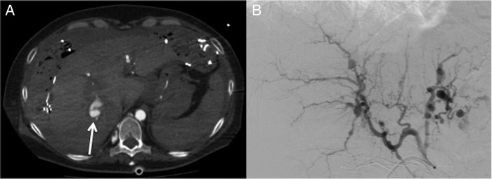
A 52-year-old woman presented with acute abdominal pain in the right upper quadrant. Her medical history revealed a laparoscopic gastric bypass some years earlier for morbid obesity. Initially, symptomatic gallstone disease was suspected. Owing to sudden haemodynamic instability, the patient underwent acute exploratory laparotomy in a large community hospital. At surgery, an extensive liver haemorrhage without clear focus was found, which could only be controlled by abdominal packing with surgical gauzes. After resuscitation, the patient was transferred to our tertiary centre. CT imaging and selective abdominal angiography showed a ruptured right hepatic artery aneurysm, which was subsequently successfully coiled and embolised. A remarkable finding was the co-existence of diffuse aneurysmatic disease involving almost all visceral arteries (figure 1).
Figure 1.

Hepatic arterial blush (arrow) at CT imaging (A) and diffuse visceral aneurysmatic disease at angiography (B).
The abdominal packs were removed 3 days later. At re-laparotomy, segmental necrosis of the small bowel as well as of the gallbladder was found. A total length of 175 cm of small bowel was resected in combination with a cholecystectomy. Also, a liver biopsy was taken.
Owing to a persisting septic profile, CT imaging was performed the next day and was suggestive of progressive small bowel ischaemia. A second re-laparotomy revealed diffuse necrosis of nearly the entire small bowel and large parts of the colon, not amenable to surgical therapy. The patient died shortly thereafter in the intensive care unit after ceasing haemodynamic support.
Investigations
Angiography: ruptured right hepatic artery aneurysm in combination with multiple aneurysms involving almost all visceral arteries.
Transoesophageal echocardiography: no signs of endocarditis.
Laboratory results: antineutrophil cytoplasmic antibodies negative, hepatitis B virus negative. Minimally elevated prothrombin time and activated partial thromboplastin time.
Pathology report liver biopsy: no signs of vasculitis.
Pathology report short bowel segment: ischaemic necrosis in combination with arterial changes suggestive of PAN.
Differential diagnosis
Diffuse aneurysmatic disease of small-sized and medium-sized arteries is most often caused by some type of vasculitis (most commonly PAN). Connective tissue disorders such as segmental arterial mediolysis or fibromuscular dysplasia are less common, as are mycotic aneurysms.
Treatment
Treatment with high-dose steroids (1000 mg of methylprednisolone daily) was started with regard to the presumptive diagnosis of PAN. However, progressive bowel ischaemia could not be prevented in this patient.
Outcome and follow-up
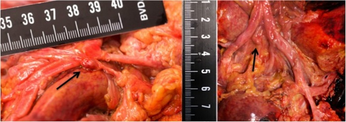
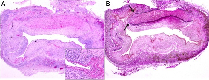
Autopsy revealed severe vasculitis of medium-sized arteries of the liver, heart and bowel. Within the liver, branches of the hepatic artery showed signs of arteritis with multiple small aneurysms, with large areas of parenchymal necrosis (figures 2 and 3). Diffuse haemorrhagic changes were also seen in the kidneys, lungs and ovaries.
Figure 2.

Macroscopic image of multiple aneurysms of medium-sized hepatic arteries (arrows).
Figure 3.

Microscopic image of a medium-sized artery (H&E, original magnification ×2) demonstrating influx of inflammatory cells and fibrinoid necrosis of the arterial wall (inset) (A) with destruction of the internal and external elastic lamina (arrows; elastic van Gieson staining) (B).
Discussion
Diffuse aneurysmatic disease of intra-abdominal medium-sized arteries is a rare presentation of PAN. Until now, only sporadic cases have been published.1–3 Most hepatic artery aneurysms are diagnosed incidentally. Only few patients have presented with acute major haemorrhage, which is associated with significant mortality.
When PAN is suspected, early treatment with high-dose steroids should be initiated. In case of life-threatening haemorrhage, the availability of expert interventional radiologists is essential.4
Learning points.
Polyarteritis nodosa (PAN) is a systemic necrotising vasculitis of medium-sized arteries and is usually characterised by segmental transmural inflammation and necrosis.
Although rare, PAN may present with diffuse aneurysmatic disease of intra-abdominal medium-sized arteries.
Initial presentation of undiagnosed PAN may be a life-threatening haemorrhage caused by a ruptured abdominal aneurysm.
Interventional radiologists are crucial to control major haemorrhage from visceral arteries.
In case of diffuse aneurysmatic disease of medium-sized arteries, the diagnosis of PAN should always be suspected.
Footnotes
Competing interests: None.
Patient consent: Obtained.
Provenance and peer review: Not commissioned; externally peer reviewed.
References
- 1.Battula N, Tsapralis D, Morgan M et al. Spontaneous liver haemorrhage and haemobilia as initial presentation of undiagnosed polyarteritis nodosa. Ann R Coll Surg Engl 2012;94:163–5. 10.1308/003588412X13171221590737 [DOI] [PMC free article] [PubMed] [Google Scholar]
- 2.Parent BA, Cho SW, Buck DG et al. Spontaneous rupture of hepatic artery aneurysm associated with polyarteritis nodosa. Am Surg 2010;76:1416–19. [DOI] [PubMed] [Google Scholar]
- 3.Stambo GW, Guiney MJ, Cannella XF et al. Coil embolization of multiple hepatic artery aneurysms in a patient with undiagnosed polyarteritis nodosa. J Vasc Surg 2004;39:1122–4. 10.1016/j.jvs.2004.01.018 [DOI] [PubMed] [Google Scholar]
- 4.Herskowitz MM, Flyer MA, Sclafani SJ. Percutaneous transhepatic coil embolization of a ruptured intrahepatic aneurysm in polyarteritis nodosa. Cardiovasc Intervent Radiol 1993;16:254–6. 10.1007/BF02602973 [DOI] [PubMed] [Google Scholar]


