Description
A pre-menopausal woman aged 40 years was diagnosed with locally advanced carcinoma of the breast. Since her malignancy was strongly positive for oestrogen receptors, after completion of treatment with surgery (modified radical mastectomy), chemotherapy (four cycles of doxorubicin-cyclophosphamide, followed by four cycles of paclitaxel) and radiotherapy (50 Gy in 25 fractions, delivered to the chest wall, and the ipsilateral supraclavicular and axillary nodal stations), she was initiated on oral tamoxifen at a dose of 20 mg once daily.
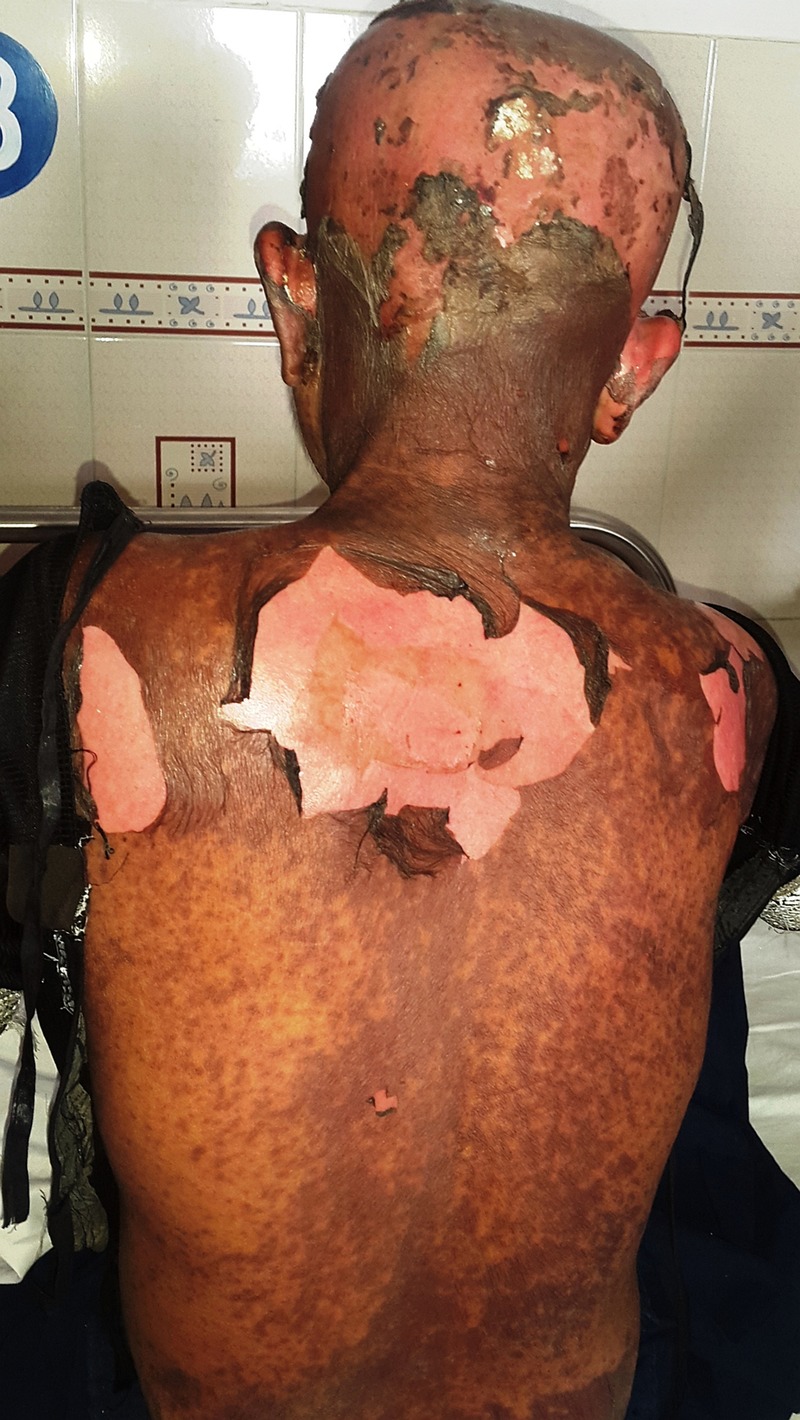
Two weeks after the initiation of tamoxifen, the patient returned to the emergency department with a history of skin over a large area of her body peeling off as she tried to dry her skin with a towel after she took a bath (figure 1). On questioning, she recalled that there were no other symptoms other than that of generalised malaise and fatigue over the preceding week. On examination, there was absence of epidermis on approximately 40% of her body surface area, involving the back, the abdominal wall, the scalp and the sacral region. She was assigned the clinical diagnosis of toxic epidermal necrolysis (TEN) and was immediately admitted and treated in the burns treatment unit under strict asepsis, antibiotic prophylaxis and intravenous fluid support. The patient made a full recovery after 24 days.
Figure 1.

The patient presented with a history of skin over a large portion of her body having unexpectedly and painlessly separated away while drying herself with a towel after bathing.
The patient had previously completed a total of eight cycles of chemotherapy uneventfully. During the occurrence of TEN, the patient was not on any drug other than tamoxifen. Since antihormonal therapy is crucial in the management of breast cancer and because tamoxifen could no longer be considered in this patient, a consensus was reached to continue antioestrogen therapy using letrozole after ovarian ablation (using a gonadotropin-releasing hormone analogue).
The entities TEN and Steven-Johnson syndrome (SJS) exist as part of a continuum. The nomenclature is TEN when >30% of body surface area is involved, and SJS when <10% of body surface area is involved. The intermediate state is called ‘SJS/TEN overlap’.1
The main reported cause behind TEN is drug reactions, which are implicated in 80–95% of TEN cases.2 The commonly implicated drugs include sulfonamides, β-lactams, allopurinol, methotrexate, anti-epileptics, anxiolytics and, occasionally, non-steroidal anti-inflammatory drugs. Non-drug causes are rare, and include contrast agents and infectious agents such as mycoplasma pneumonia. Immunocompromised patients, such as organ transplant recipients, or those with HIV infection, are at an increased risk of TEN.3
The current understanding regarding the aetiopathogenesis of TEN points to an immunological mechanism. It is hypothesised that CD8+ T cells are hyperstimulated by drugs or their metabolites to release molecules (such as perforin, granzyme B, granulysin, tumour necrosis factor (TNF) α and fas ligand), which leads to keratinocyte death. Since keratinocytes are responsible for holding surrounding skin cells together, their death causes skin detachment.1 2
Despite being rare, with an incidence of 0.4–1.9 cases per million per year, TEN warrants early diagnosis given its life-threatening potential.2 The definitive diagnosis of TEN depends on histopathological confirmation, as revealed by scattered necrotic keratinocytes (in early cases), or full-thickness epidermal necrosis with subepidermal split (in advanced cases). However, early clinical diagnosis is crucial for prompt treatment. The presence of mucositis (oral/ocular/genital) is found in nearly all patients with TEN, and is helpful in identifying early cases. Important signs include the Nikolsky sign (gentle lateral pressure leading to separation of the papillary dermis layer from the basal layer) and the Asboe-Hansen sign (pressure on bullae leading to lateral extension).3
Learning points.
Toxic epidermal necrolysis is a potentially life-threatening condition with an approximate mortality rate of 30%.4
The ideal management for toxic epidermal necrolysis includes treatment under aseptic conditions akin to the management of patients with burns.5
The preferred drug for the hormonal therapy of pre-menopausal breast cancer happens to be tamoxifen. However, if aromatase inhibitors need to be used, it is important that a state of menopause be induced by the use of gonadotrophin-releasing hormone analogues.6
Footnotes
Contributors: IM prepared the manuscript. SR identified the case for publication and performed the literature review. AP and AA reviewed the literature and finalised the manuscript.
Competing interests: None declared.
Patient consent: Obtained.
Provenance and peer review: Not commissioned; externally peer reviewed.
References
- 1.Harr T, French LE. Toxic epidermal necrolysis and Stevens-Johnson syndrome. Orphanet J Rare Dis 2010;5:39 10.1186/1750-1172-5-39 [DOI] [PMC free article] [PubMed] [Google Scholar]
- 2.Schwartz RA, McDonough PH, Lee BW. Toxic epidermal necrolysis: part I. Introduction, history, classification, clinical features, systemic manifestations, etiology, and immunopathogenesis. J Am Acad Dermatol 2013;69:173.e1–13. 10.1016/j.jaad.2013.05.003 [DOI] [PubMed] [Google Scholar]
- 3.Schwartz RA, McDonough PH, Lee BW. Toxic epidermal necrolysis: part II. Prognosis, sequelae, diagnosis, differential diagnosis, prevention, and treatment. J Am Acad Dermatol 2013;69:187.e1–16. 10.1016/j.jaad.2013.05.002 [DOI] [PubMed] [Google Scholar]
- 4.Majumdar S, Mockenhaupt M, Roujeau J et al. Interventions for toxic epidermal necrolysis. Cochrane Database Syst Rev 2002:CD001435. [DOI] [PMC free article] [PubMed] [Google Scholar]
- 5.Mahar PD, Wasiak J, Hii B et al. A systematic review of the management and outcome of toxic epidermal necrolysis treated in burns centres. Burns 2014;40:1245–54. 10.1016/j.burns.2014.02.006 [DOI] [PubMed] [Google Scholar]
- 6.Montagna E, Cancello G, Colleoni M. The aromatase inhibitors (plus ovarian function suppression) in premenopausal breast cancer patients: ready for prime time? Cancer Treat Rev 2013;39:886–90. 10.1016/j.ctrv.2013.04.007 [DOI] [PubMed] [Google Scholar]


