

Abstract
Henoch-Schönlein purpura (HSP) is the most common vasculitis found in children. It usually affects the small vessels of the skin, joints, gastrointestinal tract and, more rarely, kidneys, brain, lungs and genitalia. Apart from classical presentation with purpuric rashes around buttocks and lower extremities, features such as arthralgia, abdominal pain, haematuria and proteinuria as well as scrotal swelling have been described in the literature. Penile involvement is rare and is not commonly described. We describe a child with HSP who developed penile involvement. We review the literature of all the cases reported in detail, in order to highlight useful clinical presentation, management and prognosis of this rare manifestation.
Background
Henoch-Schönlein purpura (HSP) is the commonest vasculitis in children. It is a systemic leucocytoclastic vasculitis of the small vessels with perivascular deposition of immunoglobulin A (IgA) immune-complexes leading to inflammation and necrosis of arterioles, capillaries and postcapillary venules. It mainly affects the small vessels of the skin, joints, gastrointestinal tract and, more rarely, kidneys, brain, lungs and genitalia. Clinically it is characterised by a palpable purpuric rash with a typical distribution on the buttocks, thighs and lower legs.
Scrotal involvement, consisting mainly of scrotal swelling and tenderness, may occur in HSP, usually unilaterally, with a mean prevalence of 11.6% (ranging from 2% to 38%).1 2 Penile involvement is much rarer, with few cases reported in detail in the literature, as far as we could ascertain.3–9 It may occur in isolation or in association with scrotal involvement.1
We describe a child with HSP who developed penile involvement. We review the literature of all the cases reported in detail, in order to highlight useful features of the clinical presentation, management and prognosis of this rare manifestation.
Case presentation
Six months following an episode of Henoch-Schnölein purpura (HSP), a 4-year-old boy presented with an acute swelling of the penis over the previous few hours, with no preceding illnesses or other symptoms. There was no history of trauma or penile discharge, the swelling was painless and there was no difficulty in passing urine, and the scrotum looked normal. With the exception of a palpable erythaematous rash on the elbows and abdomen, he was otherwise well and had no symptoms in his joints.
Although on examination he had a widespread palpable erythaematous maculopapular rash on his elbows and abdomen, and marked swelling of the uncircumcised penis associated with discolouration of the overlying skin, he appeared well and was apyrexial, haemodynamically stable and well perfused. Over the next 24 h, the maculopapular rash became purpuric and the boy developed a purpuric rash with swelling, a bruise and tenderness over the penis, the upper scrotum and the suprapubic area (figure 1), associated with dysuria. It was not possible to elicit specific testicular tenderness as the whole genital area was swollen and tender.
Figure 1.
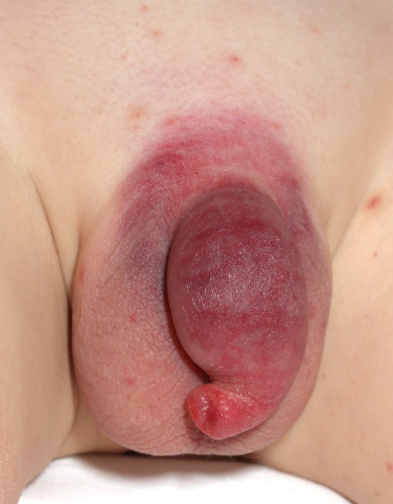
Swelling, erythaema and purpura of the penis, upper scrotum and suprapubic area seen 24 h after presentation.
Investigations
Doppler ultrasound studies demonstrated increased blood flow to the testicles, with normal flow velocity and normal resistive indices in the intratesticular arteries, strongly excluding the possibility of testicular torsion and supporting the diagnosis of orchitis instead.
Urinalysis showed only traces of blood and leucocytes, but no proteinuria or nitrites. Serum C reactive protein, full blood count, coagulation tests, urea, creatinine and electrolytes were all within normal limits and urinary tract infection was excluded the following day by a negative urine culture.
The diagnostic points are summarised as follows:
Progressive development of the suggestive vasculitic rash;
Diffuse swelling and tenderness over the whole scrotum and suprapubic area without localisation of the symptoms to a particular testicle;
Absence of fever or urethral discharge;
Sterile urine culture;
Normal testicular Doppler ultrasound findings.
Differential diagnosis
Based on the history of HSP, acute presentation with generalised rashes, generalised tenderness and a bruise on the genital area, and normal ultrasound findings, a diagnosis of orchitis and balanitis secondary to HSP was made.
In a child presenting with swelling and tenderness in the genital area, before the appearance of the vasculitic rash, the other differential diagnoses to consider are:
Testicular torsion: It typically presents with acute severe unilateral scrotal pain; there are no purpuric rashes or bruises present.
Urethritis: Dysuria and urethral discharge may occur, but there are no purpuric rashes or bruises present.
Orchi-epididymitis: There is usually localised scrotal swelling and pain, with no purpuric rashes or bruises.
Sexual abuse: It is a diagnosis of exclusion based on a careful history and examination of findings.
Treatment
The patient was started on intravenous prednisolone, co-amoxiclav, followed 2 days later by a daily dose of 30 mg of oral prednisolone for a week.
Outcome and follow-up
Dysuria, swelling and pain improved considerably within 24 h. The patient was discharged but continued to intermittently develop purpuric rash and swelling of the ankles but without genital involvement. All symptoms and abnormal urinary findings resolved over a 12-month period.
Discussion
Although testicular and scrotal involvements in HSP are very rare, involvement of the penis is even less well described. We compare our patient with seven affected children described in detail in the literature (table 1).3–9 Another 10 affected children were excluded because of lack of clinical description.1 10–13 In one series, genital involvement occurred in 10 of 155 boys with HSP (11.6%) with only 3 of the 10 affected children having concomitant penile involvement.1 This complication of HSP vasculitis most likely results from the deposition of immune complexes in the small vessels of an end organ with a complex microvascular structure, such as the penis.
Table 1.
Characteristic of Henoch-Schönlein purpura patients with penile swelling
| Reference | Age (years) | Circumcised | Onset of penile swelling after rash | Purpura | Erythaema | Oedema | Local tenderness | Other abnormalities | Therapy | Outcome |
|---|---|---|---|---|---|---|---|---|---|---|
| Caliskan et al4 | 2 | No | 2 weeks | Yes, 1 day after treatment | Yes | Yes | Yes | No rash at presentation, painful micturition symptoms for 2 weeks | Warm water bath | Improved within 2 days |
| Ferrara et al6 | 3.3 | No | 4 days after resolution of rash | Yes | Yes | Yes | Yes | Joints symptoms 4 days earlier, mastoiditis 6 days earlier | None | 2 days |
| Our patient | 4 | No | Same day | Yes | Yes | Yes | Yes | No joints symptoms, scrotal swelling, dysurea | Prednisolone | Improved within 24 h |
| Sandell10 | 4 | No | 5 days | Yes | Yes | Yes | No | – | Methyl prednisolone then prednisolone | Improved within 12 h |
| David et al5 | 4 | No | A few days? | Yes | Yes | Yes | Yes | Scrotum involvement | None | Improved after? |
| Pennesi9 | 4.9 | No | 3 days | No | Yes | Yes | Yes | – | Corticosteroids | 2 days |
| Lind8 | 9 | Unknown | 1 day | Yes | No | No | Yes | Arthritis 4 days earlier, haematuria and proteinuria Priapism 5 days later |
Caudal anaesthesia | 9 h |
| Burrows et al3 | 19 | No | 2 months | Yes | No | No | No | – | None | Settled within 2 months |
Involvement of genitalia in females has rarely been reported in the literature. This includes one report of tubo-ovarian abscess complicating the vasculitis in a pubertal 15-year-old girl, ovarian cystic changes in a prepubertal girl, and occasional vulvar involvement.14–16
Our patient's presentation has some unique clinical features: as the penile swelling preceded the typical HSP purpuric rash, which was absent from the skin and the penis for over 24 h and as scrotal involvement also occurred later, the correct diagnosis was not made immediately at presentation. The presence of dysuria and tenderness might also have led to the erroneous diagnosis of urinary tract infection, balanitis or urethritis. The association with genital purpura also had the potential to falsely lead to the suspicion of child sexual abuse with all its implications. Testicular torsion has also been reported in HSP.17 18 In most cases of testicular torsion with absence of HSP features, Doppler ultrasound is not always indicated; and immediate surgical exploration is indicated instead when there is strong clinical suspicion. However, Doppler ultrasound is still indicated when there is a low index of suspicion of testicular torsion in HSP or when there is a strong suspicion of an alternate diagnosis such as orchitis.18 The ultrasound findings associated with the rapid spontaneous resolution of scrotal swelling, and the development of the vasculitis rash following the initial presentation, made the diagnosis of testicular torsion extremely unlikely in our patient.
Table 1 summarises the characteristics of our patient and of the other seven reports that have documented this manifestation in children. Six of eight children were under 5 years of age and none of them were circumcised. Excluding our patient, for whom the penile swelling developed before the typical purpuric rash of HSP, in all other cases it developed within a few days of the typical rash. There were also two patients in whom penile swelling appeared 2 weeks and 2 months later. There was no vasculitis rash in two of the cases when penile involvement occurred, and in one of them the penile symptoms occurred 4 days after resolution of the rashes. The penile manifestations consisted of purpura in all but one case, and six children had erythaema or oedema and seven had tenderness. Scrotal involvement occurred in only two children, and one had priapism. There were two children with painful micturition and one child had proteinuria with haematuria. Three children had preceding symptoms of joint involvement that began a few days to 2 weeks earlier. Corticosteroid therapy was administered to three children, one with associated priapism had caudal anaesthesia, one was treated with warm water bath and three children were managed only symptomatically. There was improvement in six children between 9 and 48 h of management. The 19-year-old patient only showed improvement after 2 weeks. All children made complete recovery with no lasting sequelae.
Recognition of penile involvement in children with HSP, with or without testicular or scrotal involvement, is important if diagnosis is to be made early and unnecessary investigations, procedures or therapy are to be avoided. Parents can be reassured that complete resolution is the norm with no long-lasting sequelae. The role of steroid therapy remains controversial with few anecdotal reports of clinical improvement with or without their use. There are no results from randomised trials to clarify their role.
Learning points.
Penile swelling is a rare manifestation of the Henoch-Schönlein purpura.
When it occurs before the typical vasculitic rash, the aetiological diagnosis may be difficult.
Diagnosis should be based on exclusion of other serious pathology in such cases.
Acknowledgments
Clare Glendining: Medical Illustration.
Footnotes
Competing interests: None.
Patient consent: Obtained.
Provenance and peer review: Not commissioned; externally peer reviewed.
References
- 1.Mintzer CO, Nussinovitch M, Danziger Y et al. Scrotal involvement in Henoch-Schonlein purpura in children. Scand J Urol Nephrol 1998;32:138–9. 10.1080/003655998750014530 [DOI] [PubMed] [Google Scholar]
- 2.Byrn JR, Fitzgerald JF, Northway JD et al. Unusual manifestations of Henoch-Schonlein syndrome. Am J Dis Child 1976;130:1335–7. [DOI] [PubMed] [Google Scholar]
- 3.Burrows NP, Sonnex C, Roberts SO et al. Cutaneous vasculitis presenting on the penis. Genitourin Med 1993;69:301–2. [DOI] [PMC free article] [PubMed] [Google Scholar]
- 4.Caliskan B, Guven A, Atabek C et al. Henoch-Schonlein purpura presenting with symptoms mimicking balanoposthitis. Pediatr Rep 2009;1:e5 10.4081/pr.2009.e5 [DOI] [PMC free article] [PubMed] [Google Scholar]
- 5.David S, Schiff JD, Poppas DP. Henoch-Schonlein purpura involving the glans penis. Urology 2003;61:1035 10.1016/S0090-4295(03)00025-6 [DOI] [PubMed] [Google Scholar]
- 6.Ferrara P, Marrone G, Nicoletti A et al. Penile involvement in Henoch-Schonlein purpura with good prognosis. Scand J Urol Nephrol 2007;41:567–9. 10.1080/00365590701365487 [DOI] [PubMed] [Google Scholar]
- 7.Lind J, Mackay A, Withers SJ. Henoch-Schonlein purpura and priapism. J Paediatr Child Health 2002;38:526–7. 10.1046/j.1440-1754.2002.00028.x [DOI] [PubMed] [Google Scholar]
- 8.Pennesi M, Biasotto E, Saccari A. Schonlein-Henoch purpura involving the penis. Arch Dis Child 2006;91:603 10.1136/adc.2005.072652 [DOI] [PMC free article] [PubMed] [Google Scholar]
- 9.Sandell J, Ramanan R, Shah D. Penile involvement in Henoch-Schonlein purpura. Indian J Pediatr 2002;69:529–30. 10.1007/BF02722659 [DOI] [PubMed] [Google Scholar]
- 10.Joseph PR. Penile edema. Clin Pediatr (Phila) 2002;41:547 10.1177/000992280204100721 [DOI] [PubMed] [Google Scholar]
- 11.Kanakrieh M, Madanat F. A review of Henoch-Schonlein purpura at the Jordan University Hospital. Jordan Med J 1982;16:35–40. [Google Scholar]
- 12.Habashneh M, Quar M, Aqrabawi H. Henoch-Schonlein purpura: presentation and complications. Jordan Med J 2004;38:38–43. [Google Scholar]
- 13.Al-Hermi BE, AAal A. A retrospective study of Henoch-Schonlein purpura in Bahrain. Kidney Forum 2000;2:306. [Google Scholar]
- 14.Pomeranz A, Korzets Z, Eliakim A et al. Relapsing Henoch-Schonlein purpura associated with a tubo-ovarian abscess due to Morganella morganii. Am J Nephrol 1997;17:471–3. 10.1159/000169155 [DOI] [PubMed] [Google Scholar]
- 15.Nader NS, Matsumoto JM, Lteif A. Cystic changes in the ovaries of a pre-pubertal girl with Henoch-Schonlein purpura. J Pediatr Endocrinol Metab 2010;23:517–19. [DOI] [PubMed] [Google Scholar]
- 16.Fischer G, Rogers M. Vulvar disease in children: a clinical audit of 130 cases. Pediatr Dermatol 2000;17:1–6. 10.1046/j.1525-1470.2000.01701.x [DOI] [PubMed] [Google Scholar]
- 17.Eadie DG, Higgins PM. Apparent torsion of the testicle in a case of Henoch-Schoenlein purpura. Br J Surg 1964;51:634–5. 10.1002/bjs.1800510822 [DOI] [PubMed] [Google Scholar]
- 18.Loh HS, Jalan OM. Testicular torsion in Henoch-Schonlein syndrome. BMJ 1974;2:96–7. 10.1136/bmj.2.5910.96 [DOI] [PMC free article] [PubMed] [Google Scholar]