Abstract
Yersinia species display a tropism for lymphoid tissues during infection, and the bacteria select innate immune cells for delivery of cytotoxic effectors by the type III secretion system. Yet the mechanism for target cell selection remains a mystery. Here we investigate the interaction of Yersinia pestis with murine splenocytes to identify factors that participate in the targeting process. We find that interactions with primary immune cells rely on multiple factors. First, the bacterial adhesin Ail is required for efficient targeting of neutrophils in vivo. However, Ail does not appear to directly mediate binding to a specific cell type. Instead, we find that host serum factors direct Y. pestis to specific innate immune cells, particularly neutrophils. Importantly, specificity towards neutrophils was increased in the absence of bacterial adhesins due to reduced targeting of other cell types, but this phenotype was only visible in the presence of mouse serum. Addition of antibodies against complement receptor 3 and CD14 blocked target cell selection, suggesting that a combination of host factors participate in steering bacteria toward neutrophils during plague infection.
Keywords: Yersinia, Virulence, Microbial-cell interaction
Introduction
Many bacteria assemble nanomachines on their surfaces to deliver protein cargo to specific destinations. In Gram-negative bacteria, type III secretion systems (T3SS) are often engaged to deliver bacterial proteins into eukaryotic cells in a process that involves assembly of a needle-like structure on the bacterial surface (Buttner, 2012). The pathogenic Yersinia species (Y. pestis, Y. pseudotuberculosis, and Y. enterocolitica) rely on a plasmid-encoded T3SS to deliver a suite of effectors, known as Yops, into immune cells (Dewoody et al., 2013). The T3SS is an essential virulence factor for Yersinia spp. and many of the Yops function to disarm the host defenses by inhibiting phagocytosis, inducing apoptosis, and modulating the inflammatory response (Viboud et al., 2005, Ruckdeschel et al., 2008). Despite sharing a highly conserved T3SS and set of Yop effectors, the three pathogenic Yersinia spp. manifest distinctly different diseases. Y. enterocolitica and Y. pseudotuberculosis are found in soil and water, and upon ingestion they typically cause self-limiting gastroenteritis (Galindo et al., 2011). In the mouse model of infection, these bacteria will colonize Peyer's patches and mesenteric lymph nodes, followed by dissemination to the spleen and liver. On the other hand, Y. pestis is the causative agent of bubonic plague, which is a vector-borne disease that is maintained in rodent reservoirs (Perry et al., 1997, Gage et al., 2005). Once delivered to a mammalian host by fleabite, the bacteria colonize the draining lymph node where bacterial replication leads to the characteristic inflamed, painful bubo. Subsequently, bacteria disseminate to more distant lymph nodes as well as spleen and liver (Sebbane et al., 2005).
Although disease progression is different for enteric Yersinia compared to Y. pestis, all three species share a tropism for lymphoid tissue. Investigating the nature of bacterial interaction with primary cells in vivo has been made possible by recent technological advancements. Using β-lactamase fused to a Yop effector, which is delivered in a T3SS-dependent manner, Yop injection into host cells can be detected by flow cytometry (Charpentier et al., 2004, Marketon et al., 2005, Koberle et al., 2009, Durand et al., 2010). With this approach, it was shown that Y. pestis selects certain immune cells for injection of Yops in vivo, particularly innate immune cells (neutrophils, dendritic cells, and macrophages) (Marketon et al., 2005, Pechous et al., 2013). A similar preference for phagocytes was observed in spleens from mice infected with Y. enterocolitica and Y. pseudotuberculosis (Koberle et al., 2009, Durand et al., 2010). More recently, a biphasic response was demonstrated in lung tissue, whereby alveolar macrophages are targeted early in infection followed by preferential targeting of neutrophils in the later stages of infection by both Y. pestis and Y. pseudotuberculosis (Pechous et al., 2013, Paczosa et al., 2014). At present, the mechanism that promotes Yersinia preference for neutrophils and other phagocytes in vivo is unknown; however, at least for the enteric Yersinia this preference appears to be maintained during in vitro infections of homogenized tissues (Koberle et al., 2009, Durand et al., 2010, Paczosa et al., 2014), which indicates that cell preference is not simply dictated by bacteria becoming surrounded and confined in tissues by infiltrating phagocytes. Rather, the data suggest that some type of receptor-ligand interaction should mediate the bacterial preference for certain cell types.
Several bacterial adhesins have been shown to mediate attachment of Yersinia to mammalian host cells. In the enteric Yersinia, Ail, YadA and invasin play a prominent role in adhesion, and correspondingly mutants lacking these genes manifest defects in colonization and dissemination during in vivo infections (Miller et al., 1988, Pepe et al., 1995, Marra et al., 1997, Handley et al., 2005, Durand et al., 2010, Uliczka et al., 2011, Paczosa et al., 2014). Adhesion to host cells is necessary for injection of Yop effectors, and in the absence of these adhesins, Yop delivery is compromised (Kapperud et al., 1985, Kapperud et al., 1987, Bliska et al., 1993, Durand et al., 2010, Maldonado-Arocho et al., 2013, Paczosa et al., 2014). In particular, Ail and YadA are required for Yop translocation in lungs of infected mice (Paczosa et al., 2014); whereas YadA, invasin, and Ail each contribute to translocation in spleens of infected mice in a strain-dependent manner, depending on the relative expression levels of each adhesin (Maldonado-Arocho et al., 2013). In addition to their roles as adhesins, YadA and Ail also facilitate resistance to serum-mediated bacterial lysis (Pierson et al., 1993, Biedzka-Sarek et al., 2005, Leo et al., 2011, Ho et al., 2012a, Schindler et al., 2012). Furthermore, Ail and YadA counteract the effects of complement, probably by inhibiting opsonophagocytosis, to allow dissemination and growth in the spleen (Maldonado-Arocho et al., 2013, Paczosa et al., 2014).
Despite their importance to pathogenesis of enteric Yersinia, neither YadA nor invasin is expressed by Y. pestis. Instead, the predominant Y. pestis adhesins appear to be Ail, Pla, and PsaA (pH6 antigen) (Felek et al., 2009, Felek et al., 2010). Ail and Pla are both outer membrane proteins, while the psa operon encodes a fimbrial structure. Pla, which is absent from the enteric Yersinia, is an outer membrane adhesin and protease that facilitates degradation of extracellular matrix proteins and promotes bacterial dissemination (Sodeinde et al., 1988, Sodeinde et al., 1992). PsaA, also called MyfA, is present in all three Yersinia spp., but its expression is restricted to slightly acidic conditions at temperatures above 36°C (Ben-Efraim et al., 1961, Iriarte et al., 1993, Yang et al., 1996). Though Ail is present in all three Yersinia spp., its expression pattern and functionality differs. In Y. pestis, Ail is expressed at lower temps, but expression is optimal at 37°C at which point Ail makes up 20-30% of the outer membrane protein content (Kolodziejek et al., 2007, Bartra et al., 2008, Felek et al., 2009, Pieper et al., 2009). Besides conferring serum resistance, Y. pestis Ail mediates adhesion via binding to the extracellular matrix proteins laminin and fibronectin (Kolodziejek et al., 2007, Bartra et al., 2008, Tsang et al., 2010, Yamashita et al., 2011). This adhesive activity is important for the T3SS, and Y. pestis strains lacking Ail suffer from severely reduced Yop translocation (Felek et al., 2009, Felek et al., 2010). It is thought that the severe attenuation of Δail mutants in the rat model stems from the combined roles for Ail in coordinating serum resistance and cell adhesion (Hinnebusch et al., 2011).
Given that adhesins function to mediate interactions between bacteria and host cells, recent work has investigated the contribution of adhesins to Yersinia target cell preference. It was demonstrated that Y. pseudotuberculosis lacking the three major adhesins YadA, invasin, and Ail was still capable of T3SS-dependent Yop injection, and that the preference for neutrophils among splenocytes was maintained, suggesting the possibility of adhesin-independent interactions (Maldonado-Arocho et al., 2013). Moreover, Maldonado et al. showed that addition of serum to ex vivo splenocyte infections enhanced bacterial specificity for neutrophils (Maldonado-Arocho et al., 2013).
Here we have attempted to identify the factors that direct Y. pestis targeting of innate immune cells for Yop injection. We find that target cell preference is maintained during ex vivo infection of splenocytes, and that neutrophils are still the preferred cell type in the absence of organ architecture. In agreement with previous work using tissue culture infections (Felek et al., 2010), we find that Ail is the predominant adhesin mediating efficient injection of murine splenocytes. In an attempt to identify factors conferring specificity to neutrophil targeting, we investigated the role of serum. Surprisingly, we found that mouse serum suppressed overall levels of Yop injection, which appears to be a consequence of increased specificity toward myeloid cells, neutrophils in particular. Notably, we show that neutrophil targeting is mediated by complement receptor 3 and, to a lesser extent, CD14.
Results
Yops are translocated by the ail mutant in vivo
In light of the observations that Ail is important for mediating Yop delivery, and that the Δail mutant is severely attenuated in vivo (Felek et al., 2009), we wondered whether the virulence defect could be attributed to a lack of Yop injection in vivo. To address this, we employed the β-lactamase (Bla) reporter system that we previously developed for measuring T3SS injection of primary immune cells (Marketon et al., 2005, Houppert et al., 2013). In this assay, translocation (injection) of the YopM-Bla reporter is monitored via cleavage of the fluorescent dye CCF2-AM. CCF2-AM-stained cells fluoresce green upon excitation with violet light (405nm). In the presence of Bla, the dye is cleaved resulting in a shift from green to blue fluorescence, depending on the amount of dye that is cleaved. Cells that have been injected with YopM-Bla will fluoresce blue. On the other hand, uninfected cells or cells infected with Y. pestis expressing GST-Bla (negative control) will fluoresce green. In combination with the immunophenotyping strategy outlined in Figure S1, flow cytometry is used to identify cell types and determine which cells have been injected.
Mice were infected with wild type KIM5, the Δail mutant, or a ΔyscU mutant (T3SS-) carrying either YopM-Bla or Gst-Bla. Importantly, the LD50 of the mutants are ∼1.7 × 10⁵ CFU for Δail and ≥ 107 CFU for ΔyscU compared to ∼10 CFU for the KIM5 parent (Perry et al., 1997, Felek et al., 2009). To compensate for the virulence defects of the mutants and ensure recovery of similar bacterial loads from spleens during a 3-day time course, we used 100-fold higher inocula for the mutants (106 CFU compared to 104 CFU for WT). Infected spleens were then harvested, and the bacterial load was quantified as well as the percentage of splenocytes that were injected with the YopM-Bla reporter. Previous work indicated that while the parent KIM5 strain could be detected in the spleen one day post-infection, YopM-Bla injection was below the limit of detection (Marketon et al., 2005). We therefore began monitoring KIM5-infected mice on day 2 post-infection. As shown in Figure 1A, the mean bacterial burden from KIM5-infected mice was 105.35 CFU per spleen on day 2, and by day 3 there was a 1.5-log increase in the bacterial load (P<0.001). Looking at the number of splenocytes injected with YopM-Bla during wild type infection, about 1.1% of cells appeared blue on day 2 (Figure 1B). These blue cells are indicative of T3SS-dependent injection of YopM-Bla, rather than phagocytosis of bacteria, because infection with either WT expressing the Gst-Bla negative control (data not shown) or with the T3SS mutant (yscU -) expressing YopM-Bla each yielded only ∼0.08% blue cells. Despite the fact that bacterial burden of wild type Y. pestis increased between days 2 and 3 of infection, the average number of YopM-Bla positive cells tended toward a slight reduction, though, this was not a statistically significant decrease. In contrast to wild type, the Δail mutant showed a trend of increasing amounts of Yop injection as the infection progressed. The colonization levels of the Δail mutant were relatively stable with only a slight increase in the average bacterial burden from day 1 to day 3 (104 CFU vs 105 CFU, respectively, P<0.05). In fact the bacterial loads of spleens from mice infected with the ail or yscU mutants on days 1 and 2 were not statistically different. Yet despite the fairly low bacterial load for the Δail mutant, there was a dramatic increase in the percentage of injected splenocytes between days 1 and 2, and this level of injection was maintained through day 3.
Figure 1. The Δail mutant translocates Yops in vivo.

Mice were infected with the indicated Y. pestis strains expressing either YopM-Bla or Gst-Bla. Spleens were harvested and homogenized at days 1, 2, and 3 post-infection. A portion of each homogenate was used to enumerate bacterial load (Panel A). The remaining splenocyte suspension was stained with CCF2-AM and analyzed by flow cytometry to quantify the number of blue (injected cells) (Panel B). Data was analyzed by one-way ANOVA with Tukey post-hoc test (* P<0.05, ** P<0.01, *** P<0.001). Asterisks reflect indicated pair-wise comparisons. The stars indicate that WT colonization on day 3 was significantly higher than all other samples. While panel B shows % injection in infected spleens according to day post-infection, panel C shows the same data according to bacterial loads. ND indicates “not determined” since only one mouse infected with the WT strain had a bacterial load below 104 cfu, and no mice infected with the Δail mutant had a bacterial load above 107 cfu (see panel A).
Because there was a broad range of bacterial load in infected mice, particularly on day 3 post-infection, we chose to pool the data from all days and then categorize according to the bacterial loads using 1-log increments (Fig. 1C). In this way, the ability of the ail mutant to target immune cells can be compared to that of WT using data from comparable bacterial burdens. When viewed this way, the data indicates that the two strains delivered Yops into immune cells to a similar extent. Though not statistically significant, there appeared to be a trend of more blue cells as the bacterial load increased until a peak is reached at about 106 cfu, above which the number of blue cells decreased. When taken together, the data indicate that when the Δail mutant's colonization defect is compensated by a higher inoculum to achieve similar bacterial burdens, Yop delivery into splenocytes is comparable to wild type Y. pestis.
We also evaluated whether recruitment of immune cells to the spleen is altered during infection by the Δail mutant. The data from WT infected animals (Fig. S2A) indicate that as the infection progressed and bacterial numbers increased, there was a corresponding increase in B cells within spleens. In contrast, dendritic cells, macrophages, and NK cells were significantly reduced as bacterial burden increased. As expected, there was an initial increase in the neutrophil population, but above 106 cfu the number of neutrophils declined. As shown in Figure S2B, infection by the Δail mutant yielded similar trends in the total splenocyte populations. Although the activation status of these cell types could differ, it appears that under these infection conditions, the Δail mutant encounters a similar overall distribution of splenocytes as does WT Y. pestis.
Ail contributes to efficient targeting of neutrophils in vivo
To analyze targeting of specific cell types for Yop injection, we evaluated three parameters: frequency of injection, distribution of injected cells, and targeting efficiency. Figure 2 shows the analysis for mice infected with the WT strain; however the general strategy for this analysis is described in Figure S3 using a simpler data set. Frequency of injection refers to the percentage of each cell type that is positive for the YopM-Bla reporter. As expected, all myeloid cell types were injected by WT at higher frequencies than were the lymphoid populations (Fig. 2A). In fact, relatively few lymphoid cells were targeted until late in infection (i.e. >107 cfu). Importantly, the percentage of injected monocytes and neutrophils increased dramatically and significantly by about 106 cfu and then remained high as bacterial burden increased (Fig. 2A). Despite this persistent targeting of neutrophils at the highest bacterial loads, neutrophils represented only a small fraction of the total population of blue cells (Fig. 2B). This also corresponds to a drop in total neutrophils (Fig. S2A). Altogether, the trend shifted from injection of myeloid cells at lower bacterial loads toward injection of B cells at >107 cfu (Fig. 2B).
Figure 2. Target cell selection during infection with WT Y. pestis.
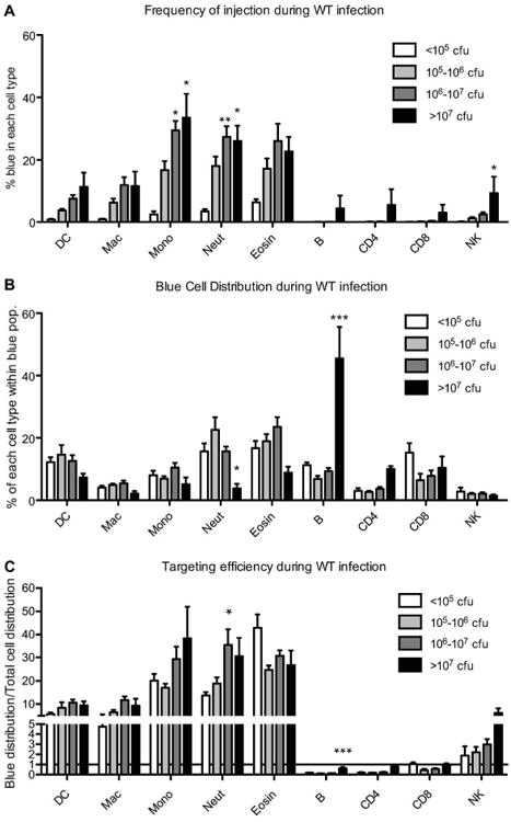
Mice described in Fig. 1 were infected with WT Y. pestis, and splenocytes were analyzed by flow cytometry (described in Fig. S1) to identify each cell type as well as blue injected cells. A) Frequency of injection: After cell types are identified from total live cells, the % of blue cells in each population was determined. B) Blue cell distribution: Blue cells were first identified from total live cells and then subjected to the myeloid and lymphoid analysis to identify cell types within the blue population. C) Targeting efficiency: The blue cell distribution was compared to the total cell distribution. The fold-difference for each cell type is the “targeting efficiency”, and these values are graphed here (horizontal line at Y=1 indicates no cell preference). Data were analyzed by 2-way ANOVA with Bonferroni multiple comparisons test to determine significant differences between the lowest cfu load compared to higher cfu loads for each cell type. (* P<0.05, ** P<0.01, *** P<0.001) DC= dendritic cells; Mac= macrophages; Mono= monocytes; Neut= neutrophils; Eosin= eosinophils; CD4= T helper cells; CD8= T cytotoxic cells; NK= NK cells.
Because the myeloid cell types are a minor portion of the splenocyte population, and because the cell distribution changes during infection, we normalized the distribution data by dividing the distribution of injected cell types by the distribution of total cell types (injected + uninjected), resulting in a metric that we term “targeting efficiency” (described in Fig. S3). Using this metric, we avoid bias in apparent target cell preferences that may stem from any fluctuations in the total numbers of individual cell types as a result of mouse-to-mouse variability or as a byproduct of infection and cell death. A targeting efficiency value of 1 indicates no preference because the relative proportion of injected cells for a particular cell type represents the same proportion of that type within the total splenocyte mixture. Targeting efficiencies greater than one indicate an enrichment of the cell type within the injected cell population compared to the total cell population (a preference for those cells). Targeting efficiencies less than one indicate that those cell types are under-represented within the injected population, which could reflect active exclusion or avoidance of those cells by the bacteria. Importantly, B and T cells were not efficiently targeted during WT infection (Fig. 2C). In contrast, targeting efficiencies were extremely high for the myeloid populations, particularly neutrophils, monocytes, and eosinophils.
The data in Figure 2 indicates that neutrophils are a major target of Yop injection during WT infections. In contrast, targeting of neutrophils was less prominent during infections with the Δail mutant (Fig. 3). To facilitate the comparison of mutant vs WT data, Figure 4 shows a side-by-side depiction of the data for the 105 -106 cfu and 106 -107 cfu categories for both strains. Most notable is the dramatic reduction in neutrophil targeting efficiency by the mutant compared to WT for both bacterial burden categories (Fig. 4B). Neutrophils as well as other myeloid cells were injected at frequencies higher than that of lymphoid cells (Fig. 3A). However, after an initial increase in injection levels, the percentages of injected cells for each myeloid cell type decreased as the bacterial loads increased beyond 106 cfu. Further distinguishing the mutant from WT is the distribution of the blue cell population (Fig. 3B). While myeloid cells represented a major portion of injected cells until very high bacterial loads during WT infection (Fig. 2B), there is no clear major population in the distribution of injected cells from mice infected with the Δail mutant (Fig. 3B). Moreover, the targeting efficiencies for the myeloid cell types, particularly neutrophils, tended to be reduced compared to WT infections (Fig. 3C and 4B).
Figure 3. Target cell selection during infection with Δail Y. pestis.
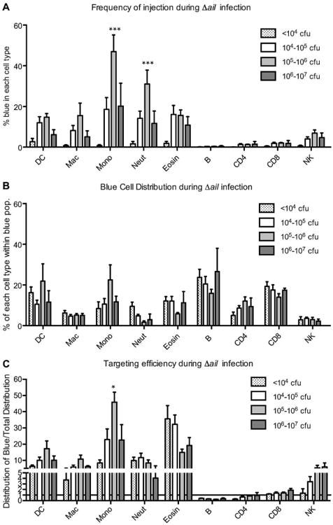
Mice described in Fig. 1 were infected with Δail Y. pestis, and splenocytes were analyzed as described in Fig. S1. A) Frequency of injection. B) Blue cell distribution. C) Targeting efficiency. Data were analyzed by 2-way ANOVA with Bonferroni multiple comparisons test to determine significant differences between the lowest cfu load compared to higher cfu loads for each cell type. (* P<0.05, ** P<0.01, *** P<0.001) DC= dendritic cells; Mac= macrophages; Mono= monocytes; Neut= neutrophils; Eosin= eosinophils; CD4= T helper cells; CD8= T cytotoxic cells; NK= NK cells.
Figure 4. Comparison of target cell selection during WT and Δail infections.
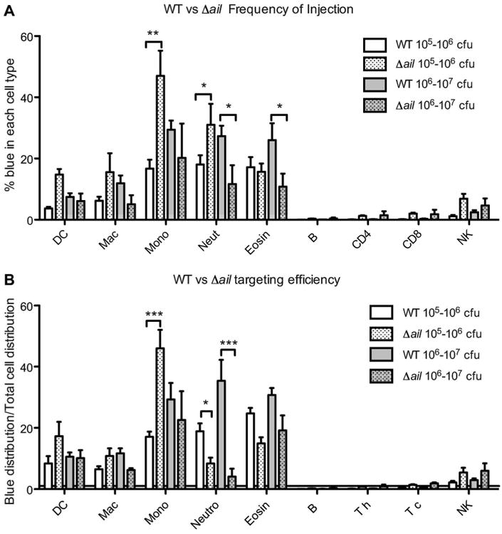
Data shown in Figures 2 and 3 were graphed together to compare injection capabilities for each bacterial strain when bacterial loads were similar. Shown here are data from the 105-106 and 106-107 cfu categories. A) Frequency of injection (% of each cell type that is blue). B) Targeting efficiency (fold difference between the blue cell distribution vs the total distribution for each cell type). Data were analyzed by 2-way ANOVA with Bonferroni multiple comparisons test to determine significant differences. (* P<0.05, ** P<0.01, *** P<0.001) DC= dendritic cells; Mac= macrophages; Mono= monocytes; Neut= neutrophils; Eosin= eosinophils; CD4= T helper cells; CD8= T cytotoxic cells; NK= NK cells.
Ex vivo analysis of target cell selection during splenocyte infection
The above data clearly indicate that Ail contributes to efficient Yop delivery into immune cells in vivo; however, interpretation of the role of Ail is complicated by the host immune response during infection. Therefore, to specifically address the contribution of Ail's adhesin activity to target cell selection, we used an ex vivo assay in which splenocyte suspensions from naive mice are used as a source of immune cells for in vitro infections (Houppert et al., 2013). In this way, wild type and mutant strains will have equal opportunity to interact with the same cell types. We identified the cell types using the same strategy described in Figure S1. Again, we determined the frequency of injection, distribution of injected cells, and targeting efficiency for each set of infections, as described in Fig. S4. When splenocytes were infected with WT Y. pestis, the preference for myeloid cell types is readily apparent (Fig. S4A). More than 90% of the neutrophils had been injected, as well as more than 70% of dendritic cells and monocytes. Our data show that relatively few macrophages were injected (∼17%), but that may be due to the fact that we excluded cells that are positive for the MHC Class II marker in our analysis to facilitate identification of other cell types. It is possible that macrophages may become activated during the short ∼1.5 hr. infection, which may lead to MHC Class II gene expression and a corresponding under-representation of macrophages in our assay. Interestingly, 46% of eosinophils were positive for YopM-Bla injection. The significance of possible eosinophil targeting during infection has not been addressed previously and may warrant future investigation. We also found that the majority of B cells were also injected, but very few T cells or NK cells were injected. The observation that B cells can be injected agrees with previous data (Marketon et al., 2005) showing that B cells can be targeted by Y. pestis in vitro, but this is in contrast to in vivo data (Fig. 2A and (Marketon et al., 2005)). Nevertheless, similar patterns of target cell preference were observed during ex vivo infections using Y. enterocolitica and Y. pseudotuberculosis (Koberle et al., 2009, Durand et al., 2010). Therefore, the Yersinia spp. appear to share the ability to target myeloid cells and B cells ex vivo.
Although nearly all neutrophils (and other myeloid cells) were injected during Y. pestis infection, this represents only a small percentage of the total population of injected cells (Fig. S4B). In contrast, because B cells are a major portion of the spleen, they correspondingly represent a major portion of the total population of injected cells. When targeting efficiency is evaluated for WT infection (Fig. S4E), it is clear that myeloid cells and B cells are preferred targets, while T cells and NK cells appear to be excluded.
Mouse serum influences Yop injection and reveals a role for Ail
We next wanted to address the potential role of bacterial adhesins, particularly Ail, in target cell selection. Toward that end, ex vivo assays were performed with wild type, Δail, Δpla/psaA, or the Δail/pla/psaA triple mutant. To our surprise, initial experiments revealed no profound differences between wild type and any mutant (Fig. 5 A-B and Fig. S5). However, we wondered if the conditions in the ex vivo assays might preclude observation of a targeting defect by the adhesin mutants. Since splenocytes are homogenized and rinsed prior to infection, there could be a soluble serum factor that is required for adhesin-mediated specificity and is missing or in low abundance. We therefore asked whether adding mouse serum to our ex vivo infection assay would influence targeting and would thereby reveal mutant deficiencies. Figure 5C shows the results of infecting murine splenocytes in the presence of either 10% normal mouse serum (NMS) or heat-inactivated mouse serum (IMS). Contrary to our expectations, addition of NMS significantly reduced overall injection. The suppressive effect of NMS appears to rely on heat-labile factors because adding IMS to the infections had no effect. Moreover, in the presence of normal mouse serum, the Δail mutant displayed reduced levels of injection compared to wild type (Fig. 5D). To our surprise, the Δpla/psaA mutant gave rise to significantly more injected cells compared to the wild type infection. That phenotype seems to be dependent on Ail because with the triple mutant (Δail/pla/psaA), the frequency of injection was identical to that of the single Δail mutant. Together these experiments indicate that Ail appears to be necessary for efficient Yop injection into splenocytes in the presence of serum, and suggest the possibility that Pla opposes Ail's adhesin activity.
Figure 5. A role for adhesins in the presence of mouse serum.
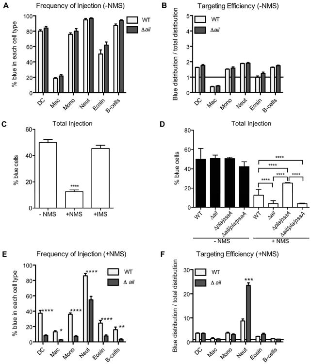
Splenocytes from naive mice were infected at an MOI of 1 with the indicated Y. pestis strains expressing YopM-Bla or Gst-Bla (not shown). Infections were performed in RPMI containing FBS and cytochalasin D for 1.5 hours. Where indicated, the medium was supplemented with 10% normal mouse serum (NMS) or heat-inactivated mouse serum (IMS). Cells were stained with CCF2-AM and then analyzed by flow cytometry. A) The percentage of blue cells for individual cell types is shown for infections lacking NMS. B) The targeting efficiency for each cell type is shown for infections lacking NMS. Horizontal line at Y=1 indicates no cell preference. C) Infections with WT Y. pestis were performed in the presence or absence of NMS or IMS. The percentage of blue cells in the total splenocyte population for each infection is shown. Data were analyzed using 1-way ANOVA with Dunnett multiple comparisons test (control= no serum). D) The percentage of blue cells in the total splenocyte population for each infection is shown. Data were analyzed using 1-way ANOVA with Bonferroni multiple comparisons test (**** P<0.0001). E) The percentage of blue cells for individual cell types is shown for infections containing NMS. F) The targeting efficiency for each cell type is shown for infections containing NMS. Horizontal line at Y=1 indicates no cell preference. Data in panels A-B and E-F were analyzed by 2-way ANOVA with Bonferroni multiple comparisons test to determine significant differences for each cell type infected with WT compared to the mutant. (* P<0.05, ** P<0.01, **** P<0.0001) DC= dendritic cells; Mac= macrophages; Mono= monocytes; Neut= neutrophils; Eosin= eosinophils.
Mouse serum enhances neutrophil targeting
Since addition of mouse serum reduced the total number of injected splenocytes, we investigated whether the phenomenon correlated with any changes to target cell selection. When splenocytes in the presence of 10% NMS were infected with WT Y. pestis, we observed significantly fewer (P<0.0001) injected B cells, dendritic cells, monocytes, and eosinophils (Fig. 5E, white bars) compared to infections performed in the absence of NMS (Fig. 5A, white bars). The frequency of injection was reduced ∼50% for dendritic cells, monocytes, and eosinophils, whereas for B cells the frequency of injection was reduced by ∼75%. In contrast, neutrophil injection was unaffected as nearly all neutrophils were targeted for Yop injection in both conditions. This maintenance of neutrophil targeting in the midst of reduced targeting of other cell types correlates with ∼10-fold higher targeting efficiency for neutrophils in the presence of NMS (Fig. 5F, white bars) compared to conditions lacking NMS (Fig. 5B, white bars). Thus, it appears that one or more heat-labile serum factors facilitate neutrophil targeting.
Ail is required for efficient injection of splenocytes
To investigate the nature of the injection defect for the Δail mutant seen in Figure 5D, we analyzed the immune cell populations to determine whether there was a change in target cell selection. In the presence of serum, there were significant decreases in the frequency of injection by the Δail mutant for B cells and all myeloid populations except macrophages, though even macrophages followed the same trend (Fig. 5E, black bars). Injection of T cells and NK cells was not significantly affected, since they were still not targeted to any appreciable extent (data not shown). Although there were fewer neutrophils injected by the Δail mutant, the magnitude of this decrease was less than that of other cell types. Therefore neutrophils represent a larger fraction of the total injected population in Δail mutant infection, which correlates to a dramatic increase in the targeting efficiency of neutrophils for the Δail mutant compared to wild type (Fig. 5F). Importantly, none of the targeting defects were observed for the Δail mutant in the absence of NMS (Fig. 5B). Therefore, we conclude that the adhesin-dependent phenotype is also serum-dependent.
Pla counteracts Ail activity
The data in Figure 5D indicate that the Δpla/psaA mutant injected YopM-Bla into more splenocytes than did wild type Y. pestis. This is unexpected because Pla contributes to efficient injection of various tissue culture cell lines (Felek et al., 2010). To determine whether the target cell specificity is altered by the lack of Pla, we analyzed the pattern of translocation into immune cell populations during infection with the Δpla/psaA mutant. Figure S6A shows that most of the increase stems from injection of ∼50% more B cells by the mutant (black bars) compared to wild type (white bars). Injection of dendritic cells and monocytes was also significantly increased. Notably, there was still no significant increase in targeting of T cells or NK cells. Moreover, neutrophil targeting remained unaffected. Because the amount of injected neutrophils did not change, while other populations experienced higher frequencies of injection by the Δpla/psaA mutant, this means that neutrophils represent less of the total population of injected cells. Correspondingly, there was a significant reduction in the neutrophil targeting efficiency for the Δpla/psaA mutant compared to WT (Fig. S6B). However, these phenotypes were completely reversed when ail was deleted, as the triple Δail/pla/psaA mutant phenotype resembled that of the single Δail mutant (Fig. S6 C-D). These data suggest that when only Ail is present (as in the Δpla/psaA mutant), neutrophil targeting still occurs but there appears to be more interaction with the other preferred cell types (other myeloid cells and B cells), thereby detracting from neutrophil interactions. However, in the absence of Ail, the preference for B cells and myeloid cells over T cells and NK cells is maintained. Moreover, this target cell selection is maintained during infection with a triple mutant lacking all three major adhesins, Ail, Pla, and PsaA. These observations raise the possibility that both adhesin-dependent and -independent processes mediate target cell selection.
Bacterial adhesion to splenocytes
Throughout our experiments, we have consistently seen that T cells and NK cells are not selected as targets for Yop injection, regardless of the experimental conditions. Moreover, we have seen fluctuations in the extent to which the bacteria target the preferred cell types. Therefore, we investigated whether these observations correlate with bacterial binding to host cells. To that end, we introduced a plasmid expressing a GFP variant (GFPuv) into Y. pestis wild type and Δail mutant strains and then performed standard ex vivo infections of splenocytes in the presence or absence of serum. Using flow cytometry, we analyzed bacterial adhesion by measuring the percentage of each cell type that was positive for GFP. Importantly, cytochalasin D was included in all ex vivo assays to prevent phagocytosis of attached bacteria. As shown in Figure S7A, there was ≥ 50% bacterial adherence under all conditions. In the absence of serum, the Δail mutant was associated with more cells than was wild type Y. pestis (61% compared to 53%). Addition of serum led to slightly reduced adhesion for both strains, and this effect was abrogated by heat inactivating the serum. To determine whether target cell selection for Yop injection correlates with bacterial adhesion, we identified which cells were GFP positive (Fig. S7 B-D). Surprisingly we found that most cell types had equivalent levels of GFP+ cells (∼50% of the population), though there was a higher frequency of GFP+ cells for neutrophils, monocytes, and eosinophils, and this was true for both wild type and Δail mutant infections (Fig. S7B). The effect of adding NMS to infections (Fig. S7A) correlated with reduced adhesion to B cells for both WT and the ail mutant (Fig. S7 C-D). Taking into account all the data in Fig. S7, the fact that nearly 100% of neutrophils were GFP+ agrees well with the fact that nearly all neutrophils become targets for Yop injection. However, the correlation does not extend to other cell types. For example, T cells and NK cells show equivalent bacterial adhesion compared to B cells, yet they do not appear to become T3SS targets. Thus, a lack of Yop injection by Y. pestis is not simply due to a lack of bacterial adhesion.
Contribution of host factors to target cell selection
Because there is an adhesin-independent phenomenon that contributes to neutrophil targeting (Fig. 5 E-F and Fig. S6 C-D), we investigated serum factors as potential specificity determinants. Since neutrophil targeting is compromised by heat inactivating serum, we assumed that some component of complement might be involved. We therefore attempted to rule out a role for antibody-mediated opsonization in target cell selection using two approaches. First, we used Protein A/G beads to deplete antibodies from serum, and then analyzed whether target cell selection was affected. We did not see any differences in targeting during infection with either the wild type or Δail mutant (data not shown). Second, we used Fc Block (anti-CD16/CD32) to block antibody Fc receptors on splenocytes prior to infection. Once again, this treatment had no affect on targeting during ex vivo infections (data not shown).
We then considered whether complement and/or other serum components were responsible for neutrophil targeting, keeping in mind that multiple factors may be at play. For example, LPS binding protein (LBP) is a soluble serum factor known to opsonize bacteria and mediate interactions with CD14 (Wright et al., 1989, Grunwald et al., 1996). Moreover, LcrV, a secreted T3SS component, has reported interactions with CD14 and TLR2 (Sing et al., 2002, Abramov et al., 2007), and recently Ail was shown to bind C4b-binding protein (C4BP), Factor H, and the complement proteins C4, C4b, C3b (Ho et al., 2012a, Ho et al., 2012b, Ho et al., 2014). In addition, the alternative pathway for complement activation could yield C3b opsonization. Concomitant Ail binding to Factor H would accelerate Factor I-mediated cleavage of C3b to iC3b, which is a primary ligand for CR3 and to a lesser extent CR4 (Sengelov, 1995, Blom et al., 2003, Ho et al., 2012b, Ho et al., 2014). Therefore, it is possible that target cell selection arises from a combination of interactions between the bacteria and host cells.
Since neutrophils are primary targets during Y. pestis infection, we attempted to narrow down the candidates by first considering which receptors might be enriched on neutrophils. To that end, we surveyed the expression of CD11b (for CR3), CD11c (for CR4), CD14, and TLR2 on splenocytes incubated in medium containing NMS (Fig. 6). As expected, most of the myeloid cells expressed CD11b, in contrast to B cells (frequency indicated by white bars); however the expression level (mean fluorescence intensity, MFI, indicated by black bars) was highest in neutrophils (Fig. 6A). On the other hand, expression of CD11c appeared to be most frequent and strongest in dendritic cells (Fig. 6B). Interestingly we found that CD14 was highly enriched in neutrophils, as ∼60% of the population was CD14+ and also expressed high levels per cell (Fig. 6C). TLR2 was found on most of the neutrophils, and to a lesser extent on other myeloid cells and B cells, however expression level per cell was highest in neutrophils (Fig. 6D).
Figure 6. Potential receptors are enriched on neutrophils.
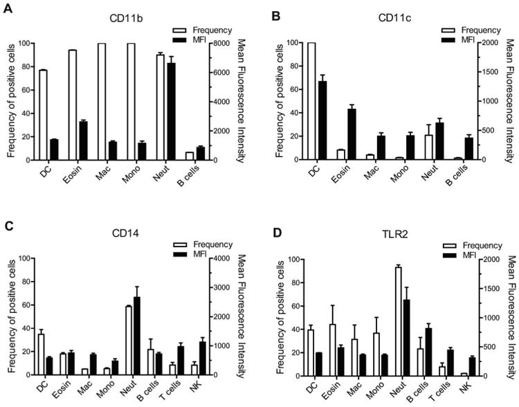
Splenocytes were suspended in RPMI containing FBS and NMS and stained with antibodies to identify cell types and fluorescently-labeled A) anti-CD11b, B) anti-CD11c, C) anti-CD14, or D) anti-TLR2 was added to quantify expression of receptors on each cell type. The percentage of each cell type positive for the indicated marker is shown on the left y-axis. The Mean Fluorescence Intensity (MFI) for those cells positive for each marker is shown on the right y-axis. DC= dendritic cells; Mac= macrophages; Mono= monocytes; Neut= neutrophils; Eosin= eosinophils; CD4= T helper cells; CD8= T cytotoxic cells; NK= NK cells.
Neutrophil targeting is mediated by host factors
To investigate the possibility of multiple or complex interactions between Y. pestis and target cells, we attempted to block target cell selection by pre-treating splenocytes with several antibodies, either alone or in combination, prior to infection. We then performed ex vivo infections in the presence or absence of NMS using wild type Y. pestis and the Δail/pla/psaA mutant. If the pre-treatments inhibit serum-dependent binding, then targeting should be adhesin dependent and should be observed during infection with wild type but not the triple adhesin mutant. We chose to use anti-CD11b and anti-CD11c to block CR3 and CR4, respectively. We chose anti-CD14 to block multiple possible routes of interaction (via LBP, TLR2, TLR4), since CD14 is a common facilitator of those pathways. As expected, pre-treatment with the antibody cocktail in the absence of NMS had no effect on target cell selection during infection with either wild type or the triple adhesin mutant (Fig. S8). In contrast, the antibody cocktail led to a dramatic reduction of neutrophil targeting in the presence of NMS (Fig. S8). We then compared the ability of either wild type Y. pestis or the triple adhesin mutant to target neutrophils in the presence of both NMS and the antibody cocktail (Fig. 7). We found that the antibodies significantly reduced the percentage of both monocytes and neutrophils that were injected by wild type (Fig. 7A), and this led to a corresponding drop in targeting efficiency for both cell types (Fig. 7B). For the adhesin mutant, antibody pre-treatment nearly abolished injection of splenocytes (Fig. 7 A-B). Notably, non-preferred cell types did not become substitute targets for the T3SS when the normal targets (neutrophils) were neutralized via the antibody cocktail. We interpret these results to mean that the adhesin-independent targeting of neutrophils is specific and is mediated by at least one of the receptors blocked by the antibody cocktail.
Figure 7. Host receptors mediate target cell specificity.
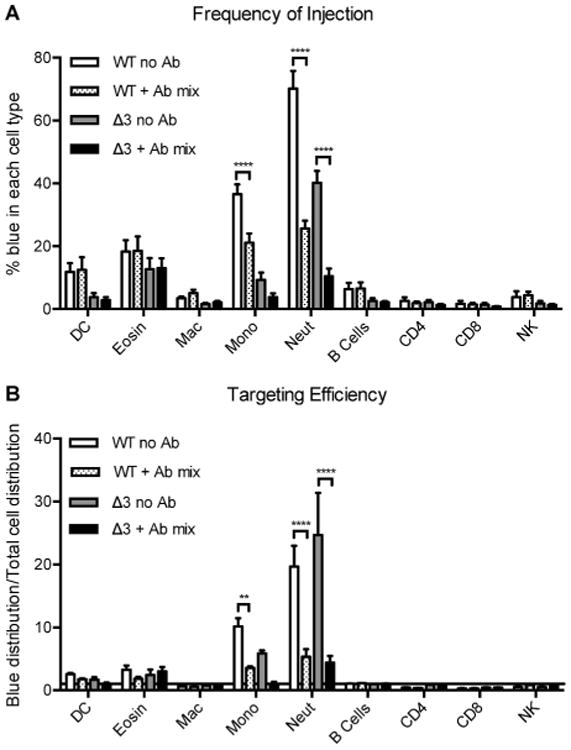
Splenocytes were left untreated (no Ab) or incubated with antibodies against CD14, CD11b, and CD11c (+ Ab mix) prior to infecting with either WT or the Δail/pla/psaA mutant (Δ3) expressing YopM-Bla or Gst-Bla (not shown). Infections were performed in RPMI containing FBS, cytochalasin D, and NMS. Cells were stained with CCF2-AM and antibodies, and then analyzed by flow cytometry. A) The percentage of blue cells for individual cell types is shown. B) The targeting efficiency for each cell type is shown. Horizontal line at Y=1 indicates no cell preference. 2-way ANOVA with Bonferroni multiple comparisons test to determine significant differences for each cell type. (** P<0.01, **** P<0.0001) DC= dendritic cells; Mac= macrophages; Mono= monocytes; Neut= neutrophils; Eosin= eosinophils; CD4= T helper cells; CD8= T cytotoxic cells; NK= NK cells.
We then assessed the ability of individual antibodies to block neutrophil targeting. We found that anti-CD14 treatment yielded only slight effects on targeting by either wild type or the adhesin mutant (Fig. 8). In striking contrast, anti-CD11b severely reduced monocyte and neutrophil targeting (Fig. 8), similar to the blockade observed with the antibody cocktail (Fig. 7). Thus it appears that CR3 is a major mediator of target cell specificity during plague infection.
Figure 8. CR3 is the primary mediator of neutrophil specificity.
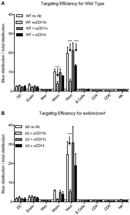
Splenocytes were left untreated (no Ab) or incubated with individual antibodies against CD14, CD11b, or CD11c prior to infecting with either WT or the Δail/pla/psaA mutant (“Δ3”) expressing YopM-Bla or Gst-Bla (not shown). Infections were performed in RPMI containing FBS, cytochalasin D, and NMS. Cells were stained with CCF2-AM and antibodies, and then analyzed by flow cytometry. A) The targeting efficiency for each cell type infected with WT is shown. B) The targeting efficiency for each cell type infected with the Δail/pla/psaA mutant is shown. Horizontal lines at Y=1 indicate no cell preference. Data were analyzed using 2-way ANOVA with Bonferroni multiple comparisons test (* P<0.05, ** P<0.01, **** P<0.0001).
Discussion
The tropism of Yersinia spp. for lymphoid tissue is well established, and recent work describing the targeting of innate immune cell populations for Yop injection during infection provides a satisfying correlation to tropism (Marketon et al., 2005, Koberle et al., 2009, Durand et al., 2010). The professional phagocytes of the myeloid lineage are shared targets across the Yersinia spp., and this has been observed in lungs as well as spleens (Pechous et al., 2013, Paczosa et al., 2014). Of the myeloid cells, it seems that neutrophils are a particularly important target for the T3SS, since a failure to inactivate these potent mediators of inflammation compromises virulence, and correspondingly depletion of neutrophils can rescue virulence defects or alter the course of disease (Echeverry et al., 2007, Ye et al., 2009, Laws et al., 2010, Ye et al., 2011, Thorslund et al., 2013). Moreover, targeting of neutrophils is steadfast, and even when neutrophils are removed target cell specificity is not altered dramatically (Durand et al., 2010, Pechous et al., 2013, Paczosa et al., 2014). Given the conserved preference of neutrophil targeting across the Yersinia spp., how then is specificity mediated? Since receptor-ligand interactions had been demonstrated for Yersinia interactions with cultured cell lines (Kapperud et al., 1987, Isberg et al., 1990, Kienle et al., 1992, Young et al., 1992, Payne et al., 1998, Dersch et al., 1999, Isberg et al., 2000, Lahteenmaki et al., 2001, Wiedemann et al., 2001, Eitel et al., 2002, Lobo, 2006, Tsang et al., 2010, Yamashita et al., 2011), initial forays were focused logically on bacterial adhesins. However the repertoire and functionality of adhesins is not conserved between Yersinia, and it remained to be determined if the same receptor-ligand interactions observed in cell culture infections contribute to specific targeting of primary immune cells in vivo. Indeed, the recent work on Y. pseudotuberculosis has begun to unravel the mystery of target cell selection by showing that bacterial adhesins only partially contribute to the process, while also revealing the additional complexities that host factors bring to the picture (Durand et al., 2010, Maldonado-Arocho et al., 2013, Paczosa et al., 2014).
Using an attenuated strain in a mouse model of septicemic plague, it was demonstrated that Y. pestis targets several phagocytic, myeloid cell types (neutrophils, macrophages, and dendritic cells) in the spleens of infected mice (Marketon et al., 2005). This general target cell preference is maintained in lung tissue using a fully-virulent strain in a pneumonic plague model (Pechous et al., 2013). Interestingly, the dominant target population in lungs during the first few hours of infection was alveolar macrophages, but by 12 hours post-infection the targeting preference clearly transitioned over to neutrophils. A similar trend was observed during Y. pseudotuberculosis infection of lungs, and Ail and YadA were shown to mediate neutrophil targeting in that model (Paczosa et al., 2014). Furthermore, Y. pseudotuberculosis Ail, YadA, and invasin contribute to target cell selection in spleens (Maldonado-Arocho et al., 2013). Though the factors mediating in vivo target cell specificity had not been characterized for Y. pestis yet, prior work demonstrated that Ail was the major adhesin required for T3SS-mediated delivery of Yop effectors into culture cell lines (Felek et al., 2009, Felek et al., 2010). Moreover, the Δail mutant was extremely attenuated in the septicemic plague model (Felek et al., 2009). We therefore set out to determine if Ail (or the other major adhesins) played a role in immune cell targeting.
We were surprised to find that the Δail mutant was capable of injecting Yops into host cells in vivo (Fig. 1). In fact, when spleens containing similar bacterial loads were compared, the total number of injected splenocytes did not differ between WT and the Δail mutant. However, further investigation revealed that the ability of the mutant to specifically target neutrophils for Yop injection in vivo was dramatically reduced (Fig. 4). Prior work already showed that Ail is required for resistance to complement-mediated killing and that Ail somehow inhibits a pro-inflammatory response to infection (Bartra et al., 2008, Felek et al., 2009, Hinnebusch et al., 2011, Ho et al., 2014). Since mouse serum is not bactericidal, and since an ail mutant is more attenuated in rats than in mice (Bartra et al., 2008, Kolodziejek et al., 2010, Hinnebusch et al., 2011), Ail likely has additional functions beyond preventing bacterial lysis due to membrane attack complex formation. Our data add to those observations regarding Ail's role as a virulence factor by demonstrating its requirement for proper target cell selection in vivo.
To explore the nature of the targeting defect for the Δail mutant, we developed an ex vivo assay to measure target cell selection. As was shown for the enteric Yersinia (Koberle et al., 2009, Durand et al., 2010), Y. pestis preference for innate immune cells was observed in the ex vivo assay (Fig. 5). Interestingly, B cells were also targets of the T3SS; however this observation was only true in the absence of mouse serum (Fig. 5 A-B). When considered with the in vivo data (Marketon et al., 2005) (Fig. 2), the results indicate that B cells are not preferred targets for Yop injection in spleens of Y. pestis infected mice. That B and T cells are not preferred is not due to a lack of bacterial interaction. In fact bacteria interacted with these lymphocytes as well as myeloid populations, such as dendritic cells and macrophages (Fig. S7). It could be that some adhesion events do not bring the bacteria in direct contact with the cell surface, or it is possible that either the nature of the interaction or the quantity of interactions along an interface influence whether adhesion leads to injection. Regardless, the observations that myeloid cells, particularly neutrophils, are selected as preferred targets for Yop injection appears to be a general phenomenon, as it applies to targeting in lungs as well as spleen, and it is true for both Y. pestis and Y. pseudotuberculosis (Pechous et al., 2013, Paczosa et al., 2014).
During the course of our investigation into the role of bacterial adhesins during target cell selection, we were surprised to find no dramatic phenotype associated with any of the adhesin mutants during splenocyte infection in the absence of NMS (Fig. 5 A-B and Fig. S5). This is in contrast to observations from infections of a variety of cultured cell lines (Felek et al., 2009, Felek et al., 2010). However, a role for Ail as a major adhesin was revealed once NMS was added to the ex vivo assays (Fig. 5 E-F and Fig. S5 C-D). Given that neutrophils were still preferentially targeted amidst overall reduced injection levels, and that the Δail and Δail/pla/psaA mutants behaved similarly, we conclude that target cell selection in the spleen is a combination of adhesin-dependent and -independent processes. The idea that adhesins serve a general role as facilitators of efficient host interaction is in agreement with Y. pseudotuberculosis observations, whereby adhesin mutants still demonstrated cell preference during splenocyte infection (Durand et al., 2010). In contrast, the preference for neutrophils in lung tissue was dependent upon the presence of either Ail or YadA during Y. pseudotuberculosis pneumonia (Paczosa et al., 2014). The role of Y. pestis adhesins in neutrophil targeting during pneumonic plague has not been addressed, but these data suggest that the use of adhesins during Yersinia infections may differ depending on the locale.
The observation that the Y. pestis Δail phenotype was only observed upon addition of NMS to splenocyte infections suggested a serum factor mediates Ail binding to host cells. This dependence on NMS for adhesin activity in Y. pestis is a notable point of difference compared to Y. pseudotuberculosis, whose adhesins do mediate host interactions even in serum-free medium (Maldonado-Arocho et al., 2013). Nonetheless, serum factors such as complement clearly impact the adhesin-dependent colonization of spleens during Y. pseudotuberculosis infection (Paczosa et al., 2014). Thus, while targeting in the lung may rely primarily on adhesins, targeting in the spleen may arise from a combination of adhesin and serum-dependent interactions.
Understanding the role of any potential serum factor in Y. pestis adhesin-dependent events is complicated by the observations that one or more heat-labile components of NMS reduced overall injection levels while also increasing the targeting efficiency for neutrophils (Fig. 5 D&F), suggesting that there are likely multiple host factors acting in different ways. Since our assays already contained heat-inactivated FBS in the cell culture medium, existing heat-stabile components could potentially mediate the general targeting of myeloid cells and B cells in an adhesin-independent manner. Addition of NMS to the mixture would then contribute heat-labile factors to enhance neutrophil targeting.
One factor that is at least partially retained in heat-inactivated FBS is LBP (Meszaros et al., 1995). LBP binds to either free LPS or to bacterial surfaces and catalyzes an interaction with CD14 and TLR4 (Wright et al., 1989, Wright et al., 1990, Kitchens, 2000, da Silva Correia et al., 2001). Notably, TLR4 is highly expressed by mouse myeloid cells and B cells, but not T cells (Applequist et al., 2002), supporting the idea that TLR4 may participate in an adhesin-independent interaction with Y. pestis and may lead to the broad targeting of B cells and myeloid cells. CD14 is present as both a membrane receptor and as a soluble serum component, though it is heat-labile (Landmann et al., 1996a, Landmann et al., 1996b). Considering our data that show membrane CD14 is not present on the majority of murine splenic B cells (Fig. 6), along with the fact that plasma from normal healthy mice does not contain high levels of soluble CD14 (Fearns et al., 1995), we would predict that the Y. pestis-TLR4 interaction on B cells is direct, rather than facilitated by a CD14 intermediate. That type of interaction potentially could be outcompeted by myeloid cells, such as monocytes and neutrophils that have better expression of CD14 (Fig. 6). Furthermore, addition of NMS would provide an abundance of fresh LBP that could coat the bacteria and potentially obscure direct LPS-TLR4 binding, thereby suppressing Yop injection into B cells. It is interesting to note that TLR4 knockout mice infected with Y. pestis show a faster time to death (Hajjar et al., 2012). That observation, as well as our data, suggest the possibility that in the absence of TLR4, there is less B cell injection and instead the targeting of neutrophils is enhanced.
The serum factor(s) that mediate Y. pestis adhesin-dependent interactions and neutrophil targeting are heat-labile, and therefore we considered complement proteins as candidates. The complement cascade can be initiated via three pathways: classical, MB-lectin, and alternative, all of which culminate in the deposition of the opsonin C3b on the pathogen surface followed by formation of membrane-attack complexes as well as binding to complement receptors (Walport, 2001). However, many pathogens circumvent this process by recruiting regulators or inactivators of complement proteins to the bacterial surface (Lambris et al., 2008). Importantly, Ail from Y. pseudotuberculosis and Y. pestis are nearly identical (only differing by up to 2 amino acids depending on the strain (Ho et al., 2012a, Kolodziejek et al., 2012, Tsang et al., 2013)) and both bind C4BP, which recruits Factor I to inactivate C4 to C4b (Ho et al., 2012a, Ho et al., 2014). Furthermore Y. pseudotuberculosis Ail (and possibly Y. pestis Ail) binds to Factor H, which is a cofactor for Factor I and facilitates degradation of C3b to iC3b (Ho et al., 2012b, Ho et al., 2014). Additionally, Y. pestis Ail binds to C4, C4b, and C3b (Ho et al., 2014). Therefore, it is possible that in the presence of NMS, Y. pestis becomes coated with ligands for complement receptors that contribute to target cell preference. When Ail is present, deposition of iC3b may predominate and direct the targeting of neutrophils via CR3.
In vivo, bacterial infection leads to high amounts of circulating opsonins (LBP, CD14, and complement proteins), which we hypothesize are likely contributors to efficient neutrophil targeting during Y. pestis infection. In the absence of Ail, interactions with cell types other than neutrophils dominate (Fig. 3 and 4). This may be the result of the inability to bind iC3b directly (via Ail) or indirectly (via Ail-mediated recruitment of Factor I and Factor H) (Ho et al., 2014). However, LBP and CD14 interactions would be unaffected, which might allow interactions with TLRs to prevail. In contrast to in vivo experiments (Fig. 4B), the ex vivo experiments (Fig. 5F) reveal a different trend for the ail mutant. Whereas the mutant was deficient for neutrophil targeting compared to WT in vivo, it was better than WT at targeting neutrophils ex vivo. There are a number of possible explanations for this difference. First, the activation state of cells may differ and thereby affect expression of TLRs and CRs and subsequently the interactions with Y. pestis. Secondly, we expect that the potential opsonins would effectively be in unlimited supply during in vivo infections, since they are constantly being produced and production would likely be increased during infection. In contrast, serum used in ex vivo infections comes from naive mice and would not contain large amounts of C3 or CD14 (Triglia et al., 1980, Fearns et al., 1995). Therefore, Ail may effectively capture the existing components to present iC3b as a CR3 ligand, but those components would be in limiting supply, and the Ail-mediated sequestration of iC3b and its regulators may result in further processing to yield C3dg or other inactive products. Additionally, the opposing Pla activity (Fig. 5D and S6) may arise from Pla-mediated degradation of these limited complement components into inactive fragments (Sodeinde et al., 1992). In the absence of Pla, it may be that Ail levels and/or activity may be higher, leading to increased Factor I recruitment and subsequent degradation of iC3b into C3dg, which is recognized by CR2 on B cells (Weis et al., 1984), or into fragments that are not recognized by complement receptors. In contrast, when Ail is absent, spontaneous generation of C3b via the alternative pathway from the low levels of C3 in naive serum will provide limited deposition of C3b and this may be further degraded into small amounts of iC3b by Pla. However, while Pla has been reported to degrade C3 (Sodeinde et al., 1992), it is not known whether Pla interacts with fluid-phase C3 products vs. those that have been deposited on the bacterial surface. It also is not known whether Pla degradation of C3 leads to products that can interact with CRs.
CR3 is the main receptor for iC3b (Ross et al., 1982, Wright et al., 1986). Furthermore, there is evidence that CR3 binding is enhanced by concerted interactions with CD14/TLR2 (Troelstra et al., 1999, Sendide et al., 2005). Since CR3 is highly expressed by neutrophils, along with CD14 and TLR2 (Fig. S6) (Sengelov, 1995), these proteins have the potential to be clustered in micro-domains on neutrophils. Interestingly, CD14 knockout mice show partial protection from plague infection, in terms of bacterial burden as well as percent survival (Depaolo et al., 2008), and this may reflect a supportive role for CD14 in enhancing neutrophil targeting in vivo. Future investigations will need to examine directly the roles of putative opsonins (C3/iC3b, LBP, soluble CD14) and their receptors during plague infection in order to determine the exact nature of the receptor-ligand interactions and their contributions toward target cell selection.
Materials and Methods
Bacterial strains and media
Yersinia pestis KIM D27, an attenuated variant of the Y. pestis mediaevalis strain KIM lacking the 102-kb pgm locus (Brubaker, 1969), was used as the parent strain. Y. pestis CHI30 (yscU∷Mu) was generated by random mutagenesis of KIM D27 using MuAphP1 (Marketon et al., 2005, Houppert et al., 2012). The streptomycin-resistant adhesin mutant Δail (KIM5-3001 Δail) has been described (Felek et al., 2010) and was used for mouse infections. Unmarked adhesin mutants (Δail, ΔplaΔpsaA and ΔailΔplaΔpsaA) in Y. pestis strain KIM D27 were constructed as described previously for KIM5-3001 using lambda-RED recombineering (Felek et al., 2010). The Bla reporter plasmids pMM83 (YopM-Bla) and pMM91 (Gst-Bla) have been described and were introduced into all Y. pestis strains to measure T3SS activity (Marketon et al., 2005). Y. pestis strains were propagated on Heart Infusion Agar (HIA) plates at 26°C for two days. Overnight cultures were grown in Heart Infusion Broth (HIB) at 26°C. Antibiotics were added as appropriate to a final concentration of 20 μg/ml chloramphenicol and 50 μg/ml ampicillin.
Ethics Statement
This study was carried out in accordance with the recommendations in the Guide for the Care and Use of Laboratory Animals of the National Institutes of Health. The work was approved by the Institutional Animal Care and Use Committee at Indiana University. Our approved protocol number is 12-051. All efforts were made to minimize suffering of the mice.
In vivo Infections
Y. pestis overnight cultures were washed and diluted to appropriate densities in PBS. Female 6-7 week old C57BL/6 mice (Harlan Laboratories, Indianapolis, Indiana) were anesthetized with isoflurane and retro-orbitally injected with 104 CFU of Y. pestis KIM5 carrying pMM83 (YopM-Bla) or pMM91 (GST-Bla), 106 CFU of Y. pestis yscU- (pMM83), or 106 CFU of Δail carrying pMM83 or pMM91. These doses were chosen based on preliminary experiments that monitored bacterial colonization resulting from a range of doses. We chose doses that were sufficient to ensure 100% of mice were colonized and would yield similar ranges of bacterial loads for KIM5 and the Δail mutant without causing mortality within the three-day time frame. At daily intervals, mice were euthanized via CO2 inhalation followed by cervical dislocation. Mice were perfused with 30mL of sterile 1× PBS, and spleens were harvested and homogenized. A portion of the homogenate was serially diluted into PBS and plated onto HIA to enumerate the bacterial load. To assess injection of the Bla reporter, the remainder of the spleen suspension was processed for flow cytometry as follows. Cells were strained through a 70μm nylon filter. Red blood cells were lysed with Red Blood Cell Lysis Buffer (0.13 mM NH4Cl, 0.017 mM Tris, 500 ml H2O pH 7.2) for 5 minutes at room temperature. Splenocytes were pelleted at 1500 × g for 3 minutes and resuspended in RPMI-1640 (Cellgro 10-040-CV) + 10% FBS. Cells were then stained with CCF2-AM (Invitrogen) at 0.2× for 1 hour at room temperature. Cells were pelleted at 1500 × g for 3 minutes and the supernatant was removed. Cells were then incubated with Fc Block (eBiosciences, Clone: 93) for 25 minutes on ice and then pelleted at 1500 × g for 3 minutes. Splenocytes were then stained for 35 minutes on ice using the lymphoid and myeloid antibody panels described below. Splenocytes were then centrifuged and resuspended in HBSS Flow (1× HBSS, 0.5 mM EDTA, 25 mM HEPES, 2% BSA, pH 7.4) with propidium iodide (PI) Solution (BD Pharmingen 556463) at 0.01 μg/ml. Flow cytometry was performed on a BD AriaII or a BD LSRII flow cytometer in the Indiana University Flow Cytometry Core Facility (IU FCCF), and data was analyzed using FlowJo (Tree Star, Inc) and Prism 5 (GraphPad Software, Inc). The data are a compilation of three independent trials for wild type infections (n=18), two independent trials for Δail infections (n=11) and one trial for yscU- infections (n=5). The data are graphed showing mean and standard error with statistical analysis as indicated in figure legends.
Ex vivo infections
Ex vivo injection assays were performed as described by Houppert et al. (Houppert et al., 2013) with minor modifications indicated below. Briefly, spleens from naïve female 6-7 week old C57BL/6 mice were homogenized in 1 ml of PBS. The homogenate was incubated for 5 min at room temperature with Red Blood Cell Lysis Buffer, pelleted by centrifugation at 1500×g for 3 min, and resuspended in RPMI supplemented with 10% heat-inactivated FBS (HyClone) and strained through a 70μm filter. Cells were incubated with 1 μg/ml cytochalasin D (Sigma) for 30 minutes prior to infection to inhibit phagocytosis. Mid-exponential-phase Y. pestis cultures were incubated for 1.5 h at 37°C to induce the T3SS. 1×10⁶ splenocytes were infected with indicated strains of Y. pestis carrying plasmids expressing YopM-Bla (or GST-Bla as a negative control) in 100μl RPMI+FBS with or without 10% mouse serum (MP Biomedicals). Where indicated, heat-inactivated (56°C, 30 min.) mouse serum was used. Bacteria were added to splenocytes at MOI of 1, and the infection mixture was centrifuged at 500 × g for 5 min to facilitate cell contact. Infections were incubated at 37°C, 5%CO2 for 1.5 hours. Infected splenocytes were collected by centrifugation and resuspended in fresh medium supplemented with 50 μg/ml kanamycin to stop bacterial growth and injection. Cells were stained with 1× CCF2-AM for 1 h at room temperature. Cells were pelleted at 1,500 × g for 3 min, and resuspended in HBSS-Flow. Next, the cells were incubated with Fc Block for 20 min on ice. Cells were pelleted at 1,500 × g for 3 min, resuspended in HBSS-Flow. Splenocytes were then stained for 35 minutes on ice using the lymphoid and myeloid antibody panels described below. Following the staining, cells were pelleted and resuspended in HBSS Flow containing PI (0.075 μg/ml). Flow cytometry was performed on a BD LSRII flow cytometer in the IU FCCF, and data was analyzed using FlowJo and GraphPad Prism. The data are graphed showing mean and standard error with statistical analysis as indicated in figure legends.
Identification of immune cell populations
To identify the major splenocyte immune cell populations, the cell suspensions were subjected to staining by the following two antibody cocktails. Lymphoid panel: CD19-PeCy7 (BD Pharmingen; clone 1D3), CD4-APC-eFluor 780 (eBiosciences; clone RM4-5), CD8-PerCP-eFluor 710 (eBiosciences; clone 53-6.7), and NK1.1-APC (BD Pharmingen; clone PK136). Myeloid panel: CD11b-PeCy7 (BD Pharmingen; clone 1D3), CD11c-APC (BD Pharmingen; clone HL3), major histocompatibility complex class II (MHC-II)-PerCP-eFluor 710 (eBiosciences; clone M5/114.15.2), and GR-1-APC-Cy7 (BD Pharmingen; clone RB6-8C5). The flow cytometry gating strategy is described in Figure S2 and is based on previous work (Geddes et al., 2007, Rose et al., 2012). Forward and side scatter were used to separate and discard debris and doublets. Live cells were identified by propidium iodide exclusion and forward scatter. Live events were then gated to identify B cells (CD4− CD8− CD19+ NK1.1−), T-helper cells (CD4+ CD8−), T cytotoxic cells (CD4− CD8+), NK cells (CD4− CD8− CD19− NK1.1+), neutrophils (SSCInt GR1Hi), eosinophils (SSCHi/Int GR1Int), dendritic cells (CD11c+), macrophages (MHC II− CD11c− CD11b+ GR1−), and monocytes (MHC II− CD11c− CD11b+ GR1Int). The percentage of blue (injected) cells was determined for the total population and for each individual cell type cells (termed “frequency of injection” for each cell type). The distribution of all cell types was also determined, as well as the distribution of cell types within the blue (injected) population. To calculate the “targeting efficiency” the relative proportion of a cell type in the blue population was divided by the relative proportion of the cell type in the total splenocyte population (distribution in blue cells/distribution in total cells) as described in Figure S5.
Surface Marker Profiles
Spleens from naïve 6-7 week old female mice were harvested and processed as described above for ex vivo infections. Cells were incubated with 1μg/ml cytochalasin D (Sigma) for 1.5 hr at 37°C, 5% CO2 to be consistent with our infection conditions. The splenocytes were pelleted by centrifugation at 1,500 × g for 3 min, resuspended in HBSS-Flow, and incubated with Fc Block for 20 min on ice. Then, the cells were pelleted at 1,500 × g for 3 min and resuspended in HBSS-Flow containing the relevant antibody cocktail. Two different antibody panels were used to test the prevalence and intensity of CD14 and TLR2 markers on myeloid cells. Myeloid TLR2 panel: TLR2-PeCy7 (eBiosciences; clone: T2.5), CD11b-PeCy5 (eBiosciences; clone: M1/70), CD11c-AlexaFluor 700 (eBiosciences; clone: N418), GR-1-PeCy5.5 (eBiosciences; clone: RB6-8C5), MHC-II-APC-eFluor 780 (eBiosciences; clone: M5/114.15.12). Myeloid CD14 panel: a similar panel was used with CD14-APC (eBiosciences; clone: Sa2-8) and CD11b-PeCy7 (BD Pharmingen; clone: M1/70) replacing TLR2-PeCy7 and CD11b-PeCy5. For investigation of the lymphoid cells, three panels of three antibodies were used: [Lymphoid panel 1] CD19-AlexaFluor 700 (BD Pharmingen; clone: 1D3), TLR2-PeCy7, and CD14-APC; [Lymphoid panel 2] CD3-AlexaFluor 700 (BD Pharmingen; clone: 17A2), TLR2-PeCy7, and CD14-APC; and [Lymphoid panel 3] NK1.1-AF700 (BD Pharmingen; clone: PK136), TLR2-PeCy7, and CD14-APC. Staining was done for 35 min on ice and then, the cells were pelleted at 1500 × g for 3 min. Following staining, the cells were resuspended in HBSS-Flow containing propidium iodide (0.075 μg/ml). The flow cytometry gating strategy outlined in Figure S2 was used with the following modifications for identifying lymphoid cells. Live events were gated to identify B cells positive for TLR2 or CD14 (CD19+ TLR2+) or (CD19+ CD14+) respectively. Similarly, T cells (CD3+ TLR2+) or (CD3+ CD14+) and NK cells (NK1.1+ TLR2+) or (NK1.1+ CD14+) were gated to identify those positive for the surface markers. The percentage of TLR2 and CD14 was determined for each individual cell type along with the mean fluorescence intensity (MFI) of either TLR2 or CD14. In addition, the percentage of CD11b or CD11c was determined for each of the myeloid cell populations, as well as the corresponding MFIs. MFI was calculated by finding the mean intensity for TLR2+, CD14+, CD11b+, or CD11c+ cells within each population. Flow cytometry was performed on a BD LSRII and data was analyzed using FlowJo and GraphPad Prism. The data are graphed showing mean and standard error. Statistical analysis was done using one-way ANOVA with Dunnett's multiple comparison test.
Serum depletion or blocking experiments
Ex vivo infections were performed as described above with the following exceptions. Depletion of IgG: IgG was depleted from serum by incubating 0.5 ml Protein A/G Agarose (Pierce) with 1 ml of mouse serum at 4°C, overnight. The beads were centrifuged and serum was removed and stored on ice until used. Receptor blocking: For blocking surface receptors on splenocytes, cells were incubated with FcBlock for 20 minutes at 37 °C and then with the following antibodies (1:50 dilution) for 35 minutes at 37 °C prior to infection: anti-CD21/35 (Biolegend, clone 7E9), anti-CD11b (same as above), anti-CD11c (same as above), anti-CD14 (Biolegend, clone Sa-14-2). After incubation with antibodies, infections were carried out according to our ex vivo protocol. Fibronectin receptor blocking: In a separate blocking experiment, cells were pre-treated with anti-fibronectin (Sigma, F3648) prior to infection.
Adhesion assays
The indicated Y. pestis strains were transformed with pGFPuv (Clontech), so the strains constitutively expressed a GFP variant that can be excited with UV or violet light. To detect bacterial adhesion to specific cell types, ex vivo infection assays were performed as described above using Y. pestis strains expressing GFPuv. Flow cytometry was performed using the BD LSRII cytometer. GFPuv fluorescence was detected from the violet laser using a FITC detector and the immunophenotyping panel was the same as described above for ex vivo assays. Data shown represent means with standard deviation. Statistical analysis was done using one-way ANOVA with Tukey post-hoc test.
Supplementary Material
Figure S1. Flow cytometry gating scheme. All cells were stained with CCF2-AM and propidium iodide (PI). To identify lymphoid populations, cells were stained with a cocktail anti-CD19, anti-CD4, anti-CD8, and anti-NK1.1. To identify myeloid populations, cells were stained with a cocktail of anti-GR1, anti-CD11c, anti-CD11b, and anti-MHC Class II. Analysis began by gating on whole cells in a FSC-A vs. SSC-A plot. Whole cells were sent to a FSC-A vs. FSC-H plot to separate doublets and aggregates. Single cells were sent to a PI vs. FSC-A plot to separate live and dead cells. Live cells were then used for further analysis. For the lymphoid panel, live cells were sent to a CD8 vs. CD4 plot to identify T cells. The double-negative population was then sent to a NK1.1 vs. CD19 gate to identify B cells and NK cells. For the myeloid panel, live cells were sent to a GR1 vs. SSC-A plot to identify neutrophils and eosinophils. The remaining cells were then sent to a CD11c vs. MHC II gate to identify dendritic cells. The double-negative populations was sent to a GR1 vs. CD11b plot to identify macrophages and monocytes. To calculate which cells had been injected (“frequency of injection”), either the total live cell population or the individual cell types were then sent to a blue vs. green plot. To determine the proportion of each cell type within the blue population (“blue cell distribution”), live cells were sent to the blue vs. green plot, and then blue cells were subjected to the myeloid or lymphoid analysis to identify cell types within the blue population.
Figure S2. Analysis of total splenocytes from infected mice. Mice described in Fig. 1 were infected with either WT (Panel A) or Δail (Panel B) Y. pestis, and splenocyte populations were identified (described in Fig. S1). Changes in splenocyte populations according to bacterial burden are shown. Data were analyzed by 2-way ANOVA with Bonferroni multiple comparisons test to determine significant differences between uninfected controls compared to cfu categories for each cell type. (* P<0.05, ** P<0.01, *** P<0.001) DC= dendritic cells; Mac= macrophages; Mono= monocytes; Neut= neutrophils; Eosin= eosinophils; CD4= T helper cells; CD8= T cytotoxic cells; NK= NK cells.
Figure S3. Representative analysis of flow cytometry data from in vivo infections. The data shown are from in vivo experiments with WT Y. pestis shown in Fig. 1 and 2, but only data from the 107 cfu category are shown here. The flow cytometry data analysis is described in Figure S1. A) After cell types are identified from total live cells, the % of blue cells in each population is determined. We refer to this as the “Frequency of injection”. B) Blue cells are first identified from total live cells and then subjected to the myeloid and lymphoid analysis to identify cell types within the blue population. We refer to this as the “Blue cell distribution”. C) Total live cells are subjected to the myeloid and lymphoid analysis to identify cell types within the total live cell population. This include both uninjected (green) and injection (blue cells). We refer to this as the “Total cell distribution”. D) The blue cell distribution is compared to the total cell distribution. The fold-difference for each cell type is shown above the pairs of bars. We refer to this difference as “targeting efficiency”. E) The targeting efficiency values are graphed to visualize which cells are enriched in the blue population compared to their proportion in the total splenocyte mixture (horizontal line at Y=1 indicates no cell preference).
Figure S4. Representative analysis of flow cytometry data from ex vivo infections. The data shown are from an infection of splenocytes with WT Y. pestis at an MOI of 1 in the absence of mouse serum. The flow cytometry data analysis is described in Figure S2. A) After cell types are identified from total live cells, the % of blue cells in each population is determined. We refer to this as the “Frequency of injection”. B) Blue cells are first identified from total live cells and then subjected to the myeloid and lymphoid analysis to identify cell types within the blue population. We refer to this as the “Blue cell distribution”. C) Total live cells are subjected to the myeloid and lymphoid analysis to identify cell types within the total live cell population. This include both uninjected (green) and injection (blue cells). We refer to this as the “Total cell distribution”. D) The blue cell distribution is compared to the total cell distribution. The fold-difference for each cell type is shown above the pairs of bars. We refer to this difference as “targeting efficiency”. E) The targeting efficiency values are graphed to visualize which cells are enriched in the blue population compared to their proportion in the total splenocyte mixture (horizontal line at Y=1 indicates no cell preference).
Figure S5. Adhesin mutants have no defect in translocation in standard ex vivo infections. Splenocytes from naive mice were infected at an MOI of 1 with the indicated Y. pestis strains expressing YopM-Bla or Gst-Bla (not shown). Infections were performed in standard RPMI containing FBS and cytochalasin D for 1.5 hours. Cells were stained with CCF2-AM and antibodies and then analyzed by flow cytometry. The targeting efficiency for each cell type is shown (horizontal bar at Y=1 indicates no cell preference). Data were analyzed using 2-way ANOVA with Bonferroni multiple comparisons test to determine significant differences for each cell type infected with WT compared to the mutant. (* P<0.05, ** P<0.01, *** P<0.001, **** P<0.0001) DC= dendritic cells; Mac= macrophages; Mono= monocytes; Neut= neutrophils; Eosin= eosinophils; CD4= T helper cells; CD8= T cytotoxic cells; NK= NK cells.
Figure S6. Target cell preference is adhesin independent. Splenocytes from naive mice were infected at an MOI of 1 with the indicated Y. pestis strains expressing YopM-Bla or Gst-Bla (not shown). Infections were performed in standard RPMI containing FBS, cytochalasin D, and NMS for 1.5 hours. Cells were stained with CCF2-AM and antibodies, and then analyzed by flow cytometry. A&C) The percentage of blue cells for individual cell types is shown. B&D) The targeting efficiency for each cell type is shown (horizontal line at Y=1 indicates no cell preference). Data were analyzed using 2-way ANOVA with Bonferroni multiple comparisons test to determine significant differences for each cell type infected with WT compared to the mutant. (* P<0.05, *** P<0.001, **** P<0.0001) DC= dendritic cells; Mac= macrophages; Mono= monocytes; Neut= neutrophils; Eosin= eosinophils; CD4= T helper cells; CD8= T cytotoxic cells; NK= NK cells.
Figure S7. Target cell selection is not determined by bacterial adhesion. Splenocytes were infected with indicated Y. pestis strains expressing GFPuv. Infections were performed in RPMI containing FBS, cytochalasin D, and where indicated NMS or IMS was added. Cells were stained with antibodies and analyzed by flow cytometry to identify cell types and determine which cells had associated bacteria (GFP+). A) The percentage of total splenocytes that are GFP+ is shown. B) The comparison between WT and Δail infections in the presence of NMS is shown. C) The effect of NMS on WT infection is shown. D) The effect of NMS on Δail infection is shown. Data were analyzed using 2-way ANOVA with Bonferroni multiple comparisons test to determine significant differences for each cell type (white bars compared to black bars). (* P<0.05, **** P<0.0001) DC= dendritic cells; Mac= macrophages; Mono= monocytes; Neut= neutrophils; Eosin= eosinophils; CD4= T helper cells; CD8= T cytotoxic cells; NK= NK cells.
Figure S8. Blocking of host receptors requires serum factors. Splenocytes were left untreated (no Ab) or incubated with antibodies against CD14, CD11b, and CD11c (+ Ab mix) prior to infecting with WT Y. pestis expressing YopM-Bla or Gst-Bla (not shown). Infections were performed in RPMI containing FBS, cytochalasin D, and with or without 10% NMS. Cells were stained with CCF2-AM and antibodies to identify cell types, and then analyzed by flow cytometry. A) The targeting efficiency for wild type infection is shown B) The targeting efficiency for Δail/pla/psaA mutant infection is shown. Horizontal line at Y=1 indicates no cell preference. 2-way ANOVA with Bonferroni multiple comparisons test to determine significant differences for each cell type pre-treated with Ab mix compared to the untreated control. (**** P<0.0001) DC= dendritic cells; Mac= macrophages; Mono= monocytes; Neut= neutrophils; Eosin= eosinophils.
Acknowledgments
We would like to thank Dr. William DePaolo for many helpful discussions and technical advice, Dr. Joan Mecsas for helpful discussions and critical insight, and Christiane Hassel in the IU Flow Cytometry Core Facility for technical training and guidance. E.S.K. acknowledges support from NIH/NIAID grants R03 AI092318 and R21 AI090194. M.M.M acknowledges support through startup funds from Indiana University-Bloomington, from the Office of the Vice Provost of Research at IU-B through the Faculty Research Support Program, and from NIH/NIAID grant R01 AI07055, as well as membership within and support from the Region V “Great Lakes” Regional Center of Excellence in Biodefense and Emerging Infectious Diseases Consortium (NIH Award 1-U54-AI-057153).
References
- Abramov VM, Khlebnikov VS, Vasiliev AM, Kosarev IV, Vasilenko RN, Kulikova NL, et al. Attachment of LcrV from Yersinia pestis at dual binding sites to human TLR-2 and human IFN-gamma receptor. Journal of proteome research. 2007;6:2222–2231. doi: 10.1021/pr070036r. [DOI] [PubMed] [Google Scholar]
- Applequist SE, Wallin RP, Ljunggren HG. Variable expression of Toll-like receptor in murine innate and adaptive immune cell lines. International immunology. 2002;14:1065–1074. doi: 10.1093/intimm/dxf069. [DOI] [PubMed] [Google Scholar]
- Bartra SS, Styer KL, O'Bryant DM, Nilles ML, Hinnebusch BJ, Aballay A, Plano GV. Resistance of Yersinia pestis to complement-dependent killing is mediated by the Ail outer membrane protein. Infect Immun. 2008;76:612–622. doi: 10.1128/IAI.01125-07. [DOI] [PMC free article] [PubMed] [Google Scholar]
- Ben-Efraim S, Aronson M, Bichowsky-Slomnicki L. New antigenic component of Pasteurella pestis formed under specific conditions of pH and temperature. J Bacteriol. 1961;81:704–714. doi: 10.1128/jb.81.5.704-714.1961. [DOI] [PMC free article] [PubMed] [Google Scholar]
- Biedzka-Sarek M, Venho R, Skurnik M. Role of YadA, Ail, and Lipopolysaccharide in Serum Resistance of Yersinia enterocolitica Serotype O:3. Infect Immun. 2005;73:2232–2244. doi: 10.1128/IAI.73.4.2232-2244.2005. [DOI] [PMC free article] [PubMed] [Google Scholar]
- Bliska JB, Copass MC, Falkow S. The Yersinia pseudotuberculosis adhesin YadA mediates intimate bacterial attachment to and entry into HEp-2 cells. Infect Immun. 1993;61:3914–3921. doi: 10.1128/iai.61.9.3914-3921.1993. [DOI] [PMC free article] [PubMed] [Google Scholar]
- Blom AM, Kask L, Dahlback B. CCP1-4 of the C4b-binding protein alpha-chain are required for factor I mediated cleavage of complement factor C3b. Mol Immunol. 2003;39:547–556. doi: 10.1016/s0161-5890(02)00213-4. [DOI] [PubMed] [Google Scholar]
- Brubaker RR. Mutation rate to non-pigmentation in Pasteurella pestis. J Bacteriol. 1969;98:1404–1406. doi: 10.1128/jb.98.3.1404-1406.1969. [DOI] [PMC free article] [PubMed] [Google Scholar]
- Buttner D. Protein export according to schedule: architecture, assembly, and regulation of type III secretion systems from plant- and animal-pathogenic bacteria. Microbiol Mol Biol Rev. 2012;76:262–310. doi: 10.1128/MMBR.05017-11. [DOI] [PMC free article] [PubMed] [Google Scholar]
- Charpentier X, Oswald E. Identification of the secretion and translocation domain of the enteropathogenic and enterohemorrhagic Escherichia coli effector Cif, using TEM-1 beta-lactamase as a new fluorescence-based reporter. J Bacteriol. 2004;186:5486–5495. doi: 10.1128/JB.186.16.5486-5495.2004. [DOI] [PMC free article] [PubMed] [Google Scholar]
- da Silva Correia J, Soldau K, Christen U, Tobias PS, Ulevitch RJ. Lipopolysaccharide is in close proximity to each of the proteins in its membrane receptor complex. transfer from CD14 to TLR4 and MD-2. J Biol Chem. 2001;276:21129–21135. doi: 10.1074/jbc.M009164200. [DOI] [PubMed] [Google Scholar]
- Depaolo RW, Tang F, Kim I, Han M, Levin N, Ciletti N, et al. Toll-like receptor 6 drives differentiation of tolerogenic dendritic cells and contributes to LcrV-mediated plague pathogenesis. Cell Host Microbe. 2008;4:350–361. doi: 10.1016/j.chom.2008.09.004. [DOI] [PMC free article] [PubMed] [Google Scholar]
- Dersch P, Isberg RR. A region of the Yersinia pseudotuberculosis invasin protein enhances integrin-mediated uptake into mammalian cells and promotes self-association. EMBO J. 1999;18:1199–1213. doi: 10.1093/emboj/18.5.1199. [DOI] [PMC free article] [PubMed] [Google Scholar]
- Dewoody R, Merritt PM, Houppert AS, Marketon MM. YopK regulates the Yersinia pestis type III secretion system from within host cells. Mol Microbiol. 2011;79:1445–1461. doi: 10.1111/j.1365-2958.2011.07534.x. [DOI] [PMC free article] [PubMed] [Google Scholar]
- Dewoody RS, Merritt PM, Marketon MM. Regulation of the Yersinia type III secretion system: traffic control. Frontiers in cellular and infection microbiology. 2013;3:4. doi: 10.3389/fcimb.2013.00004. [DOI] [PMC free article] [PubMed] [Google Scholar]
- Durand EA, Maldonado-Arocho FJ, Castillo C, Walsh RL, Mecsas J. The presence of professional phagocytes dictates the number of host cells targeted for Yop translocation during infection. Cell Microbiol. 2010;12:1064–1082. doi: 10.1111/j.1462-5822.2010.01451.x. [DOI] [PMC free article] [PubMed] [Google Scholar]
- Echeverry A, Schesser K, Adkins B. Murine neonates are highly resistant to Yersinia enterocolitica following orogastric exposure. Infect Immun. 2007;75:2234–2243. doi: 10.1128/IAI.01681-06. [DOI] [PMC free article] [PubMed] [Google Scholar]
- Eitel J, Dersch P. The YadA protein of Yersinia pseudotuberculosis mediates high-efficiency uptake into human cells under environmental conditions in which invasin is repressed. Infect Immun. 2002;70:4880–4891. doi: 10.1128/IAI.70.9.4880-4891.2002. [DOI] [PMC free article] [PubMed] [Google Scholar]
- Fearns C, Kravchenko VV, Ulevitch RJ, Loskutoff DJ. Murine CD14 gene expression in vivo: extramyeloid synthesis and regulation by lipopolysaccharide. J Exp Med. 1995;181:857–866. doi: 10.1084/jem.181.3.857. [DOI] [PMC free article] [PubMed] [Google Scholar]
- Felek S, Krukonis ES. The Yersinia pestis Ail protein mediates binding and Yop delivery to host cells required for plague virulence. Infect Immun. 2009;77:825–836. doi: 10.1128/IAI.00913-08. [DOI] [PMC free article] [PubMed] [Google Scholar]
- Felek S, Tsang TM, Krukonis ES. Three Yersinia pestis adhesions facilitate Yop delivery to eukaryotic cells and contribute to plague virulence. Infect Immun. 2010;78:4134–4150. doi: 10.1128/IAI.00167-10. [DOI] [PMC free article] [PubMed] [Google Scholar]
- Gage KL, Kosoy MY. Natural history of plague: perspectives from more than a century of research. Annu Rev Entomol. 2005;50:505–528. doi: 10.1146/annurev.ento.50.071803.130337. [DOI] [PubMed] [Google Scholar]
- Galindo CL, Rosenzweig JA, Kirtley ML, Chopra AK. Pathogenesis of Y. enterocolitica and Y. pseudotuberculosis in Human Yersiniosis. J Pathog. 2011;2011:182051. doi: 10.4061/2011/182051. [DOI] [PMC free article] [PubMed] [Google Scholar]
- Geddes K, Cruz F, Heffron F. Analysis of cells targeted by Salmonella type III secretion in vivo. PLoS Pathog. 2007;3:e196. doi: 10.1371/journal.ppat.0030196. [DOI] [PMC free article] [PubMed] [Google Scholar]
- Grunwald U, Fan X, Jack RS, Workalemahu G, Kallies A, Stelter F, Schutt C. Monocytes can phagocytose Gram-negative bacteria by a CD14-dependent mechanism. J Immunol. 1996;157:4119–4125. [PubMed] [Google Scholar]
- Hajjar AM, Ernst RK, Fortuno ES, 3rd, Brasfield AS, Yam CS, Newlon LA, et al. Humanized TLR4/MD-2 mice reveal LPS recognition differentially impacts susceptibility to Yersinia pestis and Salmonella enterica. PLoS Pathog. 2012;8:e1002963. doi: 10.1371/journal.ppat.1002963. [DOI] [PMC free article] [PubMed] [Google Scholar]
- Handley SA, Newberry RD, Miller VL. Yersinia enterocolitica invasin-dependent and invasin-independent mechanisms of systemic dissemination. Infect Immun. 2005;73:8453–8455. doi: 10.1128/IAI.73.12.8453-8455.2005. [DOI] [PMC free article] [PubMed] [Google Scholar]
- Hinnebusch BJ, Jarrett CO, Callison JA, Gardner D, Buchanan SK, Plano GV. Role of the Yersinia pestis Ail protein in preventing a protective polymorphonuclear leukocyte response during bubonic plague. Infect Immun. 2011;79:4984–4989. doi: 10.1128/IAI.05307-11. [DOI] [PMC free article] [PubMed] [Google Scholar]
- Ho DK, Riva R, Kirjavainen V, Jarva H, Ginstrom E, Blom AM, et al. Functional recruitment of the human complement inhibitor C4BP to Yersinia pseudotuberculosis outer membrane protein Ail. J Immunol. 2012a;188:4450–4459. doi: 10.4049/jimmunol.1103149. [DOI] [PubMed] [Google Scholar]
- Ho DK, Riva R, Skurnik M, Meri S. The Yersinia pseudotuberculosis outer membrane protein Ail recruits the human complement regulatory protein factor H. J Immunol. 2012b;189:3593–3599. doi: 10.4049/jimmunol.1201145. [DOI] [PubMed] [Google Scholar]
- Ho DK, Skurnik M, Blom AM, Meri S. Yersinia pestis Ail recruitment of C4b-binding protein leads to factor I-mediated inactivation of covalently and noncovalently bound C4b. European Journal of Immunology. 2014:n/a–n/a. doi: 10.1002/eji.201343552. [DOI] [PubMed] [Google Scholar]
- Houppert AS, Bohman L, Merritt PM, Cole CB, Caulfield AJ, Lathem WW, Marketon MM. RfaL is required for Yersinia pestis type III secretion and virulence. Infect Immun. 2013;81:1186–1197. doi: 10.1128/IAI.01417-12. [DOI] [PMC free article] [PubMed] [Google Scholar]
- Houppert AS, Kwiatkowski E, Glass EM, DeBord KL, Merritt PM, Schneewind O, Marketon MM. Identification of chromosomal genes in Yersinia pestis that influence type III secretion and delivery of Yops into target cells. PLoS One. 2012;7:e34039. doi: 10.1371/journal.pone.0034039. [DOI] [PMC free article] [PubMed] [Google Scholar]
- Iriarte M, Vanooteghem JC, Delor I, Diaz R, Knutton S, Cornelis GR. The Myf fibrillae of Yersinia enterocolitica. Mol Microbiol. 1993;9:507–520. doi: 10.1111/j.1365-2958.1993.tb01712.x. [DOI] [PubMed] [Google Scholar]
- Isberg RR, Hamburger Z, Dersch P. Signaling and invasin-promoted uptake via integrin receptors. Microbes Infect. 2000;2:793–801. doi: 10.1016/s1286-4579(00)90364-2. [DOI] [PubMed] [Google Scholar]
- Isberg RR, Leong JM. Multiple beta 1 chain integrins are receptors for invasin, a protein that promotes bacterial penetration into mammalian cells. Cell. 1990;60:861–871. doi: 10.1016/0092-8674(90)90099-z. [DOI] [PubMed] [Google Scholar]
- Kapperud G, Namok E, Sarpeid HJ. Temperature-inducible surface fibrillae associated with the virulence plasmid of Yersinia enterocolitica and Yersinia pseudotuberculosis. Infect Immun. 1985;47:561–566. doi: 10.1128/iai.47.2.561-566.1985. [DOI] [PMC free article] [PubMed] [Google Scholar]
- Kapperud G, Namork E, Skurnik M, Nesbakken T. Plasmid-mediated surface fibrillae of Yersinia pseudotuberculosis and Yersinia enterocolitica: relationship to the outer membrane protein Yop1 and possible importance for pathogenesis. Infect Immun. 1987;55:2247–2254. doi: 10.1128/iai.55.9.2247-2254.1987. [DOI] [PMC free article] [PubMed] [Google Scholar]
- Kienle Z, Emody L, Svanborg C, O'Toole PW. Adhesive properties conferred by the plasminogen activator of Yersinia pestis. Journal of general microbiology. 1992;138 Pt 8:1679–1687. doi: 10.1099/00221287-138-8-1679. [DOI] [PubMed] [Google Scholar]
- Kitchens RL. Role of CD14 in cellular recognition of bacterial lipopolysaccharides. Chemical immunology. 2000;74:61–82. doi: 10.1159/000058750. [DOI] [PubMed] [Google Scholar]
- Koberle M, Klein-Gunther A, Schutz M, Fritz M, Berchtold S, Tolosa E, et al. Yersinia enterocolitica targets cells of the innate and adaptive immune system by injection of Yops in a mouse infection model. PLoS Pathog. 2009;5:e1000551. doi: 10.1371/journal.ppat.1000551. [DOI] [PMC free article] [PubMed] [Google Scholar]
- Kolodziejek AM, Hovde CJ, Minnich SA. Yersinia pestis Ail: multiple roles of a single protein. Frontiers in cellular and infection microbiology. 2012;2:103. doi: 10.3389/fcimb.2012.00103. [DOI] [PMC free article] [PubMed] [Google Scholar]
- Kolodziejek AM, Schnider DR, Rohde HN, Wojtowicz AJ, Bohach GA, Minnich SA, Hovde CJ. Outer membrane protein X (Ail) contributes to Yersinia pestis virulence in pneumonic plague and its activity is dependent on the lipopolysaccharide core length. Infect Immun. 2010;78:5233–5243. doi: 10.1128/IAI.00783-10. [DOI] [PMC free article] [PubMed] [Google Scholar]
- Kolodziejek AM, Sinclair DJ, Seo KS, Schnider DR, Deobald CF, Rohde HN, et al. Phenotypic characterization of OmpX, an Ail homologue of Yersinia pestis KIM. Microbiology. 2007;153:2941–2951. doi: 10.1099/mic.0.2006/005694-0. [DOI] [PubMed] [Google Scholar]
- Lahteenmaki K, Kukkonen M, Korhonen TK. The Pla surface protease/adhesin of Yersinia pestis mediates bacterial invasion into human endothelial cells. FEBS Lett. 2001;504:69–72. doi: 10.1016/s0014-5793(01)02775-2. [DOI] [PubMed] [Google Scholar]
- Lambris JD, Ricklin D, Geisbrecht BV. Complement evasion by human pathogens. Nat Rev Microbiol. 2008;6:132–142. doi: 10.1038/nrmicro1824. [DOI] [PMC free article] [PubMed] [Google Scholar]
- Landmann R, Knopf HP, Link S, Sansano S, Schumann R, Zimmerli W. Human monocyte CD14 is upregulated by lipopolysaccharide. Infect Immun. 1996a;64:1762–1769. doi: 10.1128/iai.64.5.1762-1769.1996. [DOI] [PMC free article] [PubMed] [Google Scholar]
- Landmann R, Reber AM, Sansano S, Zimmerli W. Function of soluble CD14 in serum from patients with septic shock. J Infect Dis. 1996b;173:661–668. doi: 10.1093/infdis/173.3.661. [DOI] [PubMed] [Google Scholar]
- Laws TR, Davey MS, Titball RW, Lukaszewski R. Neutrophils are important in early control of lung infection by Yersinia pestis. Microbes Infect. 2010;12:331–335. doi: 10.1016/j.micinf.2010.01.007. [DOI] [PubMed] [Google Scholar]
- Leo JC, Skurnik M. Adhesins of human pathogens from the genus Yersinia. Adv Exp Med Biol. 2011;715:1–15. doi: 10.1007/978-94-007-0940-9_1. [DOI] [PubMed] [Google Scholar]
- Lobo LA. Adhesive properties of the purified plasminogen activator Pla of Yersinia pestis. FEMS Microbiol Lett. 2006;262:158–162. doi: 10.1111/j.1574-6968.2006.00382.x. [DOI] [PubMed] [Google Scholar]
- Maldonado-Arocho FJ, Green C, Fisher ML, Paczosa MK, Mecsas J. Adhesins and host serum factors drive Yop translocation by Yersinia into professional phagocytes during animal infection. PLoS Pathog. 2013;9:e1003415. doi: 10.1371/journal.ppat.1003415. [DOI] [PMC free article] [PubMed] [Google Scholar]
- Marketon MM, DePaolo RW, DeBord KL, Jabri B, Schneewind O. Plague bacteria target immune cells during infection. Science. 2005;309:1739–1741. doi: 10.1126/science.1114580. [DOI] [PMC free article] [PubMed] [Google Scholar]
- Marra A, Isberg RR. Invasin-dependent and invasin-independent pathways for translocation of Yersinia pseudotuberculosis across the Peyer's patch intestinal epithelium. Infect Immun. 1997;65:3412–3421. doi: 10.1128/iai.65.8.3412-3421.1997. [DOI] [PMC free article] [PubMed] [Google Scholar]
- Meszaros K, Aberle S, White M, Parent JB. Immunoreactivity and bioactivity of lipopolysaccharide-binding protein in normal and heat-inactivated sera. Infect Immun. 1995;63:363–365. doi: 10.1128/iai.63.1.363-365.1995. [DOI] [PMC free article] [PubMed] [Google Scholar]
- Miller VL, Falkow S. Evidence for two genetic loci in Yersinia enterocolitica that can promote invasion of epithelial cells. Infect Immun. 1988;56:1242–1248. doi: 10.1128/iai.56.5.1242-1248.1988. [DOI] [PMC free article] [PubMed] [Google Scholar]
- Paczosa MK, Fisher ML, Maldonado-Arocho FJ, Mecsas J. Yersinia pseudotuberculosis uses Ail and YadA to circumvent neutrophils by directing Yop translocation during lung infection. Cell Microbiol. 2014;16:247–268. doi: 10.1111/cmi.12219. [DOI] [PMC free article] [PubMed] [Google Scholar]
- Payne D, Tatham D, Williamson ED, Titball RW. The pH 6 antigen of Yersinia pestis binds to beta1-linked galactosyl residues on glycosphingolipids. Infect Immun. 1998;66:4545–4548. doi: 10.1128/iai.66.9.4545-4548.1998. [DOI] [PMC free article] [PubMed] [Google Scholar]
- Pechous RD, Sivaraman V, Price PA, Stasulli NM, Goldman WE. Early host cell targets of Yersinia pestis during primary pneumonic plague. PLoS Pathog. 2013;9:e1003679. doi: 10.1371/journal.ppat.1003679. [DOI] [PMC free article] [PubMed] [Google Scholar]
- Pepe JC, Wachtel MR, Wagar E, Miller VL. Pathogenesis of defined invasion mutants of Yersinia enterocolitica in a BALB/c mouse model of infection. Infect Immun. 1995;63:4837–4848. doi: 10.1128/iai.63.12.4837-4848.1995. [DOI] [PMC free article] [PubMed] [Google Scholar]
- Perry RD, Fetherston JD. Yersinia pestis--etiologic agent of plague. Clin Microbiol Rev. 1997;10:35–66. doi: 10.1128/cmr.10.1.35. [DOI] [PMC free article] [PubMed] [Google Scholar]
- Pieper R, Huang ST, Clark DJ, Robinson JM, Alami H, Parmar PP, et al. Integral and peripheral association of proteins and protein complexes with Yersinia pestis inner and outer membranes. Proteome science. 2009;7:5. doi: 10.1186/1477-5956-7-5. [DOI] [PMC free article] [PubMed] [Google Scholar]
- Pierson DE, Falkow S. The ail gene of Yersinia enterocolitica has a role in the ability of the organism to survive serum killing. Infect Immun. 1993;61:1846–1852. doi: 10.1128/iai.61.5.1846-1852.1993. [DOI] [PMC free article] [PubMed] [Google Scholar]
- Rose S, Misharin A, Perlman H. A novel Ly6C/Ly6G-based strategy to analyze the mouse splenic myeloid compartment. Cytometry Part A: the journal of the International Society for Analytical Cytology. 2012;81:343–350. doi: 10.1002/cyto.a.22012. [DOI] [PMC free article] [PubMed] [Google Scholar]
- Ross GD, Lambris JD. Identification of a C3bi-specific membrane complement receptor that is expressed on lymphocytes, monocytes, neutrophils, and erythrocytes. J Exp Med. 1982;155:96–110. doi: 10.1084/jem.155.1.96. [DOI] [PMC free article] [PubMed] [Google Scholar]
- Ruckdeschel K, Deuretzbacher A, Haase R. Crosstalk of signaling processes of innate immunity with Yersinia Yop effector functions. Immunobiology. 2008;213:261–269. doi: 10.1016/j.imbio.2007.11.001. [DOI] [PubMed] [Google Scholar]
- Schindler MK, Schutz MS, Muhlenkamp MC, Rooijakkers SH, Hallstrom T, Zipfel PF, Autenrieth IB. Yersinia enterocolitica YadA mediates complement evasion by recruitment and inactivation of C3 products. J Immunol. 2012;189:4900–4908. doi: 10.4049/jimmunol.1201383. [DOI] [PubMed] [Google Scholar]
- Sebbane F, Gardner D, Long D, Gowen BB, Hinnebusch BJ. Kinetics of disease progression and host response in a rat model of bubonic plague. Am J Pathol. 2005;166:1427–1439. doi: 10.1016/S0002-9440(10)62360-7. [DOI] [PMC free article] [PubMed] [Google Scholar]
- Sendide K, Reiner NE, Lee JS, Bourgoin S, Talal A, Hmama Z. Cross-talk between CD14 and complement receptor 3 promotes phagocytosis of Mycobacteria: regulation by phosphatidylinositol 3-kinase and cytohesin-1. J Immunol. 2005;174:4210–4219. doi: 10.4049/jimmunol.174.7.4210. [DOI] [PubMed] [Google Scholar]
- Sengelov H. Complement receptors in neutrophils. Critical reviews in immunology. 1995;15:107–131. [PubMed] [Google Scholar]
- Sing A, Rost D, Tvardovaskaia N, Roggenkamp A, Wiedemann A, Kirschning C, et al. Yersinia V-antigen exploits Toll-like receptor 2 and CD14 for interleukin 10-mediated immunosuppression. J Exp Med. 2002;196:1017–1024. doi: 10.1084/jem.20020908. [DOI] [PMC free article] [PubMed] [Google Scholar]
- Sodeinde OA, Sample AK, Brubaker RR, Goguen JD. Plasminogen activator/coagulase gene of Yersinia pestis is responsible for degradation of plasmid-encoded outer membrane proteins. Infect Immun. 1988;56:2749–2752. doi: 10.1128/iai.56.10.2749-2752.1988. [DOI] [PMC free article] [PubMed] [Google Scholar]
- Sodeinde OA, Subrahmanyam YV, Stark K, Quan T, Bao Y, Goguen JD. A surface protease and the invasive character of plague. Science. 1992;258:1004–1007. doi: 10.1126/science.1439793. [DOI] [PubMed] [Google Scholar]
- Thorslund SE, Ermert D, Fahlgren A, Erttmann SF, Nilsson K, Hosseinzadeh A, et al. Role of YopK in Yersinia pseudotuberculosis resistance against polymorphonuclear leukocyte defense. Infect Immun. 2013;81:11–22. doi: 10.1128/IAI.00650-12. [DOI] [PMC free article] [PubMed] [Google Scholar]
- Triglia RP, Linscott WD. Titers of nine complement components, conglutinin and C3b-inactivator in adult and fetal bovine sera. Mol Immunol. 1980;17:741–748. doi: 10.1016/0161-5890(80)90144-3. [DOI] [PubMed] [Google Scholar]
- Troelstra A, de Graaf-Miltenburg LA, van Bommel T, Verhoef J, Van Kessel KP, Van Strijp JA. Lipopolysaccharide-coated erythrocytes activate human neutrophils via CD14 while subsequent binding is through CD11b/CD18. J Immunol. 1999;162:4220–4225. [PubMed] [Google Scholar]
- Tsang TM, Felek S, Krukonis ES. Ail binding to fibronectin facilitates Yersinia pestis binding to host cells and Yop delivery. Infect Immun. 2010;78:3358–3368. doi: 10.1128/IAI.00238-10. [DOI] [PMC free article] [PubMed] [Google Scholar]
- Tsang TM, Wiese JS, Felek S, Kronshage M, Krukonis ES. Ail proteins of Yersinia pestis and Y. pseudotuberculosis have different cell binding and invasion activities. PLoS One. 2013;8:e83621. doi: 10.1371/journal.pone.0083621. [DOI] [PMC free article] [PubMed] [Google Scholar]
- Uliczka F, Pisano F, Schaake J, Stolz T, Rohde M, Fruth A, et al. Unique cell adhesion and invasion properties of Yersinia enterocolitica O:3, the most frequent cause of human Yersiniosis. PLoS Pathog. 2011;7:e1002117. doi: 10.1371/journal.ppat.1002117. [DOI] [PMC free article] [PubMed] [Google Scholar]
- Viboud GI, Bliska JB. Yersinia outer proteins: role in modulation of host cell signaling responses and pathogenesis. Annu Rev Microbiol. 2005;59:69–89. doi: 10.1146/annurev.micro.59.030804.121320. [DOI] [PubMed] [Google Scholar]
- Walport MJ. Complement. First of two parts. N Engl J Med. 2001;344:1058–1066. doi: 10.1056/NEJM200104053441406. [DOI] [PubMed] [Google Scholar]
- Weis JJ, Tedder TF, Fearon DT. Identification of a 145,000 Mr membrane protein as the C3d receptor (CR2) of human B lymphocytes. Proc Natl Acad Sci U S A. 1984;81:881–885. doi: 10.1073/pnas.81.3.881. [DOI] [PMC free article] [PubMed] [Google Scholar]
- Wiedemann A, Linder S, Grassl G, Albert M, Autenrieth I, Aepfelbacher M. Yersinia enterocolitica invasin triggers phagocytosis via beta1 integrins, CDC42Hs and WASp in macrophages. Cell Microbiol. 2001;3:693–702. doi: 10.1046/j.1462-5822.2001.00149.x. [DOI] [PubMed] [Google Scholar]
- Wright SD, Jong MT. Adhesion-promoting receptors on human macrophages recognize Escherichia coli by binding to lipopolysaccharide. J Exp Med. 1986;164:1876–1888. doi: 10.1084/jem.164.6.1876. [DOI] [PMC free article] [PubMed] [Google Scholar]
- Wright SD, Ramos RA, Tobias PS, Ulevitch RJ, Mathison JC. CD14, a receptor for complexes of lipopolysaccharide (LPS) and LPS binding protein. Science. 1990;249:1431–1433. doi: 10.1126/science.1698311. [DOI] [PubMed] [Google Scholar]
- Wright SD, Tobias PS, Ulevitch RJ, Ramos RA. Lipopolysaccharide (LPS) binding protein opsonizes LPS-bearing particles for recognition by a novel receptor on macrophages. J Exp Med. 1989;170:1231–1241. doi: 10.1084/jem.170.4.1231. [DOI] [PMC free article] [PubMed] [Google Scholar]
- Yamashita S, Lukacik P, Barnard TJ, Noinaj N, Felek S, Tsang TM, et al. Structural insights into Ail-mediated adhesion in Yersinia pestis. Structure. 2011;19:1672–1682. doi: 10.1016/j.str.2011.08.010. [DOI] [PMC free article] [PubMed] [Google Scholar]
- Yang Y, Merriam JJ, Mueller JP, Isberg RR. The psa locus is responsible for thermoinducible binding of Yersinia pseudotuberculosis to cultured cells. Infect Immun. 1996;64:2483–2489. doi: 10.1128/iai.64.7.2483-2489.1996. [DOI] [PMC free article] [PubMed] [Google Scholar]
- Ye Z, Kerschen EJ, Cohen DA, Kaplan AM, van Rooijen N, Straley SC. Gr1+ cells control growth of YopM-negative Yersinia pestis during systemic plague. Infect Immun. 2009;77:3791–3806. doi: 10.1128/IAI.00284-09. [DOI] [PMC free article] [PubMed] [Google Scholar]
- Ye Z, Uittenbogaard AM, Cohen DA, Kaplan AM, Ambati J, Straley SC. Distinct CCR2+ Gr1+ cells control growth of the Yersinia pestis ΔyopM mutant in liver and spleen during systemic plague. Infect Immun. 2011;79:674–687. doi: 10.1128/IAI.00808-10. [DOI] [PMC free article] [PubMed] [Google Scholar]
- Young VB, Falkow S, Schoolnik GK. The invasin protein of Yersinia enterocolitica: internalization of invasin-bearing bacteria by eukaryotic cells is associated with reorganization of the cytoskeleton. The Journal of cell biology. 1992;116:197–207. doi: 10.1083/jcb.116.1.197. [DOI] [PMC free article] [PubMed] [Google Scholar]
Associated Data
This section collects any data citations, data availability statements, or supplementary materials included in this article.
Supplementary Materials
Figure S1. Flow cytometry gating scheme. All cells were stained with CCF2-AM and propidium iodide (PI). To identify lymphoid populations, cells were stained with a cocktail anti-CD19, anti-CD4, anti-CD8, and anti-NK1.1. To identify myeloid populations, cells were stained with a cocktail of anti-GR1, anti-CD11c, anti-CD11b, and anti-MHC Class II. Analysis began by gating on whole cells in a FSC-A vs. SSC-A plot. Whole cells were sent to a FSC-A vs. FSC-H plot to separate doublets and aggregates. Single cells were sent to a PI vs. FSC-A plot to separate live and dead cells. Live cells were then used for further analysis. For the lymphoid panel, live cells were sent to a CD8 vs. CD4 plot to identify T cells. The double-negative population was then sent to a NK1.1 vs. CD19 gate to identify B cells and NK cells. For the myeloid panel, live cells were sent to a GR1 vs. SSC-A plot to identify neutrophils and eosinophils. The remaining cells were then sent to a CD11c vs. MHC II gate to identify dendritic cells. The double-negative populations was sent to a GR1 vs. CD11b plot to identify macrophages and monocytes. To calculate which cells had been injected (“frequency of injection”), either the total live cell population or the individual cell types were then sent to a blue vs. green plot. To determine the proportion of each cell type within the blue population (“blue cell distribution”), live cells were sent to the blue vs. green plot, and then blue cells were subjected to the myeloid or lymphoid analysis to identify cell types within the blue population.
Figure S2. Analysis of total splenocytes from infected mice. Mice described in Fig. 1 were infected with either WT (Panel A) or Δail (Panel B) Y. pestis, and splenocyte populations were identified (described in Fig. S1). Changes in splenocyte populations according to bacterial burden are shown. Data were analyzed by 2-way ANOVA with Bonferroni multiple comparisons test to determine significant differences between uninfected controls compared to cfu categories for each cell type. (* P<0.05, ** P<0.01, *** P<0.001) DC= dendritic cells; Mac= macrophages; Mono= monocytes; Neut= neutrophils; Eosin= eosinophils; CD4= T helper cells; CD8= T cytotoxic cells; NK= NK cells.
Figure S3. Representative analysis of flow cytometry data from in vivo infections. The data shown are from in vivo experiments with WT Y. pestis shown in Fig. 1 and 2, but only data from the 107 cfu category are shown here. The flow cytometry data analysis is described in Figure S1. A) After cell types are identified from total live cells, the % of blue cells in each population is determined. We refer to this as the “Frequency of injection”. B) Blue cells are first identified from total live cells and then subjected to the myeloid and lymphoid analysis to identify cell types within the blue population. We refer to this as the “Blue cell distribution”. C) Total live cells are subjected to the myeloid and lymphoid analysis to identify cell types within the total live cell population. This include both uninjected (green) and injection (blue cells). We refer to this as the “Total cell distribution”. D) The blue cell distribution is compared to the total cell distribution. The fold-difference for each cell type is shown above the pairs of bars. We refer to this difference as “targeting efficiency”. E) The targeting efficiency values are graphed to visualize which cells are enriched in the blue population compared to their proportion in the total splenocyte mixture (horizontal line at Y=1 indicates no cell preference).
Figure S4. Representative analysis of flow cytometry data from ex vivo infections. The data shown are from an infection of splenocytes with WT Y. pestis at an MOI of 1 in the absence of mouse serum. The flow cytometry data analysis is described in Figure S2. A) After cell types are identified from total live cells, the % of blue cells in each population is determined. We refer to this as the “Frequency of injection”. B) Blue cells are first identified from total live cells and then subjected to the myeloid and lymphoid analysis to identify cell types within the blue population. We refer to this as the “Blue cell distribution”. C) Total live cells are subjected to the myeloid and lymphoid analysis to identify cell types within the total live cell population. This include both uninjected (green) and injection (blue cells). We refer to this as the “Total cell distribution”. D) The blue cell distribution is compared to the total cell distribution. The fold-difference for each cell type is shown above the pairs of bars. We refer to this difference as “targeting efficiency”. E) The targeting efficiency values are graphed to visualize which cells are enriched in the blue population compared to their proportion in the total splenocyte mixture (horizontal line at Y=1 indicates no cell preference).
Figure S5. Adhesin mutants have no defect in translocation in standard ex vivo infections. Splenocytes from naive mice were infected at an MOI of 1 with the indicated Y. pestis strains expressing YopM-Bla or Gst-Bla (not shown). Infections were performed in standard RPMI containing FBS and cytochalasin D for 1.5 hours. Cells were stained with CCF2-AM and antibodies and then analyzed by flow cytometry. The targeting efficiency for each cell type is shown (horizontal bar at Y=1 indicates no cell preference). Data were analyzed using 2-way ANOVA with Bonferroni multiple comparisons test to determine significant differences for each cell type infected with WT compared to the mutant. (* P<0.05, ** P<0.01, *** P<0.001, **** P<0.0001) DC= dendritic cells; Mac= macrophages; Mono= monocytes; Neut= neutrophils; Eosin= eosinophils; CD4= T helper cells; CD8= T cytotoxic cells; NK= NK cells.
Figure S6. Target cell preference is adhesin independent. Splenocytes from naive mice were infected at an MOI of 1 with the indicated Y. pestis strains expressing YopM-Bla or Gst-Bla (not shown). Infections were performed in standard RPMI containing FBS, cytochalasin D, and NMS for 1.5 hours. Cells were stained with CCF2-AM and antibodies, and then analyzed by flow cytometry. A&C) The percentage of blue cells for individual cell types is shown. B&D) The targeting efficiency for each cell type is shown (horizontal line at Y=1 indicates no cell preference). Data were analyzed using 2-way ANOVA with Bonferroni multiple comparisons test to determine significant differences for each cell type infected with WT compared to the mutant. (* P<0.05, *** P<0.001, **** P<0.0001) DC= dendritic cells; Mac= macrophages; Mono= monocytes; Neut= neutrophils; Eosin= eosinophils; CD4= T helper cells; CD8= T cytotoxic cells; NK= NK cells.
Figure S7. Target cell selection is not determined by bacterial adhesion. Splenocytes were infected with indicated Y. pestis strains expressing GFPuv. Infections were performed in RPMI containing FBS, cytochalasin D, and where indicated NMS or IMS was added. Cells were stained with antibodies and analyzed by flow cytometry to identify cell types and determine which cells had associated bacteria (GFP+). A) The percentage of total splenocytes that are GFP+ is shown. B) The comparison between WT and Δail infections in the presence of NMS is shown. C) The effect of NMS on WT infection is shown. D) The effect of NMS on Δail infection is shown. Data were analyzed using 2-way ANOVA with Bonferroni multiple comparisons test to determine significant differences for each cell type (white bars compared to black bars). (* P<0.05, **** P<0.0001) DC= dendritic cells; Mac= macrophages; Mono= monocytes; Neut= neutrophils; Eosin= eosinophils; CD4= T helper cells; CD8= T cytotoxic cells; NK= NK cells.
Figure S8. Blocking of host receptors requires serum factors. Splenocytes were left untreated (no Ab) or incubated with antibodies against CD14, CD11b, and CD11c (+ Ab mix) prior to infecting with WT Y. pestis expressing YopM-Bla or Gst-Bla (not shown). Infections were performed in RPMI containing FBS, cytochalasin D, and with or without 10% NMS. Cells were stained with CCF2-AM and antibodies to identify cell types, and then analyzed by flow cytometry. A) The targeting efficiency for wild type infection is shown B) The targeting efficiency for Δail/pla/psaA mutant infection is shown. Horizontal line at Y=1 indicates no cell preference. 2-way ANOVA with Bonferroni multiple comparisons test to determine significant differences for each cell type pre-treated with Ab mix compared to the untreated control. (**** P<0.0001) DC= dendritic cells; Mac= macrophages; Mono= monocytes; Neut= neutrophils; Eosin= eosinophils.


