Abstract
Human cystic echinococcosis is a zoonosis caused by the larval cestode Echinococcus granulosus. Hepatocellular carcinoma is one of the most common types of cancer in the whole world including China. A few reports about cystic echinococcosis concurrent with hepatocellular carcinoma were noted until now. In addition, the association between these two diseases is still not well defined as the case with cystic echinococcosis with hepatocellular carcinoma is rare. In this case report, we presented a female herdsman living in Xinjiang Uyghur Autonomous region, China, which may raise the possibility that echinococcosis may play a role in the development of hepatocellular carcinoma.
Keywords: Echinococcosis, hepatocellular carcinoma, liver
Introduction
Human cystic echinococcosis (CE), or designated as hydatid cyst disease, refers to a zoonosis caused by the larval cestode Echinococcus granulosus [1,2]. Currently, the disease remains endemic in sheep-raising areas of the world as the sheep are the major intermediate host. Residents of the sheep-raising area can be infected incidentally. The most frequent site for the cystic lesions observed in the cystic echinococcosis is liver, followed by lung, brain and other organs [3]. Hepatocellular carcinoma (HCC), one of the most common types of cancer in China including Xinjiang, is always secondary to either a viral hepatitis infection (i.e. hepatitis B or C) or cirrhosis [4]. To date, the association between these two diseases was still not fine defined due to lack of clinical evidences. In this case report, we presented a patient with CE accompanied by HCC in a female herdsman living in Xinjiang, China. This study further raises the possibility that echinococcosis could play a role in the development and metastasis of HCC.
Case report
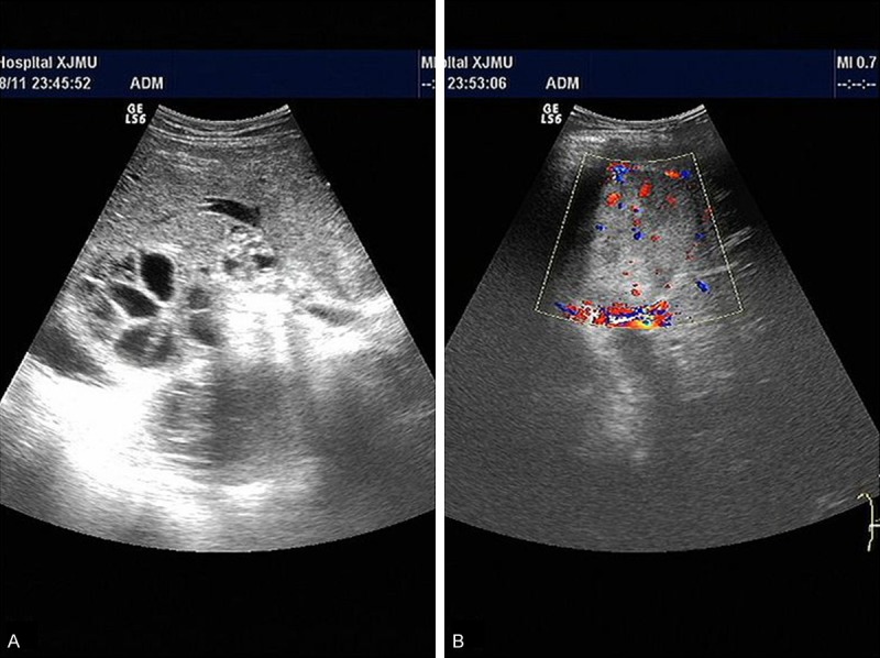
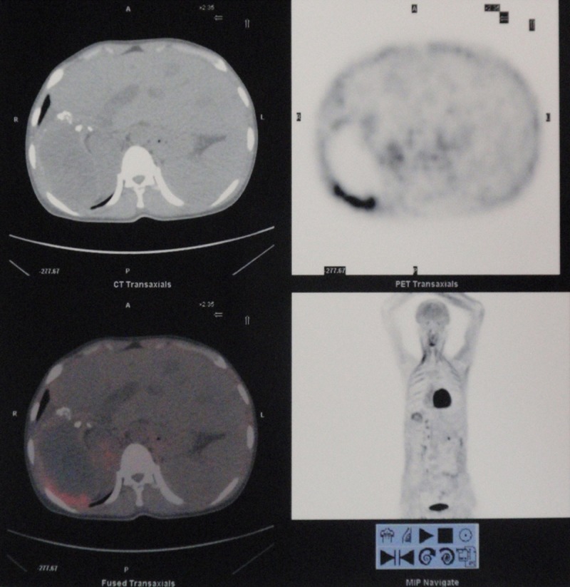
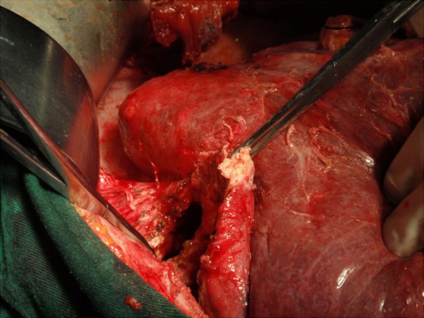
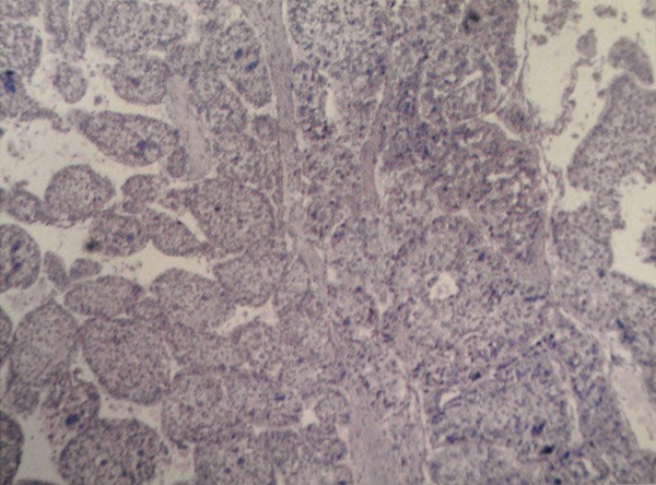
The patient was a 27-year-old female herdsman living in Xinjiang Uyghur Autonomous region, China. She came to our department due to sudden pain in the right upper quadrant of the abdomen. No decreased blood pressure and clinical manifestations of shock were observed. No vomiting, constipation, diarrhea, fever, shivering, night sweat, itch of skin and icterus was reported. No alcohol and viral hepatitis history was noted. The pain extended to the whole superior belly without radiating pain later. Ultrasound imaging indicated Echinococcus granulosus in the right lobe of liver. In addition, the E. granulosuss ruptured into abdominal cavity and biliary tract (Figure 1). For the positron emission tomography-computed tomography (PET/CT) scanning, a huge mass of E. granulosus with a size of 93.9×58.6 mm was noted in the right posterior lobe of the liver. A significant condense of radiopharmaceuticals were noted in the position near the peritoneum, indicating the E. granulosus may invaded into the abdominal cavity (Figure 2). With regards to the serum testing, significant increase of leucocytes and neutrophilic granulocytes was observed. Slight increase of total bilirubin was noted. Serologic testing for the viral hepatitis was negative. Then removal of hydatid cyst and exploration of common bile duct was performed. During the surgery, E multilocularis was noted in abdominal cavity and biliary tract. An E. granulosus mass and solid tumor mass was observed (Figure 3). Pathological testing indicated diffuse CE infection accompanied by hepatocellular carcinoma (Figure 4). For the treatment, drug administration (Albendazole) and hepatic arterial infusion chemotherapy was performed. The patient died for metastases of hepatic carcinoma after one year.
Figure 1.

A. E. granulosus indicated by ultrasound imaging. B. A solid mass was observed in left medial lobe of the liver, raising the possibility for hepatocellular carcinoma.
Figure 2.

Cyst rupture indicated by the PET/CT scanning. No tumor mass was noted.
Figure 3.

E. granulosus and tumor mass was observed during the removal of hydatid cyst and exploration of common bile duct.
Figure 4.

Physiological testing confirmed HCC under an Olympus BX41 microscope (Olympus Corp., Tokyo, Japan) with a magnification of ×100.
Discussion
Both CE and HCC are chronic disease with no typical clinical manifestations [5]. The identification of EC or HCC was comparatively convenient even though some complications may occur at the same time. However, the identification of CE accompanied by HCC was difficult in clinical practices as CE can induce tumour-like, infiltrative growth of parasite lesions in liver through the extensive proliferation of metacestodes. For the association between CE and HCC, previous report indicated that no correlation has been confirmed as few reports were noted in patients with CE accompanied by HCC [5]. In addition, the pathogenesis of each disease seems different: Human CE was considered an infection of by the larval cestode E. granulosus, which can affect liver, lung, brain and other organs [3]. However, HCC was mainly caused by hepatitis virus, finally lead to canceration and metastasis [4].
According to our knowledge, the parasites can reside within the liver of their hosts and remain clinically unnoticed for an extended period. Therefore, it is speculated that the metacestode must have acquired some means of modulating the human immune response, by counteracting adverse reactions of the host and influencing the physiology of the peri-parasitic area to its own advantage. Moreover, human cancer is strongly associated with abnormalities of immune system [6]. Recently, Stadelmann et al reported that the E. multilocularis phosphoglucose isomerase (EmPGI), a component of laminated layer of metacestodes, showed a sequence similarity of 86% with human PGI at the terms of animo acid sequence [7]. Mammalian PGI is a multi-functional protein which can act as a cytokine, growth factor and inducer of angiogenesis, and played a role in tumor development and metastasis [8]. Based on this, our study may raise the possibility that human CE may induce the development and metastasis of HCC in human beings.
Acknowledgements
All authors had contribution for the final report. This work was supported by National Clinical Key Subject- General Surgery Construction Project, and National Science Foundation of China (No. 81260220).
Disclosure of conflict of interest
None.
References
- 1.Brunetti E, Kern P, Vuitton DA. Expert consensus for the diagnosis and treatment of cystic and alveolar echinococcosis in humans. Acta Trop. 2010;114:1–16. doi: 10.1016/j.actatropica.2009.11.001. [DOI] [PubMed] [Google Scholar]
- 2.Eckert J, Conraths FJ, Tackmann K. Echinococcosis: an emerging or re-emerging zoonosis? Int J Parasitol. 2000;30:1283–94. doi: 10.1016/s0020-7519(00)00130-2. [DOI] [PubMed] [Google Scholar]
- 3.Smego RA Jr, Bhatti S, Khaliq AA, Beg MA. Percutaneous aspiration-injection-reaspiration drainage plus albendazole or mebendazole for hepatic cystic echinococcosis: a meta-analysis. Clin Infect Dis. 2003;37:1073–83. doi: 10.1086/378275. [DOI] [PubMed] [Google Scholar]
- 4.Nash KL, Woodall T, Brown AS, Davies SE, Alexander GJ. Hepatocellular carcinoma in patients with chronic hepatitis C virus infection without cirrhosis. World J Gastroenterol. 2010;16:4061–5. doi: 10.3748/wjg.v16.i32.4061. [DOI] [PMC free article] [PubMed] [Google Scholar]
- 5.Zold E, Barta Z, Zeher M. Hydatid disease of the liver and associated hepatocellular carcinoma. Clin Gastroenterol Hepatol. 2005;3:xxxv. doi: 10.1016/s1542-3565(05)00288-0. [DOI] [PubMed] [Google Scholar]
- 6.Reiche EM, Nunes SO, Morimoto HK. Stress, depression, the immune system, and cancer. Lancet Oncol. 2004;5:617–25. doi: 10.1016/S1470-2045(04)01597-9. [DOI] [PubMed] [Google Scholar]
- 7.Stadelmann B, Spiliotis M, Muller J, Scholl S, Muller N, Gottstein B, Hemphill A. Echinococcus multilocularis phosphoglucose isomerase (EmPGI): a glycolytic enzyme involved in metacestode growth and parasite-host cell interactions. Int J Parasitol. 2010;40:1563–74. doi: 10.1016/j.ijpara.2010.05.009. [DOI] [PubMed] [Google Scholar]
- 8.Yanagawa T, Funasaka T, Tsutsumi S, Watanabe H, Raz A. Novel roles of the autocrine motility factor/phosphoglucose isomerase in tumor malignancy. Endocr Relat Cancer. 2004;11:749–59. doi: 10.1677/erc.1.00811. [DOI] [PubMed] [Google Scholar]


