Abstract
OmpR is a transcriptional regulator implicated in the control of various cellular processes and functions in Enterobacteriaceae. This study was undertaken to identify genes comprising the OmpR regulon in the human gastrointestinal pathogen Yersinia enterocolitica. Derivatives of an ompR-negative strain with random transposon insertions creating transcriptional fusions with the reporter gene lacZ were isolated. These were supplied with the wild-type ompR allele in trans and then screened for OmpR-dependent changes in β-galactosidase activity. Using this strategy, five insertions in genes/operons positively regulated by OmpR and two insertions in genes negatively regulated by this protein were identified. Genetic analysis of one of these fusion strains revealed that the gene acrR, encoding transcriptional repressor AcrR is negatively regulated by OmpR. Differential analysis of membrane proteins by SDS-PAGE followed by mass spectrometry identified the protein AcrB, a component of the AcrAB-TolC multidrug efflux pump, as being positively regulated by OmpR. Analysis of the activity of the acrR and acrAB promoters using gfp fusions confirmed their OmpR-dependent repression and activation, respectively. The identification of putative OmpR-binding sites and electrophoretic mobility shift assays confirmed that this regulator binds specifically to both promoter regions with different affinity. Examination of the activity of the acrR and acrAB promoters after the exposure of cells to different chemicals showed that bile salts can act as an OmpR-independent inducer. Taken together, our findings suggest that OmpR positively controls the expression of the AcrAB-TolC efflux pump involved in the adaptive response of Y. enterocolitica O:9 to different chemical stressors, thus conferring an advantage in particular ecological niches.
Introduction
Multidrug efflux pumps are major determinants of drug resistance in bacteria. These pumps are classified into five different families according to their structure and function: MFS (major facilitator superfamily), SMR (small multidrug resistance family), MATE (multidrug and toxic compound extrusion family), ABC (ATP binding cassette superfamily), and RND (resistance nodulation cell division family) [1]. The AcrAB-TolC efflux pump belongs to the RND family, members of which are particularly effective in conferring drug resistance in Gram-negative bacteria [2,3]. The AcrAB-TolC efflux pump has a wide substrate spectrum encompassing antibiotics, dyes, detergents, bile salts, toxins and environmental compounds [4,5,6]. AcrAB-TolC is a tripartite system that mediates the expulsion of periplasmic substrates across the outer membrane. AcrB is an inner membrane efflux protein extended into the periplasm, AcrA is a periplasmic adaptor protein and TolC forms a channel in the outer membrane [5,7]. In Escherichia coli the expression of the acrAB operon is regulated by three activators, MarA, SoxS and Rob, and one repressor, AcrR [8,9,10]. In Salmonella, an additional activator, RamA is also involved in the control of acrAB transcription [11,12]. Factors that induce the AcrAB-TolC efflux pump in Salmonella and E. coli include indole, bile salts, ethanol, high osmolarity and the stationary phase [8,13]. The AcrAB-TolC efflux pump has yet to be extensively studied in yersiniae species pathogenic to humans. Comparative analysis of clinical strains of Y. enterocolitica has demonstrated AcrAB and MarA overexpression, which is associated with the fluoroquinolone and multidrug resistance phenotypes [14].
Yersinia enterocolitica causes yersiniosis, an infectious disease of the gastrointestinal tract which, after salmonellosis and campylobacteriosis, is the third most common zoonotic bacterial disease in Europe [15]. Y. enterocolitica is a heterogeneous species that encompasses many bio-serotypes displaying varying degrees of virulence. These bacteria are free-living in the environment or live in association with a mammalian host. Thus, like other enteropathogens, they are exposed to various environmental factors characterizing specific ecological niches [16,17].
Bacterial adaptation to new conditions requires efficient modulation of gene expression. Two-component signal-transduction systems (TCSs) combine signal recognition, signal transduction and gene expression [18]. The prevalence of TCSs is due to their important role in the regulation of bacterial cellular processes including competence, conjugation, sporulation, bioluminescence, antibiotic synthesis, motility, biofilm formation, pathogenesis, regulation of metabolic pathways and transport of nutrients and ions [19]. TCSs enable a rapid response to changes in environmental conditions such as pH, temperature, osmotic pressure, nutrient availability and ion concentrations [18].
The EnvZ/OmpR system of E. coli, which is responsible for regulating the synthesis of the outer membrane porins OmpF and OmpC, enables cells to survive fluctuations in the osmolarity of the environment [20]. The two components of this system are the transmembrane histidine kinase EnvZ and its cognate response regulator OmpR, a cytoplasmic transcription factor [21]. Further studies have shown that OmpR can act as a global transcriptional regulator involved in controlling the expression of a wide variety of genes in Enterobacteriaceae, including virulence genes of pathogenic strains [22,23,24]. OmpR has been shown to be the most important two-component regulator in the acid response in Salmonella enterica sv. Typhimurium, E. coli and Y. pseudotuberculosis [25,26,27]. OmpR regulates a type VI secretion system in Y. pseudotuberculosis, which permits survival in acidic environments by maintaining intracellular pH homeostasis [28].
In Y. enterocolitica, the EnvZ/OmpR system was first characterized during an analysis of the physiological consequences of the loss of the OmpR protein. An ompR mutant (AR4, ΔompR::Km) of Y. enterocolitica strain Ye9 (bio-serotype 2/O9) subspecies palearctica, showed significant sensitivity to osmolarity, oxidative, thermal and acid stress. It was found that OmpR is involved in protecting cells against adverse intracellular conditions experienced following macrophage phagocytosis [29]. OmpR participates in the expression of OmpC, OmpF and OmpX porins, and also important pathogenicity determinants like the Yop proteins, invasin, adhesin Ail and flagella [29,30,31,32,33,34]. Moreover, OmpR appears to modulate the adhesion and invasion abilities of Y. enterocolitica and promotes biofilm development [35].
In light of the available evidence we assume that OmpR acts as a global transcriptional regulator in Yersinia cells. The present study was undertaken to identify genes comprising the OmpR regulon. OmpR-dependent genes were recognized by randomly inserting the lacZ reporter gene throughout the genome of an ompR mutant of Y. enterocolitica Ye9, and then comparing β-galactosidase activity in the presence and absence of ompR expression. In parallel, proteins from outer membrane protein-enriched fractions, whose synthesis is up- or downregulated by OmpR were also identified. Our results indicate that acrR and acrAB are members of the OmpR regulon in Y. enterocolitica. OmpR appears to up-regulate expression of the AcrAB-TolC efflux pump directly by activating acrAB transcription and indirectly by inhibiting acrR (encoding repressor AcrR) transcription.
Results
Identification of OmpR-regulated genes in the genome of Y. enterocolitica
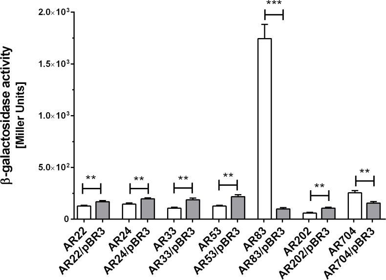
To identify genes whose expression is under the control of the EnvZ/OmpR signaling pathway in Y. enterocolitica Ye9, we obtained 960 independent chromosomal transcriptional fusions with the lacZ reporter gene following transposon mutagenesis of an ompR mutant with Tn5-B22. Next, a plasmid expressing ompR was introduced into each of these strains using a mass-mating technique. Individual transposon mutants were screened on LB agar containing X-Gal for differences in reporter gene expression with and without ompR supplied in trans. Of the 960 mutants screened, seven contained insertions in OmpR-regulated genes (Table 1). Five mutants carried insertions in genes/operons positively regulated by OmpR, and two mutants had an insertion in genes negatively regulated by OmpR. Quantitative β-galactosidase assays confirmed the mutant phenotypes and indicated that OmpR increased expression in mutants AR22, AR24, AR33, AR53 and AR202 by between 30 and 80%, while it decreased the expression level in mutant AR704 by almost 40%. In the case of strain AR83, the presence of OmpR in trans led to a significant decrease (8-fold) in the expression of the lacZ reporter gene (Fig 1).
Table 1. Characterization of the sites of Tn5-B22 transposition in Y. enterocolitica mutants.
| Mutant | site of transposon insertion a | protein (function) | regulation d |
|---|---|---|---|
| AR22 | YE105_C2078 b | TyrR (DNA-binding transcriptional | + |
| regulator) | |||
| AR24 | YE105_C1595 b | FimA (pilus assembly protein) | + |
| AR33 | YE105_C0917 b | general secretion pathway protein J | + |
| (type II secretion system) | |||
| AR53 | YE105_C3385 c | glycosyl hydrolase family protein | + |
| AR83 | YE105_C1163 b | DsrE (oxidative sulfur metabolism) | - |
| AR202 | intergenic region | hypothetical proteins (unknown | + |
| (YE105_C2160, | function) | ||
| YE105_C2160) c | |||
| AR704 | YE105_C2117 b | hypothetical protein (homolog of | - |
| tellurite resistance protein TerB) |
a sequence of region flanking transposon insertion comes from the genome of Y. enterocolitica subsp. palearctica 105.5R(r) (NC_015224) deposited in the NCBI database.
b lacZ is in the same orientation as the region with similarity to the database entry.
c lacZ is in the opposite orientation to the region with similarity to the database entry.
d + or-mean positive or negative OmpR-dependent regulation.
Fig 1. β-galactosidase activity exhibited by different transposon mutants and trans-complemented strains.
The data represent mean values (± standard deviation) from three independent experiments performed in triplicate. Asterisks indicate statistically significant differences (**—p<0.01; ***—p<0.001) according to Student’s unpaired t-test.
To identify the sites of transposon insertion in the selected mutants, the DNA regions flanking these insertions were amplified by AP-PCR and then sequenced. Analysis of these nucleotide sequences revealed that each mutant was the result of a unique insertion event (Table 2). Five transposon insertions were positioned such that the lacZ reporter gene was oriented in the same direction as an open reading frame (ORF) identified by sequence similarity. BLAST searches revealed identity between these ORFs and sequences from Y. enterocolitica subsp. palearctica 105.5R(r). In strain AR22 (positive effect of OmpR) transposition occurred at gene YE105_C2078, encoding DNA-binding transcriptional regulator TyrR. Insertion in strain AR24 (positive effect) was in gene YE105_C1595, which is probably involved in fimbrial synthesis. This gene encodes a homolog of the FimA pilus assembly protein. Strain AR33 (positive effect) had an insertion in gene YE105_C0917, which probably forms an operon with YE105_C0916. These two genes respectively encode general secretion pathway proteins J and I, which are involved in the formation of a type II secretion system. Transposition in strain AR83 (negative effect of OmpR) occurred in gene YE105_C1163, encoding a protein which displays significant similarity to DsrE, an essential factor in oxidative sulfur metabolism. Notably, gene YE105_C1162, encoding transcriptional repressor AcrR was identified immediately upstream of dsrE. In strain AR704 (negative effect) the lacZ reporter gene was transcribed from the promoter of gene YE105_C2117, encoding a hypothetical protein with homology to the tellurite resistance protein TerB. In the case of strain AR202 (positive effect of OmpR) insertion occurred in the intergenic region between genes YE105_C2160 and YE105_C2161. However, it seems that the reporter gene is transcribed from the promoter of the downstream gene YE105_C2163, encoding hypothetical protein ADZ42659.1 of unknown function. In strain AR53 (positive effect) transposition occurred in gene YE105_C3385, but in the opposite orientation to this gene promoter. Using the BPROM program for the prediction of bacterial promoters (http://linux1.softberry.com/), we identified two hypothetical promoter sequences located between genes YE105_C3382 and YE105_C3383. Thus, the OmpR-dependent regulation in mutant AR53 may be due to transcription from an OmpR-regulated promoter downstream from and oriented convergently to the lacZ gene.
Table 2. Putative OmpR-regulated proteins of Y. enterocolitica Ye9 identified by SDS-PAGE and LC-MS/MS a .
| Putative OmpR | band | protein | gene | theoretical |
|---|---|---|---|---|
| regulation | molecular | |||
| mass (kDa) b | ||||
| Induction at 27°C | p120 | AcrB (inner membrane | YE3101 Y. enterocolitica | 113.36 |
| protein, component of the | subsp. enterocolitica 8081 | |||
| AcrAB-TolC multidrug | ||||
| efflux pump) | ||||
| p110 | ferrichrome-iron receptor | Y11_35641 | 78.57 | |
| Y. enterocolitica subsp. | ||||
| palearctica Y11 | ||||
| Induction at 37°C | p100 | dehydratase (bifunctional | YE105_C0819 | 93.43 |
| aconitate hydratase 2/2- | Y. enterocolitica subsp. | |||
| methylisocitrate | palearctica 105.5R(r) | |||
| dehydratase) | ||||
| p85 | putative ABC transporter ATP-binding | Ye3942 Y. enterocolitica subsp. | 71.91 | |
| protein | enterocolitica 8081 | |||
| Repression at | p80 | 60-kDa heat shock protein | YE0354 Y. enterocolitica | 57.67 |
| 37°C | subsp. | |||
| enterocolitica 8081 | ||||
| p60 | elongation factor Tu | YE3927 Y. enterocolitica | 43.22 | |
| subsp. | ||||
| enterocolitica 8081 |
a Outer membrane protein-enriched fractions from bacteria grown to stationary phase at 27°C and 37°C were analyzed.
b Theoretical molecular masses of Y. enterocolitica proteins were calculated from their amino acid sequences deposited in the NCBI database using ExPASy proteomics tool Compute pI/Mw (http://www.expasy.org).
Identification of OmpR-regulated membrane proteins by SDS-PAGE and mass spectrometry analysis
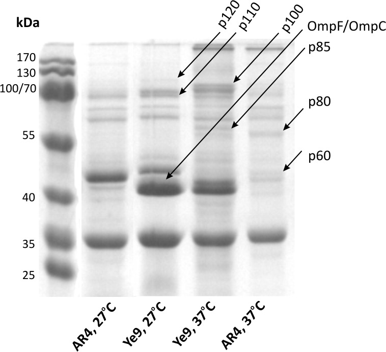
In parallel with the mutant screen described above, we analyzed membrane proteins of the wild-type Y. enterocolitica Ye9 and strain AR4 (the ompR mutant), grown under different temperature conditions, using SDS-PAGE. Changes in the membrane protein profiles due to the growth temperature and the presence and activity of the OmpR regulator were observed (Fig 2). A differential analysis of band intensities was used to select proteins for identification by LC-MS/MS. Following this procedure, we identified six putative OmpR-controlled proteins that matched Y. enterocolitica proteins present in the NCBI protein databases (Table 2). The synthesis of four proteins was higher in the wild-type strain than in mutant AR4: p120 and p110 (cells grown at 27°C), and p100 and p85 (cells grown at 37°C). Two other proteins, p80 and p60, were present at lower levels in the wild-type strain compared to AR4. The proteins that appeared to be positively controlled by OmpR were AcrB (p120; an essential component of the AcrAB-TolC multidrug efflux pump), ferrichrome-iron receptor (p110), dehydratase (p100; a bifunctional aconitate hydratase 2/2-methylisocitrate dehydratase) and a putative ABC transporter ATP-binding protein (p85). The proteins presumed to be negatively regulated by OmpR were a 60-kDa heat shock protein (p80) and elongation factor Tu (p60).
Fig 2. Influence of OmpR activity on the Y. enterocolitica membrane protein profile.
Proteins were isolated from strains Ye9 (wild type) and AR4 (ompR mutant) grown overnight in LB medium at 27°C or 37°C. In each case, 50 μg of protein were separated by SDS-PAGE and visualized by Coomassie blue staining. Putative OmpR-regulated proteins subsequently identified by LC-MS/MS are indicated by arrows. The bands were named according to their migration in the 12% polyacrylamide gel relative to the molecular weight standards.
Genes acrR and dsrE are organized in an operon
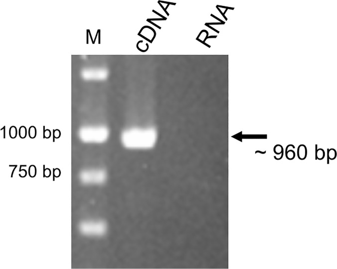
Transposition of Tn5-B22 in the strain AR83 (negative effect of OmpR) occurred in gene YE105_C1163 (encoding a DsrE homolog) located upstream of gene YE105_C1162, encoding transcriptional repressor AcrR. To determine whether these two genes are organized in an operon, RT-PCR analysis was performed (Fig 3). Our data showed that the putative acrR and dsrE genes of Y. enterocolitica Ye9 are co-transcribed as a bicistronic mRNA. Thus, the gene encoding transcriptional repressor AcrR seems to be negatively regulated by OmpR.
Fig 3. RT-PCR analysis of the acrRdsrE operon.
The analysis was performed using total RNA isolated from strain Ye9 grown in LB medium at 27°C. The size of the amplified fragment was estimated by comparison with the size marker DNAs (lane M). A negative control reaction was performed using DNase-treated RNA as the template.
Influence of OmpR activity on acrR and acrAB promoter function
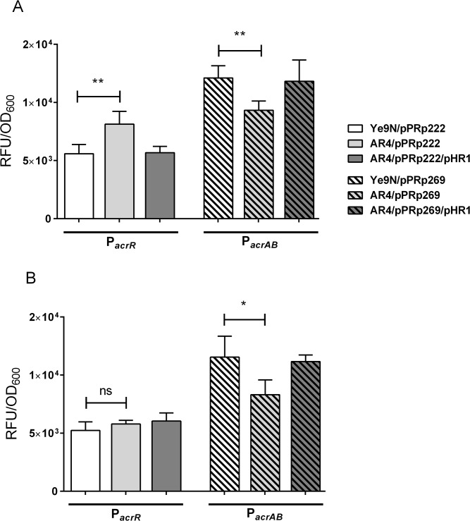
The genetic screen and the differential analysis of membrane proteins resulted in the identification of several putative members of the OmpR regulon. In one case, both of these methods suggested that OmpR might be involved in controlling expression of the AcrAB-TolC multidrug efflux pump in Y. enterocolitica Ye9. To confirm this hypothesis, we decided to construct fusions of the acrR and acrAB promoters with the gfp reporter gene. Bioinformatic analysis of the acrR and acrAB promoter regions revealed the presence of single putative OmpR binding sites, located at the respective positions -142 to -123 nt and -106 to -87 nt relative to the ATG start codons (Fig 4). These sequences exhibited 45% and 50% identity to the E. coli consensus OmpR-binding site, respectively, and both possessed the conserved motif GxxxC and the AC base pairs thought to be important for interaction with the OmpR regulator [36,37,38,39]. Taking into account the results of this in silico sequence analysis, reporter fusions of the acrR promoter (pPRp222, acrR::gfp) and acrAB promoter (pPRp269, acrAB::gfp) with gfp were constructed. The plasmid constructs carrying the appropriate fusions, i.e. pPRp222 and pPRp269, were then transformed into wild-type strain Ye9 and ompR mutant AR4, and GFP fluorescence was measured in bacteria grown in LB medium to exponential and stationary phase. Complementation analysis was performed with strains AR4/pPRp222 and AR4/pPRp269 carrying additional plasmid pHR4 encoding the active OmpR protein [29]. Data presented in Fig 5A shows that in the exponential phase of growth, the ompR mutant AR4 displayed a 30% increase in fluorescence produced by the acrR::gfp fusion (pPRp222) and a 20% decrease in fluorescence from the acrAB::gfp fusion (pPRp269), compared to the wild-type strain. In the stationary phase of growth, the lack of the OmpR protein resulted in a 30% decrease in the level of fluorescence activity of strain AR4/pPRp269, while no difference was observed for strain AR4/pPRp222, compared to the wild type (Fig 5B). These results demonstrated the involvement of OmpR in the negative regulation of acrR and the positive regulation of acrAB expression, depending on the growth phase.
Fig 4. Intergenic sequences upstream of the Y. enterocolitica acrR and acrAB operons.
The initiation codons (ATG) are in bold, and putative -35 and -10 promoter elements are underlined. The putative OmpR binding sites identified by in silico analysis (R1, R2) are shaded gray. Potential binding sites for AcrR (A1, A2) are boxed. Potential binding site elements aligned with the E. coli consensus sequences, with the percentage identities presented in parentheses.
Fig 5. Effect of OmpR and growth phase on acrR and acrAB promoter activity.
Strains Ye9 and AR4 carrying the promoter-gfp fusion constructs were cultivated in LB medium at 27°C to exponential phase (A) and stationary phase (B). Data represent mean fluorescence activity values normalized to the OD600 of the culture (± standard deviation) from three independent experiments performed in triplicate. The significance of differences between the values was calculated using Student’s unpaired t-test (ns [non significant]—p>0.05, *—p<0.05, **—p<0.01).
Effect of temperature and OmpR activity on acrR and acrAB promoter function
To study the influence of OmpR and temperature on the expression of acrR and acrAB, strains Ye9 and AR4 carrying pPRp222 or pPR269, were grown in LB medium at 27, 30 or 37°C to exponential phase, and the level of GFP fluorescence was measured (Fig 6). Compared with the level at 27°C, the GFP fluorescence produced by the acrAB::gfp fusion in wild-type strain Ye9 was increased by about 20% at 30°C, and by about 30% at 37°C (Fig 6). Thermoregulation of the acrAB::gfp fusion was also observed in ompR mutant strain AR4, although a relatively lower level of acrAB expression was noted. No differences in the level of GFP fluorescence were observed in either strain carrying the acrR::gfp fusion. These results indicated that temperature does not affect acrR expression, but is an important factor in regulating acrAB promoter activity, regardless of the presence or absence of the OmpR protein. Thus, OmpR is not involved in the observed thermoregulation.
Fig 6. The influence of growth temperature on acrR and acrAB promoter activity.
Strains Ye9 and AR4 carrying the promoter-gfp fusion constructs were cultivated in LB medium at 27°C, 30°C or 37°C to exponential phase. Data represent mean fluorescence activity values normalized to the OD600 of the culture (± standard deviation) from three independent experiments performed in triplicate. Significance of differences between the values was calculated using Student’s unpaired t-test (**—p<0.01, ***—p<0.001).
Effect of stress conditions on acrR and acrAB promoter function
Since the transcription of acrAB in E. coli and Salmonella is up-regulated by general stress factors, including the presence of bile salts, deoxycholate, indole, ethanol, paraquat, NaCl, and fluoroquinolones [8,13,40], we investigated the effect of these chemical stressors, and also low pH and the combination of high osmolarity/low pH, on acrR and acrAB expression using the gfp fusion constructs in the wild-type and ompR mutant strains (Table 3). Bile salts were found to be the most potent inducer of acrR and acrAB expression, causing an increase of approximately 3-fold in GFP fluorescence within 60 min of exposure. Interestingly, upregulation of acrAB expression by bile salts was found not to be OmpR-dependent. Deoxycholate, indole and paraquat only slightly modulated acrAB::gfp fluorescence activity in comparison to the control. None of the other tested chemicals affected the expression level of the analyzed gfp fusions. Moreover, no influence of osmolarity or pH was observed.
Table 3. Modulation of acrR and acrAB expression in strains Ye9N and AR4 by different stressors.
| Strain | without | ciprofloxacin | sodium | ethanol | indole | paraquat | NaCl | pH 5.0 | pH 5.0/ NaCl | bile salts |
|---|---|---|---|---|---|---|---|---|---|---|
| stressors | (0.01 mM) | deoxycholate | (4%) | (3 mM) | (1 mM) | (300 mM) | (300 mM) | (5%) | ||
| (0.5 mM) | ||||||||||
| Ye9N/pPRp222 a | 5592 ± 395 | 4959 ± 318 (ns) | 5913 ± 363 (ns) | 5444 ± 748 (ns) | 6086 ± 484 (ns) | 4535 ± 351 (ns) | 5270 ± 246 (ns) | 5902 ± 654 (ns) | 6232 ± 534 (ns) | 15927 ± 1680 (***) |
| AR4/pPRp222 b | 8130 ± 552 | 5674 ± 161 (*) | 6564 ± 459 (ns) | 5889 ± 266 (*) | 6609 ± 326 (ns) | 5700 ± 353 (*) | 6513 ± 405 (ns) | 6979 ± 367 (ns) | 6611 ± 345 (ns) | 33721 ± 1389 (***) |
| AR4/pPRp222/pHR4 | 5675 ± 383 | 5274 ± 221 | 5381 ± 674 | 5546 ± 138 | 5925 ± 172 | 4723 ± 443 | 5645 ± 346 | 6108 ± 344 | 6571 ± 452 | 22078 ± 1650 |
| Ye9N/pPRp269 c | 12107 ±523 | 10882 ± 921 (ns) | 15461 ± 786 (**) | 11931 ± 475 (ns) | 14562 ± 456 (*) | 14225 ± 756 (*) | 10538 ± 680 (ns) | 13493 ± 832 (ns) | 12765 ± 820 (ns) | 39100 ± 1073 (***) |
| AR4/pPRp269 d | 9329 ± 400 | 8359 ± 860 (ns) | 10007 ± 641 (ns) | 9227 ± 389 (ns) | 10445 ± 375 (ns) | 10060 ± 540 (ns) | 8440 ± 385 (ns) | 9947 ± 412 (ns) | 9174 ± 512 (ns) | 35379 ± 1016 (***) |
| AR4/pPRp269/pHR4 | 11829 ± 1051 | 10391 ± 631 | 13016 ± 502 | 11112 ± 614 | 14293 ± 363 | 13894 ± 469 | 11513 ± 832 | 14006 ± 387 | 13984 ± 784 | 39398 ± 1370 |
The GFP fluorescence (RFU/OD600) produced by the acrR::gfp (pPRp222) and acrAB::gfp (pPRp269) fusions in Ye9N and AR4 strains are presented. Data shown are mean values ± standard deviation from three independent experiments performed in triplicate. Significance was calculated with Student’s unpaired t-test [ns (non significant)—p>0.05
*—p<0.05
**—p<0.01
***—p<0.001].
a, c Results for Ye9N strains, grown in LB medium without any stressors, were compared with those for equivalent Ye9N strains, grown in the presence of different stressors.
b, d Results for AR4 strains were compared with those for equivalent Ye9N strains grown in the same conditions.
Interaction of OmpR with the acrR and acrAB promoter regions
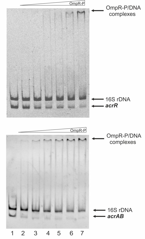
To test whether OmpR interacts with the acrR and/or acrAB promoter regions to directly control expression, we performed electrophoretic mobility shift assays (EMSAs). A recombinant OmpR-His6 protein was expressed in E. coli and purified to homogeneity. Since the affinity of OmpR for targets is dependent on its phosphorylation status, gel shift assays were performed in the presence of acetyl-phosphate to promote OmpR auto-phosphorylation. As mentioned above, one putative OmpR binding site (-142 to -123 nt from the ATG) and also one putative AcrR motif (-52 to -29 nt from the ATG) were recognized in the acrR promoter region. Moreover, a putative binding site for OmpR (-106 to -87 nt from the ATG), overlapped the putative AcrR binding motif (-116 to -93 from the ATG) and -35 promoter motif in the acrAB promoter region (Fig 4). Thus, fragments containing these putative binding sites within the acrR promoter (p222) and acrAB promoter (p269) were used as the target DNAs in EMSAs. Different amounts of the purified OmpR were incubated with these promoter fragments and these binding reactions were analyzed by electrophoresis in 6% non-denaturing polyacrylamide gels (Fig 7). Shifted complexes were clearly produced when the acrAB p269 fragment interacted with the phosphorylated OmpR protein present at the lowest concentration, i.e. 1.2 μM (Fig 7, lower panel). Furthermore, a slower migrating band also appeared when a higher amount of phosphorylated OmpR (6.2 μM) was incubated with the acrR p222 fragment (Fig 7, upper panel), but this interaction seemed weaker. The observed interactions were specific because no OmpR-P binding of a 16S rDNA control fragment was observed. Interestingly, non-phosphorylated OmpR was found to interact with the two promoter fragments with the same affinity as the phosphorylated form (data not shown). Taken together, these results demonstrated that OmpR, regardless of its phosphorylation state, can specifically bind to the acrR and acrAB promoter regions with different affinity, to inhibit and activate transcription, respectively.
Fig 7. Interaction of purified OmpR with the acrR and acrAB promoter regions examined by electrophoretic mobility shift assays.
EMSAs using Y. enterocolitica acrR (upper panel) or acrAB (lower panel) promoter fragments with in vitro phosphorylated OmpR protein (lane 1—no protein; lane 2–1.2 μM; lane 3–2.5 μM; lane 4–3.7 μM; lane 5–5 μM; lane 6–6.2 μM; lane 7–7.5 μM). A 16S rDNA fragment was included as a control in all reactions to confirm specific binding.
Discussion
The success of Y. enterocolitica as a pathogen depends on its rapid and efficient response to changing environmental conditions. TCSs play a significant role in sensing external factors and modulating the transcription of specific genes accordingly. It seems that OmpR could operate as a global regulatory protein participating in the control of many genes, including virulence determinants. To identify new members of the OmpR regulon, we performed a genetic screen and a differential analysis of membrane proteins. Initially, we employed transposon Tn5-B22 carrying a promoterless lacZ reporter gene to produce 1000 insertion mutants and studied their phenotypes in the presence and absence of OmpR. We recognize that the pool of mutants examined in this study only represents a relatively small sample of possible transposon insertions. The Y. enterocolitica subsp. palearctica 105.5R(r) genome is 4.50 Mb in size and carries 4441 genes. Together with plasmid pYV, characteristic of pathogenic Yersinia spp., which contains 110 genes, this species possesses about 4600 genes in total, so 5000 transposon mutants would correspond to 1x ORF coverage of the genome. Thus, our transposon library only covered a fraction of these ORFs, which implies that additional genes subject to regulation by OmpR remain unrecognized. On the basis of differences in β-galactosidase activity, 7 mutants were identified with insertions in genes/operons positively and negatively regulated by OmpR. A particularly noteworthy mutant was strain AR83, which exhibited an 8-fold decrease in lacZ expression in the presence of OmpR in trans. For the other mutants, these differences in β-galactosidase activity were considerably smaller. Genetic analysis of mutant AR83 revealed that transposition had occurred in a gene encoding a hypothetical protein with homology to DsrE, a protein essential to oxidative sulfur metabolism [41]. Interestingly, our in silico analysis revealed that immediately upstream of dsrE in the Y. enterocolitica genome is a gene encoding transcriptional repressor AcrR, involved in the regulation of acrAB expression in Enterobacteriaceae [42]. The AcrA and AcrB proteins are components of the multidrug efflux pump AcrAB-TolC [5]. In E. coli the acrAB operon is located upstream of the acrR gene and transcribed in the opposite orientation. In the course of the present study we demonstrated that the acrR and dsrE genes are cotranscribed from one promoter, and this gene pair constitutes an operon in Y. enterocolitica Ye9. This organization of the acrR gene seems to be characteristic only for the genus Yersinia, as judged by in silico analysis (data not shown). A differential analysis of Y. enterocolitica membrane proteins performed in parallel with the genetic screen revealed that apart from the OmpC/F porins, OmpR is required for the expression of at least six other proteins. We identified these putative OmpR-controlled proteins by SDS-PAGE followed by LC-MS/MS analysis. Among these, the protein AcrB was subject to positive control involving OmpR. This finding was consistent with the result of the genetic screen which identified dsrE and the co-transcribed acrR gene, encoding repressor AcrR, as being subject to negative OmpR control. In the light of these data, it seems that OmpR might be involved in regulating the AcrAB-TolC multidrug efflux pump directly, or indirectly via repressor AcrR. Thus, the role of OmpR in the mechanism controlling acrR and acrAB expression became the subject of subsequent experiments. We first examined the effect of the ompR mutation on the transcription of acrR and acrAB using promoter-gfp reporter fusions. By monitoring the activity of the acrR::gfp and acrAB::gfp fusions in strains Ye9 and AR4, we showed that OmpR plays an important role in the inhibition of acrR expression and the induction of acrAB expression in the exponential phase of bacterial growth. Interestingly, we observed a clear effect of temperature on acrAB expression (but not on that of acrR), which has not been observed in other enterobacteria. It is known that the EnvZ/OmpR TCS is stimulated by environmental conditions such as osmolarity or low pH, and it regulates many genes of diverse function [43]. Our results demonstrated that low pH, high osmolarity or a combination of both stress conditions did not influence acrR or acrAB expression. Of the chemical stressors tested for their influence on acrR and acrAB expression, only bile salts were found to cause the induction of both, which is in agreement with previous studies on S. Typhimurium and E. coli [8,13,40]. However, this effect was independent of the OmpR regulator.
Many bacterial operons are under the control of both global and specific regulators. It is known that apart from AcrR, the expression of acrAB is influenced by four other regulators: MarA, SoxS, Rob and RamA [8,9,10,12,13]. The induction of acrAB in Salmonella mediated by RamA occurs in response to indole and bile salts. The Rob protein is activated by bile salts and causes increased expression of the mar operon [44]. Furthermore, the superoxide generator paraquat activates acrAB via SoxS [45]. Other stress signals, including ethanol and osmolarity, also upregulate acrAB, but independently of the activity of regulators MarA, Rob or SoxS [8]. In Y. pestis, MarA mediates overexpression of acrAB, contributing to multidrug resistance [46]. MarA is a member of the XylS/AraC family of transcriptional activators like Rob and SoxS [47]. Our in silico analysis of the Y. enterocolitica genome indicated that this bacteria possesses orthologs of MarA, Rob and SoxS, but not RamA. These findings suggest the participation of MarA or other factors in acrAB and acrR expression in Y. enterocolitica in response to bile salts, and highlight the complexity of the mechanism that modulates the transcription of these operons. We have yet to identify the factor(s) that act to induce the OmpR-mediated expression of acrR or acrAB. It appears that the AcrR regulon belongs to a “stress-regulated” network, where genes are under the control of a number of apparently independent environmental stimuli. Bacteria experience the continual fluctuation of many environmental parameters, which requires fine tuning of the expression of many genes including acrR and acrAB. This would explain the difficulty in identifying a single factor mediating changes in the expression level of genes such as these. It appears that as Y. enterocolitica moves from the external environment into the intestinal tract, the changing conditions, such as the temperature shift to 37°C and raised levels of toxic compounds, especially bile salts, contribute to the induction of AcrAB.
To gain further insight into the regulatory mechanism of acrR and acrAB expression involving OmpR, we performed electrophoretic mobility shift assays. Prior in silico analysis identified single putative OmpR-binding sites in the promoters of these operons, similar to the conserved OmpR binding site reported in E. coli [36,37,38,39]. The deduced amino acid sequence of OmpR from Y. enterocolitica Ye9 is 99% identical to E. coli OmpR (data not shown) and exhibits 98% similarity to the OmpR of S. Typhimurium [48]. This suggests that binding sites for this regulator are also highly similar in these bacteria. EMSAs performed using OmpR or phosphorylated OmpR (OmpR-P) revealed specific binding of both forms to acrR and acrAB promoter fragments, with higher affinity for the latter. Previously it has been demonstrated that non-phosphorylated OmpR can bind the promoter of the fimB gene of uropathogenic E. coli (UPEC). It was also shown that the OmpR protein level could be responsible for the regulation of fimB in this bacterium [49]. Thus, it seems that apart from the phosphorylation state, the level of OmpR protein in Y. enterocolitica cells might influence acrR and acrAB expression.
The likely OmpR binding site in the acrAB promoter region overlaps the AcrR binding site, which suggests an antirepressor-type role for OmpR in the transcriptional regulation of this operon. Thus, the results of this study indicate that Y. enterocolitica acrAB might be subject to dual regulation by OmpR. We suggest that the transcription of acrR and acrAB are both directly OmpR-dependent, and OmpR also controls acrAB expression indirectly through AcrR via a locally limited feedback loop.
The molecular mechanism of OmpR-dependent regulation is not fully understood [39]. There is strong evidence that OmpR in Salmonella not only acts as a classical site-specific transcription factor, that activates both the SPI-1 and SPI-2 promoters (Salmonella pathogenicity island-1 and -2) through RNAP interaction, but it can also be considered a chromosome-structuring protein. The relaxation of DNA supercoiling can both induce and repress gene expression [50,51]. It has been shown that stress-induced changes in DNA topology recruit OmpR to the promoters of Salmonella genes ssrA, hilC and hilD, encoding transcriptional regulators of SPI-1 [52]. It is possible that this form of regulation, based on DNA topology, together with OmpR level or activity, could coordinate the expression of acrAB and/or acrR in Y. enterocolitica in response to environmental stimuli.
Materials and Methods
Bacterial strains, plasmids, media and growth conditions
The strains and plasmids used in this work are listed in Table 4. E. coli and Y. enterocolitica strains were routinely grown under aerobic conditions at 27 or 37°C in LB (Luria-Bertani) broth or on LB agar plates. When necessary, media were supplemented with the following compounds: nalidixic acid (Nal) – 30 μg ml−1; kanamycin (Km) – 50 μg ml−1; gentamicin (Gm) – 45 μg ml−1; chloramphenicol (Cm) – 25 μg ml−1; tetracycline (Tet) – 12.5 μg ml−1; X-Gal (5-bromo-4-chloro-3-indolyl galactoside) – 80 μg ml−1.
Table 4. Strains and plasmids used in this study.
| Strain or plasmid | Description | Reference or source |
|---|---|---|
| Y. enterocolitica O9 | ||
| Ye9 | pYV+, wild type | Laboratory |
| collection | ||
| AR4 | pYV+, ΔompR::Km (NalR, KmR) | [29] |
| AR22,AR24, AR33 | pTn5-B22 derivatives of AR4 (NalR, KmR, | This study |
| AR53, AR83 | GmR) | |
| AR202, AR704 | ||
| E. coli | ||
| Top10 F’ | F’{lacI q Tn10 (TetR)} mcrA Δ(mrr-hsd RMS- | Invitrogen |
| mcrBC) ϕ80lacZ ΔM15 Δ lacX74 deoR recA1 | ||
| araD139 Δ99ara-leu)7697 | ||
| Plasmids | ||
| pDrive | cloning vector (ApR, KmR) | Qiagen |
| pSUP101Tn5-B22 | mobilizable suicide vector pSUP101 derivative | [59] |
| with Tn5 element containing promoterless | ||
| lacZ (TcR, GmR) | ||
| pBR3 | pBBR1 MCS-3 with XhoI/PstI fragment | [30] |
| containing entire coding sequence of ompR | ||
| (TcR) | ||
| pHR4 | pHSG575 with 740-bp fragment of ompR (ORF | [29] |
| with rbs) (CmR) | ||
| pPROBE TT’ | broad-host-range cloning vector pBBR1MCS-3 | [57] |
| with promoterless gfp (TcR) | ||
| pPRp222 | derivative of pPROBE TT’ with EcoRI/KpnI | This study |
| fragment of acrRdsrE promoter region (TcR) | ||
| pPRp269 | derivative of pPROBE TT’ with EcoRI/KpnI | This study |
| fragment of acrAB promoter region (TcR) |
To determine the influence of different stress factors on gene expression, various compounds, including an antibiotic (ciprofoxacin), biocide (paraquat), and other chemicals (indole, deoxycholate, bile salts, ethanol), were used at sub-inhibitory concentrations [53]. Sodium chloride (300 mM NaCl) was used to induce osmotic shock. To monitor the influence of low pH on gene expression, we buffered LB medium to pH 5.0 with 0.1 M homoPIPES [homopiperazine-N,N’-bis(2-ethanesulfonic acid)].
Random insertion of the lacZ reporter gene throughout the genome of Y. enterocolitica ompR mutant strain AR4 (ΔompR::Km) followed by trans-complementation with a wild-type copy of ompR
Transposon mutagenesis of the Y. enterocolitica ompR mutant with Tn5-B22 (carrying the promoterless lacZ reporter gene) was performed using suicide plasmid pSUP101Tn5-B22 according to the procedure described by Robleto et al. [54]. Clones carrying the randomly inserted transposon were selected on LB agar plates containing appropriate antibiotics. Next, the individual transposon mutants were patched in 12 x 8 grids onto LB agar plates and then transferred, using 96-prong replicators into microtiter plates containing LB broth. Plasmid pBR3 carrying a wild-type copy of ompR was introduced in trans into each of the mutants by the following mass-mating technique. E. coli S17-1 λpir/pBR3 was grown overnight in LB broth supplemented with Tet. The cells were pelleted by centrifugation, washed twice with LB broth to remove antibiotics, and resuspended in the original volume of LB. Aliquots of this cell suspension (200 μl) were spread onto LB agar plates to form a lawn of the donor strain. The Y. enterocolitica mutants were replicated onto this lawn of E. coli S17-1 λpir/pBR3 and grown overnight at 27°C. These mating spots were then replicated onto LB agar plates containing appropriate antibiotics to select for transconjugants carrying plasmid pBR3. Collections of strains with and without pBR3 were replicated into 96-well microtiter plates containing LB broth with appropriate antibiotics and incubated overnight at 27°C. Individual transposon mutants were then screened to detect differences in reporter gene expression in the presence and absence of active ompR by growth on LB agar plates supplemented with X-Gal and antibiotics. Mutants that differed in the intensity of the blue color were identified, the corresponding patch was streaked onto a fresh plate, and a single colony was retested for β-galactosidase activity as confirmation.
β-Galactosidase assays
β-galactosidase activities were assayed by the method of Miller [55] with ONPG (o-nitrophenyl-β-D-galactopyranoside) as the substrate. Assays were routinely performed at least three times in triplicate and the data expressed as the mean ± standard deviation.
Arbitrary-primer PCR (AP-PCR) for amplification of DNA regions flanking transposon insertions
AP-PCR was carried out in two rounds, each with a different primer pair (S1 Table). In the first round, primer TN5EXT (sequence unique to the right end of the transposon) and an arbitrary primer ARB1 were used [54]. PCR was performed as follows: (i) 5 min at 95°C; (ii) six cycles of 30 s at 95°C, 30 s at 30°C, and 1 min 30 s at 72°C; (iii) 30 cycles of 30 s at 95°C, 30 s at 45°C, and 2 min at 72°C; and (iv) a final extension step of 5 min at 72°C. The second round of PCR was performed using 2 μl of the first round reaction as template with primers TN5INT (specific to the right arm of the transposon) and ARB3 (specific to the 5’ end of primer ARB1) [54]. PCR was performed as follows: (i) 1 min at 95°C; (ii) 30 cycles of 30 s at 95°C, 30 s at 52°C, and 2 min at 72°C; and (iii) a final extension step of 5 min at 72°C. The PCR products were separated by agarose gel electrophoresis and the most-intense bands were purified (Invitrogen PureLink Gel extraction kit) and sequenced using primers TN5INT and ARB3 with an ABI PRISM 377 DNA Sequencer (DNA Sequencing and Oligonucleotide Synthesis Laboratory, IBB PAN, Warsaw). The sequences were compared with sequence databases using BLAST programs from The National Center for Biotechnology Information (http://www.ncbi.nlm.nih.gov/).
DNA techniques
DNA manipulations, such as restriction digestion, ligation, transformation and conjugation were performed using standard protocols [56]. Plasmid and chromosomal DNA were purified using the BlueMATRIX Plasmid Miniprep DNA kit and Gene MATRIX Bacterial Genomic +/- DNA Purification Kit (EURx), respectively. DNA fragments were amplified by PCR using Taq DNA polymerase (Invitrogen) and oligonucleotide primers (S1 Table). PCR products were purified from the reactions using a BlueMATRIX PCR/DNA Clean-up DNA Purification Kit or following agarose gel electrophoresis with the BlueMATRIX Agarose-Out DNA Purification Kit (EURx).
Isolation of membrane proteins and SDS-PAGE analysis
Outer membrane protein-enriched fractions were obtained by treatment of Y. enterocolitica cell envelope samples with Triton X-100. These fractions, referred to henceforth as membrane proteins, were isolated from strains grown to late exponential phase at 27°C and 37°C as described previously [31]. The samples were resuspended in electrophoresis sample buffer, solubilized by boiling for 10 min and the proteins separated by electrophoresis on 12% SDS-polyacrylamide gels. Proteins were visualized by Coomassie Brilliant Blue R-250 staining.
Membrane proteins were isolated from Y. enterocolitica strains differing in their OmpR content, in three separate experiments, and SDS-PAGE of each protein preparation was performed twice. Only proteins that were clearly and reproducibly increased or decreased in relative abundance compared to the wild type strain, based on their band intensity, were further analyzed.
Protein identification by liquid chromatography-coupled tandem mass spectrometry (LC-MS/MS)
Gel slices containing the appropriate bands, visualized by Coomassie Blue staining, were excised with a clean scalpel. Prior to the LC-MS/MS analysis, the gel slices were subjected to in-gel trypsin digestion [33]. LC-MS/MS analysis of proteins was performed at the Mass Spectrometry Laboratory, IBB PAN, Warsaw.
Construction of acrR::gfp and acrAB::gfp transcriptional fusions
To obtain acrR::gfp and acrAB::gfp transcriptional fusions, appropriate DNA fragments containing the promoters of the acrR and acrAB operons were amplified by PCR using the oligonucleotide pairs FacR1/RacR222 and FacAB1/RacAB269, respectively. The promoter fragments of acrR (p222) and acrAB (p269) were cloned into the cloning vector pDrive (Qiagen). The fragments were then excised by digestion with EcoRI/KpnI and subcloned into the same sites of reporter vector pPROBE TT’ to place them adjacent to a promoterless gfp gene [57]. The resulting constructs pPRp222 and pPR269 were transferred to Y. enterocolitica strains Ye9 and AR4 by electroporation. Transformants were selected on LB agar plates supplemented with Tet (strain Ye9), or Tet and Km (strain AR4). The presence of these constructs in the Y. enterocolitica strains was confirmed by plasmid isolation and PCR with the primers used previously to amplify the promoter fragments.
Measurement of GFP fluorescence
To test the effect of different stress factors on gene expression, early log-phase bacterial cultures (OD600 ~0.2) grown in LB medium were exposed to ciprofloxacin, ethanol, indole, paraquat, bile salts, deoxycholate, 300 mM NaCl, pH 5.0 or a combination of low pH/high osmolatity at 27°C for 60 min. To monitor the influence of temperature, identical cultures were grown in LB medium at 27°C, 30°C or 37°C for 2 h. Then, 200 μl of the treated cell suspensions were transferred to wells of 96-well flat-bottomed plates (Costar) and absorbance at 600 nm and GFP fluorescence (excitation 485 nm; emission 530 nm) were measured using a TECAN infinite pro M200PRO microplate reader, according to the procedure described by Gueguen et al. [58]. The specific GFP fluorescence was expressed as the relative fluorescence intensity (RFU) divided by the OD600, after subtracting the values of a blank sample. A culture of strain Ye9 carrying vector pPROBE TT’ was used to determine background fluorescence. Individual cultures were assayed in triplicate and the reported values are the means from three independent cultures.
RNA isolation and reverse transcription
Total RNA was isolated from 107 bacterial cells from cultures grown overnight in LB medium at 27°C, using a BlueMATRIX Universal RNA Purification Kit (EURx). Following treatment with RNase-free DNase I (Sigma), RNA was reverse-transcribed with random hexamers and AMV reverse transcriptase (Sigma). The cDNA was used as the template in PCRs (RNA used as a negative control) with primers RTacR1 and RTdrE2 (S1 Table) specific for the acrRdsrE mRNA. The amplified fragment was resolved by electrophoresis on 2% agarose gels and visualized by staining with ethidium bromide.
Electrophoretic mobility shift assays (EMSAs)
OmpR binding studies were performed using purified N-terminal His-tagged OmpR protein and DNA fragments containing the acrR and acrAB control regions. The acrR promoter fragment p222 used in the transcriptional fusion with gfp was employed in the EMSA. However, a new acrAB promoter fragment p225 was amplified for this purpose by PCR with primer pair FacAB1/RacAB225 (S1 Table). OmpR-His6 synthesized in E. coli M15 was purified using Ni2+-NTA agarose as described previously [30]. The EMSA method was adapted from a previously published protocol [31]. For phosphorylation of OmpR, 20 mM acetyl phosphate (Sigma) was used. To confirm binding specificity, a 304-bp fragment of Y. enterocolitica Ye9 16S rDNA generated by PCR using primer pair 16SR1/16SR304 (S1 Table) was included as a non-specific competitor in all binding reactions. The reaction mixtures were analyzed by electrophoresis on 6% non-denaturing polyacrylamide gels run in TBE buffer (Tris-borate-EDTA) and DNA bands were stained with SYBR Green EMSA nucleic acid gel stain (Invitrogen).
Supporting Information
(PDF)
Data Availability
All relevant data are within the paper and its Supporting Information files.
Funding Statement
This research was supported by a grant from the National Science Center, Poland (grant 2011/01/B/NZ6/01845). The funders had no role in study design, data collection and analysis, decision to publish, or preparation of the manuscript.
References
- 1. Putman M, Van Veen HW, Konings WN. Molecular properties of bacterial multidrug transporters. Microbiol Mol Biol Rev. 2000;64: 672–693. [DOI] [PMC free article] [PubMed] [Google Scholar]
- 2. Murakami S, Nakashima R, Yamashita E, Yamaguchi A. Crystal structure of bacterial multidrug efflux transporter AcrB. Nature 2002;419: 587–593. [DOI] [PubMed] [Google Scholar]
- 3. Murakami S, Nakashima R, Yamashita E, Matsumoto T, Yamaguchi A. Crystal structures of a multidrug transporter reveal a functionally rotating mechanism. Nature 2006;443: 173–179. [DOI] [PubMed] [Google Scholar]
- 4. Nikaido H. Multidrug efflux pumps of gram-negative bacteria. J Bacteriol. 1996;178: 5853–5859. [DOI] [PMC free article] [PubMed] [Google Scholar]
- 5. Li XZ, Nikaido H. Efflux-mediated drug resistance in bacteria. Drugs 2004;64: 159–204. [DOI] [PubMed] [Google Scholar]
- 6. Schuldiner S. Structural biology: the ins and outs of drug transport. Nature 2006;443: 156–157. [DOI] [PubMed] [Google Scholar]
- 7. Blair JM, Piddock LJ. Structure, function and inhibition of RND efflux pumps in Gram-negative bacteria: an update. Curr Opin Microbiol. 2009;12: 512–519. 10.1016/j.mib.2009.07.003 [DOI] [PubMed] [Google Scholar]
- 8. Ma D, Alberti M, Lynch C, Nikaido H, Hearst E. The local repressor AcrR plays a modulating role in the regulation of acrAB genes of Escherichia coli by global stress signals. Mol Microbiol. 1996;19: 101–112. [DOI] [PubMed] [Google Scholar]
- 9. Randall LP, Woodward MJ. The multiple antibioticresistance (mar) locus and its significance. Res Vet Sci. 2002;72: 87–93. [DOI] [PubMed] [Google Scholar]
- 10. Rosenberg EY, Bertenthal D, Nilles ML, Bertrand KP, Nikaido H. Bile salts and fatty acids induce the expression of Escherichia coli AcrAB multidrug efflux pump through their interaction with Rob regulatory protein. Mol Microbiol. 2003;48: 1609–1619. [DOI] [PubMed] [Google Scholar]
- 11. Bailey AM, Paulsen IT, Piddock LJ. RamA confers multidrug resistance in Salmonella enterica via increased expression of acrB, which is inhibited by chlorpromazine. Antimicrob Agents Chemother. 2008;52: 3604–3611. 10.1128/AAC.00661-08 [DOI] [PMC free article] [PubMed] [Google Scholar]
- 12. Schneiders T, Amyes SG, Levy SB. Role of AcrR and RamA in fluoroquinolone resistance in clinical Klebsiella pneumoniae isolates from Singapore. Antimicrob Agents Chemother. 2003;47: 2831–2837. [DOI] [PMC free article] [PubMed] [Google Scholar]
- 13. Nikaido E, Yamaguchi A, Nishino K. AcrAB multidrug efflux pump regulation in Salmonella enterica serovar Typhimurium by RamA in response to environmental signals. J Biol Chem. 2008;283: 24245–24253. 10.1074/jbc.M804544200 [DOI] [PMC free article] [PubMed] [Google Scholar]
- 14. Fabrega A, Roca I, Vila J. Fluoroquinolone and multidrug resistance phenotypes associated with the overexpression of AcrAB and an orthologue of MarA in Yersinia enterocolitica . Int J Med Microbiol. 2010;300: 457–463. 10.1016/j.ijmm.2010.02.003 [DOI] [PubMed] [Google Scholar]
- 15. Rosner BM, Stark K, Werber D. Epidemiology of reported Yersinia enterocolitica 23 infections in Germany, 2001–2008. BMC Public Health 2010;4: 337. [DOI] [PMC free article] [PubMed] [Google Scholar]
- 16. Straley SC, Perry RD. Environmental modulation of gene expression and pathogenesis in Yersinia . Trends Microbiol. 1995;3: 310–317. [DOI] [PubMed] [Google Scholar]
- 17. Bottone EJ. Yersinia enterocolitica: the charisma continues. Clin Microbiol Rev. 1997;10: 257–276. [DOI] [PMC free article] [PubMed] [Google Scholar]
- 18. Stock JB, Ninfa AJ, Stock AM. Protein phosphorylation and regulation of adaptive responses in bacteria. Microbiol Rev. 1989;53: 450–490. [DOI] [PMC free article] [PubMed] [Google Scholar]
- 19. Albright LM, Huala E, Ausubel FM. Prokaryotic signal transduction mediated by sensor and regulator protein pairs. Annu Rev Genet. 1989;23: 311–336. [DOI] [PubMed] [Google Scholar]
- 20. Russo FD, Silhavy TJ. EnvZ controls the concentration of phosphoryled OmpR to mediate osmoregulation of the porin genes. J Mol Biol. 1990;222: 567–580. [DOI] [PubMed] [Google Scholar]
- 21. Forst SA, Roberts DL. Signal transduction by the EnvZ-OmpR phosphotransfer system in bacteria. Res Microbiol. 1994;145: 363–373. [DOI] [PubMed] [Google Scholar]
- 22. Dorman CJ, Chatfield S, Higgins CF. Characterization of porin and ompR mutants of a virulent strain of Salmonella typhimurium: ompR mutants are attenuated in vivo . Infect Immun. 1989;57: 2136–2140. [DOI] [PMC free article] [PubMed] [Google Scholar]
- 23. Bernardini MJ, Fontaine A, Sansonetti PJ. The two-component regulatory system OmpR-EnvZ controls the virulence of Shigella flexneri . J Bacteriol. 1990;172: 6274–6281. [DOI] [PMC free article] [PubMed] [Google Scholar]
- 24. Lee AK, Detweiler CS, Falkow S. OmpR regulates the two component system SsrA-SsrB in Salmonella pathogenicity island 2. J Bacteriol. 2000;182: 771–781. [DOI] [PMC free article] [PubMed] [Google Scholar]
- 25. Bang IS, Audia JP, Park YK, Foster JW. Autoinduction of the ompR response regulator by acid shock and control of the Salmonella enterica acid tolerance response. Mol Microbiol. 2002;44: 1235–1250. [DOI] [PubMed] [Google Scholar]
- 26. Stincone A, Daudi N, Rahman AS, Antczak P, Henderson I, Cole J, et al. A systems biology approach sheds new light on Escherichia coli acid resistance. Nucleic Acids Res. 2011;39: 7512–7528. 10.1093/nar/gkr338 [DOI] [PMC free article] [PubMed] [Google Scholar]
- 27. Flamez C, Ricard I, Arafah S, Simonet M, Marceau M. New insights into two-component system regulon plasticity in bacteria. Int J Med Microbiol. 2008;298: 193–207. [DOI] [PubMed] [Google Scholar]
- 28. Zhang W, Wang Y, Song Y, Wang T, Xu S, Peng Z,et al. A type VI secretion system regulated by OmpR in Yersinia pseudotuberculosis functions to maintain intracellular pH homeostasis. Environ Microbiol. 2013;15: 557–569. 10.1111/1462-2920.12005 [DOI] [PubMed] [Google Scholar]
- 29. Brzostek K, Raczkowska A, Zasada A. The osmotic regulator OmpR is involved in the response of Yersinia enterocolitica O:9 to environmental stresses and survival within macrophages. FEMS Microbiol Lett. 2003;228: 265–271. [DOI] [PubMed] [Google Scholar]
- 30. Brzostek K, Raczkowska A. The YompC protein of Yersinia enterocolitica: molecular and physiological characterization. Folia Microbiol. 2007;52: 73–80. [DOI] [PubMed] [Google Scholar]
- 31. Raczkowska A, Skorek K, Bielecki J, Brzostek K. OmpR controls Yersinia enterocolitica motility by positive regulation of flhDC expression. Antonie Van Leeuwenhoek 2010;99: 381–394. 10.1007/s10482-010-9503-8 [DOI] [PMC free article] [PubMed] [Google Scholar]
- 32. Raczkowska A, Brzóstkowska M, Kwiatek A, Bielecki J, Brzostek K. Modulation of inv gene expression by the OmpR two-component response regulator protein of Yersinia enterocolitica . Folia Microbiol. 2011;56: 313–319. 10.1007/s12223-011-0054-9 [DOI] [PubMed] [Google Scholar]
- 33. Brzóstkowska M, Raczkowska A, Brzostek K. OmpR, a response regulator of the two-component signal transduction pathway, influences inv gene expression in Yersinia enterocolitica O9. Front Cell Inf Microbiol. 2012;2(153): 1–14. [DOI] [PMC free article] [PubMed] [Google Scholar]
- 34. Skorek K, Raczkowska A, Dudek B, Miętka K, Guz-Regner K, Pawlak A, Klausa E, Bugla-Płoskońska G, Brzostek K. Regulatory protein OmpR influences the serum resistance of Yersinia enterocolitica O:9 by modifying the structure of the outer membrane. PLoS One 2013. 19;8(11):e79525 10.1371/journal.pone.0079525 [DOI] [PMC free article] [PubMed] [Google Scholar]
- 35. Raczkowska A, Skorek K, Brzóstkowska M, Łasińska A, Brzostek K. Pleiotropic effects of a Yersinia enterocolitica ompR mutation on adherent-invasive abilities and biofilm formation. FEMS Microbiol Lett. 2011;321: 43–49. 10.1111/j.1574-6968.2011.02308.x [DOI] [PubMed] [Google Scholar]
- 36. Pratt LA, Silhavy TJ. Identification of base pairs important for OmpR-DNA interaction. Mol Microbiol. 1995;17: 565–573. [DOI] [PubMed] [Google Scholar]
- 37. Huang KJ Igo MM. Identification of the bases in the ompF regulatory region, which interact with the transcription factor OmpR. J Mol Biol. 1996;262: 615–28. [DOI] [PubMed] [Google Scholar]
- 38. Egger LA, Park H, Inouye M. Signal transduction via the histidyl-aspartyl phosphorelay. Genes Cells 1997;2: 167–184. [DOI] [PubMed] [Google Scholar]
- 39. Kenney LJ. Structure/function relationships in OmpR and other winged-helix trancription factors. Curr Opin Microbiol. 2002;5: 135–141. [DOI] [PubMed] [Google Scholar]
- 40. Nikaido E, Shirosaka I, Yamaguchi A, Nishino K. Regulation of the AcrAB multidrug efflux pump in Salmonella enterica serovar Typhimurium in response to indole and paraquat. Microbiol. 2011;157: 648–655. [DOI] [PubMed] [Google Scholar]
- 41. Pott AS, Dahl C. Sirohaem sulfite reductase and other proteins encoded by genes at the dsr locus of Chromatium vinosum are involved in the oxidation of intracellular sulfur. Microbiol. 1998;144: 1881–1894. [DOI] [PubMed] [Google Scholar]
- 42. Pan W, Spratt BD. Regulation of the permeability of the gonococcal cell envelope by the Mtr system. Mol Microbiol. 1994;11: 769–755. [DOI] [PubMed] [Google Scholar]
- 43. Clarke EJ, Voigt CA. Characterization of combinatorial patterns generated by multiple two-component sensors in E. coli that respond to many stimuli. Biotechnol Bioeng. 2011;108: 666–675. 10.1002/bit.22966 [DOI] [PMC free article] [PubMed] [Google Scholar]
- 44. Jair KW, Yu X, Skarstad K, Thony B, Fujita N, Ishihama A, et al. Transcriptional activation of promoters of the superoxide and multiple antibiotic resistance regulons by Rob, a binding protein of the Escherichia coli origin of chromosomal replication. J Bacteriol. 1996;178: 2507–2513. [DOI] [PMC free article] [PubMed] [Google Scholar]
- 45. Miller PF, Gambino LF, Sulavik MC, Gracheck SJ. Genetic relationship between soxRS and mar loci in promoting multiple antibiotic resistance in Escherichia coli . Antimicrob Agents Chemother. 1994;38: 1773–1779. [DOI] [PMC free article] [PubMed] [Google Scholar]
- 46. Udani RA, Levy SB. MarA-like regulator of multidrug resistance in Yersinia pestis . Antimicrob Agents Chemother. 2006;50: 2971–2975. [DOI] [PMC free article] [PubMed] [Google Scholar]
- 47. Gallegos MT, Schleif R, Bairoch A, Hofmann K, Ramos JL. Arac/XylS family of transcriptional regulators. Microbiol Mol Biol Rev. 1997;61: 393–410. [DOI] [PMC free article] [PubMed] [Google Scholar]
- 48. Raczkowska A, Brzostek K. Identification of OmpR protein and its role in the invasion properties of Yersinia enterocolitica . Pol J Microbiol. 2004;53: 11–16. [PubMed] [Google Scholar]
- 49. Rentschler AE, Lovrich SD, Fitton R, Enos-Berlage J, Schwan WR (2013) OmpR regulation of the uropathogenic Escherichia coli fimB gene in an acidic/high osmolality environment. Microbiol. 2013;159: 316–327. 10.1099/mic.0.059386-0 [DOI] [PMC free article] [PubMed] [Google Scholar]
- 50. Galán JE, Curtiss R 3rd. Expression of Salmonella typhimurium genes required for invasion is regulated by changes in DNA supercoiling. Infect Immun. 1990;58: 1879–1885. [DOI] [PMC free article] [PubMed] [Google Scholar]
- 51. Cróinin O, Carroll RK, Kelly A, Dorman CJ. Roles for DNA supercoiling and the Fis protein in modulating expression of virulence genes during intracellular growth of Salmonella enterica serovar Typhimurium. Mol Microbiol. 2006;62: 869–882. [DOI] [PubMed] [Google Scholar]
- 52. Cameron ADS, Dorman CJ. A fundamental regulatory mechanism operating through OmpR and DNA topology controls expression of Salmonella Pathogenicity Islands SPI-1 and SPI-2. PLoS Gen. 2012;8(3):e1002615 10.1371/journal.pgen.1002615 [DOI] [PMC free article] [PubMed] [Google Scholar]
- 53. Dupont M, James CE, Chevalier J, Pages J-M. An early response to environmental stress involves regulation of OmpX and OmpF two enterobacterial outer membrane pore-forming proteins. Antimicrob Agents Chemother. 2007;51: 3190–3198. [DOI] [PMC free article] [PubMed] [Google Scholar]
- 54. Robleto EA, López-Hernández I, Silby MW, Levy SB. Genetic analysis of the AdnA regulon in Pseudomonas fluorescens: nonessential role of flagella in adhesion to sand and biofilm formation. J Bacteriol. 2003;185: 453–460. [DOI] [PMC free article] [PubMed] [Google Scholar]
- 55. Miller JH. Experiments in molecular genetics Cold Spring Harbor, NY: Cold Spring Harbor Laboratory; 1972. pp. 352–355. [Google Scholar]
- 56. Sambrook J, Russell DW. Molecular Cloning: A Laboratory Manual. 3rd ed. Cold Spring Harbor: Cold Spring Harbor Laboratory Press; 2001. [Google Scholar]
- 57. Miller WG, Leveau HJ, Lindow SE. Improved gfp and inaZ Broad-Host-Range promoter-probe vectors. MPMI 2000;13: 1243–1250. [DOI] [PubMed] [Google Scholar]
- 58. Gueguen E, Durand E, Zhang XY, d'Amalric Q, Journet L, Cascales E. Expression of a Yersinia pseudotuberculosis type VI secretion system is responsive to envelope stresses through the OmpR transcriptional activator. PLoS One 2003. June 19;8(6):e66615. [DOI] [PMC free article] [PubMed] [Google Scholar]
- 59. Simon R, Quandt J, Klipp W. New derivatives of transposon Tn5 suitable for mobilization of replicons, generation of operon fusions and induction of genes in gram-negative bacteria. Gene 1989;80: 161–169. [DOI] [PubMed] [Google Scholar]
Associated Data
This section collects any data citations, data availability statements, or supplementary materials included in this article.
Supplementary Materials
(PDF)
Data Availability Statement
All relevant data are within the paper and its Supporting Information files.


