Abstract
Background
Urinary density (UD) has been routinely used for decades as a surrogate marker for urine osmolality (Uosm). We asked if UD can accurately estimate Uosm both in healthy subjects and in different clinical scenarios of kidney disease.
Methods
UD was assessed by refractometry. Uosm was measured by freezing point depression in spot urines obtained from healthy volunteers (N = 97) and in 319 inpatients with acute kidney injury (N = 95), primary glomerulophaties (N = 118) or chronic kidney disease (N = 106).
Results
UD and Uosm correlated in all groups (p < 0.05). However, a wide range of Uosm values was associated with each UD value. When UD was ≤ 1.010, 28.4% of samples had Uosm above 350 mOsm/kg. Conversely, in 61.6% of samples with UD above 1.020, Uosm was below 600 mOsm/kg. As expected, Uosm exhibited a strong relationship with serum creatinine (Screat), whereas a much weaker correlation was found between UD and Screat.
Conclusion
We found that UD is not a substitute for Uosm. Although UD was significantly correlated with Uosm, the wide dispersion makes it impossible to use UD as a dependable clinical estimate of Uosm. Evaluation of the renal concentrating ability should be based on direct determination of Uosm.
Keywords: Kidney disease, Urinalysis, Urine density, Urine osmolality, Urine concentrating ability
Background
Measurement of urine osmolality (Uosm), the gold standard in the evaluation of urine concentrating ability, is a valuable tool for the assessment of renal function in such distinct clinical conditions as acute kidney injury (AKI) and chronic kidney disease (CKD). However, since Uosm is not routinely measured, assessment of urine density (UD) by hydrometry, refractometry or semi-quantitative colorimetric reactions has long been employed instead.
Although a correlation does exist between UD and Uosm, at least under normal physiological conditions [1-4], the assumption that UD accurately reflects Uosm, which underlies any clinical decision based on UD, has not been formally tested and, in fact, has been recently challenged [5,6].
In the present study we examined the relation between UD (measured by refractometry) and Uosm in 97 normal subjects as well as in a cohort of 319 patients with assorted renal disorders, to test the hypothesis that UD can be reliably used in routine clinical practice as a measure of Uosm.
Methods
Urine samples were consecutively obtained from 95 adult patients with AKI in intensive care unit, 118 patients with primary glomerulopathies, admitted to a Nephrology ward for investigation, 106 CKD outpatients, and 97 healthy volunteers. Urine samples were obtained from the first morning void, with no standardized water restriction. This study protocol was reviewed and approved by our Institutional Research Ethics Committee (Comissão de Ética para Análise de Projetos de Pesquisa, CAPPesq, #0045/08). A written informed consent was obtained.
UD was measured by refractometry, employing a benchtop refractometer (ATAGO CO.LTD SPR-T2, Tokyo, Japan). Results were corrected for the influence of protein and/or glucose according to conventional equations [6]: UD corrected for proteinuria = UD measured – [protein] *0.0003; UD corrected for glucosuria = UD measured – [glucose] *0.0002, where both protein and glucose in the urine are given in g/L. Uosm was measured by freezing point depression using an advanced wide-range osmometer (Model 3 W2, Advanced Instruments Inc, Needham Heights, Massachusetts). SCreat was measured with a conventional automated method (enzymatic colorimetric test, by the Jaffe reaction).
Serum sodium concentration was obtained in 203 patients (63.3%). Hypernatremia was found in 3 patients (147 to 149 mEq/l), whereas hyponatremia was observed in 8 patients (129 to 134 mEq/l). Since dysnatremia was infrequent, and never severe, this data was not further analyzed.
AKI was defined according to the current KDIGO classification [7]. In nearly all patients with glomerulopathies, hospitalization was indicated for kidney biopsy and/or clinical management of nephritic or nephrotic syndrome.
Statistical analysis
Univariate correlation analysis between single variables was performed by calculating the Spearman coefficient. Data management was performed by Prism 6.0 statistical software (Graphpad, San Diego, CA, USA). A two-tailed p value < 0.05 was considered statistically significant.
Results
Spot urine samples from 97 subjects with normal renal function (Control Group) and from 319 inpatients with acute kidney injury (AKI, N = 95), glomerulopathies (GP, N = 118) or chronic kidney disease (CKD, N = 106) were analyzed.
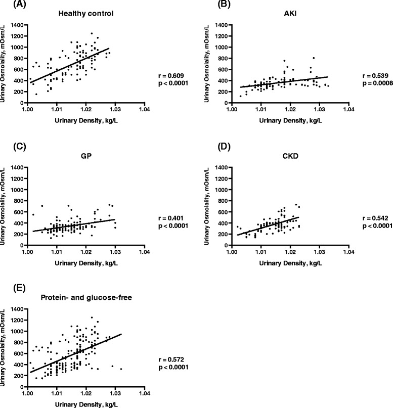
As expected, UD was consistently correlated to Uosm. Correlation was statistically significant when all groups were considered together (r = 0.462, p < 0.0001), as well as in the healthy control group (r = 0.609, p < 0.0001); in the AKI group (r = 0.539, p = 0.0008); in the GP group (r = 0.401, p < 0.0001); and in the CKD group (r = 0.542, p < 0.0001), as shown in Figure 1A, B, C and D, respectively. UD was corrected for proteinuria and glucosuria as described under Methods. Proteinuria was found in almost 60% of samples: overall, only 171 out of 416 samples were free from proteinuria or glucosuria (all 98 healthy volunteers, 35 out of 95 in the AKI group, 35 out of 106 in the CKD group, and only 3 out of 118 in the GP group). Glucosuria was found in only 5 patients from the CKD group and in 4 patients from the GP group. These 9 samples also exhibited proteinuria, and therefore both equations were used for correction. When only urine samples without proteinuria or glucosuria were analyzed, the correlation between UD and Uosm was even stronger (r = 0.572, p < 0.0001) (Figure 1E).
Figure 1.

Correlation between urinary osmolality (U osm ) and urinary density (UD) in each subgroup of patients: healthy control (A), acute kidney injury – AKI (B), glomerulopathies - GP (C), chronic kidney disease – CKD (D), and the entire group with urine free of protein and glucose (E).
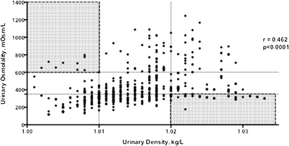
Despite the significant correlation between UD and Uosm, a wide range of Uosm values was associated with each UD value. This inconsistency was particularly striking when extreme UD values were considered (Figure 2, shaded areas): 39.8% of samples with UD lower or equal to 1.010 exhibited Uosm in excess of 350 mOsm/kg. Conversely, in 58.6% of samples with UD above 1.020, Uosm was below 600 mOsm/kg.
Figure 2.

Correlation between urinary osmolality (U osm ) and urinary density (UD) in all groups. Shaded areas are pointing to unexpected high or low UD while Uosm was the opposite.
Low, normal and high renal concentration ability was defined as UD lower or equal to 1.010, between 1.010 and 1.020, and above or equal to1.020 kg/L, and Uosm lower than 350 mOsm/kg, between 350 and 600 mOsm/kg, and higher than 600 mOsm/kg, respectively. The number of patients classified in each criterion is plotted in Table 1. Agreement between UD and Uosm is highlighted in gray.
Table 1.
Comparison between urinary density (UD) and osmolality (U osm ) classification of low and high concentration ability
| UD criteria | Osmolality criteria | ||
|---|---|---|---|
| All patients | Low (<350mOsm/kg) | Normal (350-600mOsm/kg) | High (>600mOsm/kg) |
| Low UD (≤1.010 kg/L) | 71 | 27 | 20 |
| Normal UD (1.011 – 1.019 kg/L) | 79 | 89 | 43 |
| High (UD ≥1.020 kg/L) | 26 | 25 | 36 |
| Patients with AKI | |||
| Low UD (≤1.010 kg/L) | 28 | 2 | 0 |
| Normal UD (1.011 – 1.019 kg/L) | 20 | 17 | 2 |
| High (UD ≥1.020 kg/L) | 13 | 11 | 2 |
| Patients with CKD | |||
| Low UD (≤1.010 kg/L) | 8 | 10 | 10 |
| Normal UD (1.011 – 1.019 kg/L) | 23 | 38 | 6 |
| High (UD ≥1.020 kg/L) | 3 | 5 | 3 |
| Patients with glomerulonephritis | |||
| Low UD (≤1.010 kg/L) | 28 | 7 | 1 |
| Normal UD (1.011 – 1.019 kg/L) | 36 | 21 | 2 |
| High (UD ≥1.020 kg/L) | 10 | 9 | 4 |
| Healthy individuals | |||
| Low UD (≤1.010 kg/L) | 7 | 8 | 9 |
| Normal UD (1.011 – 1.019 kg/L) | 0 | 13 | 33 |
| High (UD ≥1.020 kg/L) | 0 | 0 | 27 |
Bold numbers show agreement between UD and Uosm.
AKI, acute kidney injury; CKD, chronic kidney disease.
When analyzing healthy subjects, UD was an excellent predictor of Uosm when it was above 1.020: 100% of these samples exhibited Uosm above 600 mOsm/kg. By sharp contrast, UD failed to predict Uosm when UD was below or equal to 1.010: only 29.2% of samples had Uosm lower than 350 mOsm/kg, while Uosm was higher than 600 mOsm/kg in 37.5% of the samples.
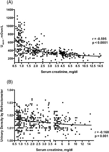
Figure 3A illustrates the relationship between Uosm and serum creatinine (Screat), for all subjects. Uosm and Screat followed a nonlinear relationship that could be fitted to a two-exponential curve (p < 0.01): Uosm was expectedly distributed across a wide range (118–1245 mOsm/kg) in subjects with Screat lower than 1.0 mg/dL and was confined to a narrow interval around 300 mOsm/kg as Screat increased. The relationship between Uosm and Screat was still significant (p < 0.05), although much less conspicuous, when UD replaced Uosm as a measure of urine concentration (Figure 3B). When analyzing subgroups, the correlation between Uosm and Screat was significant in the AKI group (r = −0.451, p < 0.001), in the GP group (r = −0.533, p = 0.0001), and in the CKD group (r = −0.546, p = 0.0001). There was no significant correlation between Uosm and Screat in the healthy control group (r = −0.108, p = 0.289). A weaker but still significant correlation between UD and Screat was shown in the AKI group (r = −0.160, p = 0.001), in the GP group (r = −0.253, p = 0.026), and in the CKD group (r = −0.209, p = 0.031). There was no correlation between UD and Screat in the healthy control group (r = −0.102, p = 0.863).
Figure 3.

Correlation between serum creatinine (S Creat ) levels and urinary osmolality (U osm ) (A), and urinary density (UD) (B).
Discussion
Urine density has long been considered as a practical surrogate marker of urine osmolality. It has even been proposed that simple equations be used in clinical practice to obtain Uosm directly from UD [8-11], whereas a website offers such calculations online [12]. In the present study, we challenged the concept that UD is a reliable marker of urine osmolality. For better accuracy, UD measurements were made utilizing a refractometer, instead of the semi-quantitative dipstick method more commonly employed. Even so, the correlation obtained between UD and Uosm, though statistically significant, was relatively weak (r = 0.462). A closer examination casts serious doubts about the clinical usefulness of UD. If an UD of 1.020 kg/L or higher were regarded as a test to detect individuals with an Uosm of at least 600 mOsm/kg [8-10], the sensitivity of such a test would be only 36%, whereas its specificity would be 81%. In other words, 64% of the subjects with concentrated urines would be missed by such test. On the other hand, good renal concentration ability might be erroneously inferred in as many as 19% of the other cases. Conversely, if an UD equal to or less than 1.010 kg/L were assumed to detect urine osmolalities below 350 mOsm/kg (isosthenuric or diluted urine), the sensitivity of the test would be only 40%, although the corresponding specificity would approach a more acceptable 80%.
The inadequacy of UD as a measure of Uosm becomes even more evident when we consider that in about 2/3 of all samples UD values were ≥1.010 and ≤1.020 kg/L, thus being irrelevant to major clinical decisions such as whether or not to administer IV saline to hypovolemic patients. When only UDs equal or higher than 1.020 kg/L were analyzed, a mere 41.4% of the samples actually exhibited Uosm above 600 mOsm/kg, whereas in 29.9% Uosm was even below 350 mOsm/kg. In the converse case, when exclusively UDs equal or lower than 1.010 kg/L were considered, 60.2% of samples exhibited Uosm values lower than 350 mOsm/kg, whereas in 16.9% Uosm even exceeded 600 mOsm/kg. These results indicate that UD cannot replace Uosm as a faithful measurement of urine concentration. Although a high Uosm may not reflect the actual volume status, such as in cases of inappropriate antidiuretic hormone secretion, in many situations it does signal the need for fluid replacement. Therefore, an erroneous interpretation of a high UD as indicative of hypovolemia may lead to inadequate and even dangerous fluid administration.
As expected, there was a significant nonlinear correlation between Uosm and Screat, reflecting the expected relationship between renal function and urine concentrating/diluting ability: individuals with normal renal function can vary urine osmolality over a wide range, while renal functional impairment is accompanied by a progressive limitation of this capacity, until urine becomes permanently isosthenuric as most of the renal function is lost. This relationship between Uosm and Screat was strongly attenuated when UD was used instead of Uosm, reinforcing the view that UD is a poor marker of renal concentrating/diluting ability.
The reasons why the relationship between UD and Uosm is less consistent than might be expected are unclear. In the present study, the effect of the possible presence of glucose and/or protein in urine was corrected by applying appropriate equations [6,13]. However, the association between UD and Uosm remained loose even after samples containing these solutes were excluded (r = 0.459, p < 0.05). It should be noted that a myriad of other solutes, commonly encountered in the urine of patients with renal disorders, such as drugs and iodinated radiocontrast agents, could increase urine density, leading to overestimation of the renal concentrating ability. Even “physiologic” solutes, such as sodium, potassium and urea, can appear in widely varying proportions in the urine of both healthy and diseased subjects, each of them exerting a different influence on urine density [9]. The unpredictability of these effects helps to explain the erratic relationship between UD and Uosm.
Conclusion
In summary, although UD correlates with Uosm, the relationship between these two parameters is largely inconsistent, even in healthy subjects, indicating that UD is a poor marker of renal concentrating/diluting capability. Direct determination of Uosm, a relatively inexpensive procedure, should be performed if reliable information about this important aspect of renal function is to be obtained from urine.
Acknowledgements
We are grateful to all patients and medical staff who collaborated to this study. There is no financial support to declare.
Footnotes
Competing interests
The authors declare that they have no competing interests.
Authors’ contributions
ACPS collected the data, participated in the design of the study and helped draft the manuscript; RZ participated in the design of the study, gave important intellectual contribution, revised and drafted the manuscript; RBO participated in the design of the study, revised and drafted the manuscript; MARS and MR performed all urinalysis and osmolality tests, revised and drafted the manuscript; JERJ participated in the in the design of the study, revised and drafted the manuscript; RME conceived the study, participated in its design, performed the statistical analysis, coordination, revised and drafted the manuscript. All authors read and approved the final manuscript.
Contributor Information
Ana Carolina P Souza, Email: anacarolinapessoa@gmail.com.
Roberto Zatz, Email: rzatz@usp.br.
Rodrigo B de Oliveira, Email: rodrigobueno.hc@gmail.com.
Mirela A R Santinho, Email: mirela.santinho@globo.com.
Marcia Ribalta, Email: marcia.mribalta@gmail.com.
João E Romão, Jr, Email: joao.egidio@uol.com.br.
Rosilene M Elias, Email: rosilenemotta@hotmail.com.
References
- 1.Czernichow P, Polak M. Testing Water regulation. 4. Basel, Switzerland: Karger; 2011. [Google Scholar]
- 2.de Buys Roessingh AS, Drukker A, Guignard JP. Dipstick measurements of urine specific gravity are unreliable. Arch Dis Child. 2001;85(2):155–7. doi: 10.1136/adc.85.2.155. [DOI] [PMC free article] [PubMed] [Google Scholar]
- 3.Bakhshandeh S, Morita Y. Comparison of urinary concentration tests: osmolality, specific gravity, and refractive index. Mich Med. 1975;74(21):399–403. [PubMed] [Google Scholar]
- 4.Imran S, Eva G, Christopher S, Flynn E, Henner D. Is specific gravity a good estimate of urine osmolality? J Clin Lab Anal. 2010;24(6):426–30. doi: 10.1002/jcla.20424. [DOI] [PMC free article] [PubMed] [Google Scholar]
- 5.Oppliger RA, Magnes SA, Popowski LA, Gisolfi CV. Accuracy of urine specific gravity and osmolality as indicators of hydration status. Int J Sport Nutr Exerc Metab. 2005;15(3):236–51. doi: 10.1123/ijsnem.15.3.236. [DOI] [PubMed] [Google Scholar]
- 6.Chadha V, Garg U, Alon US. Measurement of urinary concentration: a critical appraisal of methodologies. Pediatr Nephrol. 2001;16(4):374–82. doi: 10.1007/s004670000551. [DOI] [PubMed] [Google Scholar]
- 7.Langham RG, Bellomo R, D’ Intini V, Endre Z, Hickey BB, McGuinness S, et al. KHA-CARI guideline: KHA-CARI adaptation of the KDIGO Clinical Practice Guideline for Acute Kidney Injury. Nephrology. 2014;19(5):261–5. doi: 10.1111/nep.12220. [DOI] [PubMed] [Google Scholar]
- 8.Leech S, Penney MD. Correlation of specific gravity and osmolality of urine in neonates and adults. Arch Dis Child. 1987;62(7):671–3. doi: 10.1136/adc.62.7.671. [DOI] [PMC free article] [PubMed] [Google Scholar]
- 9.Voinescu GC, Shoemaker M, Moore H, Khanna R, Nolph KD. The relationship between urine osmolality and specific gravity. Am J Med Sci. 2002;323(1):39–42. doi: 10.1097/00000441-200201000-00007. [DOI] [PubMed] [Google Scholar]
- 10.Dorizzi R, Pradella M, Bertoldo S, Rigolin F. Refractometry, test strip, and osmometry compared as measures of relative density of urine. Clin Chem. 1987;33(1):190. [PubMed] [Google Scholar]
- 11.Medler S, Harrington F. Measuring dynamic kidney function in an undergraduate physiology laboratory. Adv Physiol Educ. 2013;37(4):384–91. doi: 10.1152/advan.00057.2013. [DOI] [PubMed] [Google Scholar]
- 12.NHS, Greater Glasgow and Clyde. Paediatrics: Clinical Guidelines. [http://www.clinicalguidelines.scot.nhs.uk/]
- 13.Rose BD, Post TW. Clinical Physiology of Acid–base and Electrolyte Disorders. 5. New York: McGraw-Hill; 2001. [Google Scholar]


