Abstract
Alveolar capillary dysplasia with misalignment of pulmonary veins (ACD/MPV) is a lethal neonatal lung disease characterised by severe pulmonary hypertension, abnormal vasculature and intractable hypoxaemia. Mechanisms linking abnormal lung vasculature with severe hypoxaemia in ACD/MPV are unknown. We investigated whether bronchopulmonary anastomoses form right-to-left shunt pathways in ACD/MVP. We studied 2 infants who died of ACD/MPV postmortem with direct injections of coloured ink into the pulmonary artery, bronchial artery and pulmonary veins. Extensive histological evaluations included serial sectioning, immunostaining and 3-dimensional reconstruction demonstrated striking intrapulmonary vascular pathways linking the systemic and pulmonary circulations that bypass the alveolar capillary bed. These data support the role of prominent right-to-left intrapulmonary vascular shunt pathways in the pathophysiology of ACD/MPV.
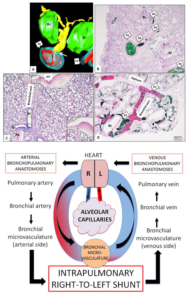
Alveolar capillary dysplasia with misalignment of pulmonary veins (ACD/MPV) is a rare and lethal neonatal lung disease associated with severe respiratory failure and pulmonary hypertension.1 Despite improved respiratory care, infants with ACD/MPV die of sever hypoxaemia after birth.2 Abnormal vascular lung histology of ACD/MPV includes dysplastic capillaries, hypertensive arterial remodelling, and prominent veins located close to distal airways and arteries (‘MPV’). Cardiac catheterisation studies have revealed absent ‘capillary blush’ phase during angiography in infants with ACD/MPV, suggesting intrapulmonary shunting.3 However, the contribution of MPV to the pathophysiology of ACD/MPV remains unknown. Preacinar anatomical communications between systemic (bronchial) and pulmonary vascular systems has been extensively described4; whether they form an alternate right-to-left vascular pathway bypassing distal lung vasculature in ACD/MPV is unclear. Postmortem injections of pulmonary arteries (PA), bronchial arteries (BA) and pulmonary veins (PV) were performed with coloured inks (green, blue and black, respectively). Histology, immunohistochemistry and three-dimensional reconstructions were done as previously described with slight modification (see supplement). Infants’ clinical course and microscopic pathology was typical of ACD/MPV. Injection of green and blue ink marked the vessels of PA, BA, bronchial vein (BV), PV including the ‘MPV’ and the bronchial microvessels (mv), while black ink was visible within the PV, BV, mv and ‘MPV’ (see online supplements 1–3). No ink filling was seen in alveolar capillaries or lymphatics (see online supplements 4 and 5). MPV bridging mv and PV were frequently noted and contained all three inks (see online supplements 1–3) suggesting that MPV are BV and may act as venous ‘shunt vessels’, as BV normally link mv associated with airways, large vessels, pleura, and lymph nodes with PV (figure 1C).5 In addition to the expanded venous bronchopulmonary anastomoses we also identified arterial bronchopulmonary anastomoses via PA-BA connections (figure 1, see online supplement 1) as part of intrapulmonary right-to-left to vascular shunt circuit (figure 1). The prominence of these vessels in ACD/MPV is likely due to enhanced flow through blood shunting from PA to systemic vessels that dilate mv and BV, which become visible as extra vessels within bronchoarterial bundles (‘MPV’). Thus, we propose that rather than representing ‘mal-developed’ vessels, MPV are BV that appear markedly dilated due to increased blood flow in PA-BA anastomotic vessels in ACD/ MPV. In summary, we report striking intrapulmonary vascular anastomoses in ACD/ MPV that could contribute to fatal hypoxaemia due to the shunting of blood away from the alveolar capillary bed.
Figure 1.
Upper panel: Main components of intrapulmonary vascular anastomotic pathways are shown. Three-dimensional image reconstruction (A) and histological sections (B) show direct connections between pulmonary arteries (PA) and bronchial arteries (BA) (white arrow) (BA-yellow, PA-red and blue, Bronchus (Br)-green). In an age-matched control lung, BV and pulmonary veins (PV) connections are present but appear very delicate, almost invisible (C, insert magnifies BV-PV connection). In alveolar capillary dysplasia with misalignment of pulmonary veins (ACD/MPV), the bronchial vessels are enlarged, especially the BV (D) appearing as dilated, thin-walled channels within the bronchovascular bundle (‘misaligned pulmonary veins’). Lower panel: Schematic of intrapulmonary right-to-left shunt in ACD/MPV lung is shown. The majority of right heart blood is shunted through the arterial bronchopulmonary anastomoses (PA, BA, mv) that drains via venous bronchopulmonary anastomoses (microvessels (mv), BV, PV) leading to significantly decreased flow within the alveolar capillary bed.
Supplementary Material
Acknowledgments
The authors thank Ashley Blair, Corina Mitchell, Jana Polzer and Eric Wartchow at Children’s Hospital Colorado for technical assistance.
Funding NIH Clinical Center (HL085703, HL68702, DK096996).
Footnotes
Contributors CG: designed experiments, performed experiments, analysed the data, drafted and revised the paper. He is guarantor. SS-L: performed, collected and analysed 3D data, reviewed manuscript. NA: collected clinical data, discussed experimental design, reviewed manuscript. JG: collected clinical data, discussed experimental design, reviewed manuscript. MKD: Performed experiments, reviewed manuscript. SHA: designed experiment, reviewed and revised manuscript.
Competing interests None.
Patient consent Obtained.
Ethics approval IRB.
Provenance and peer review Not commissioned; externally peer reviewed.
References
- 1.Bishop NB, Stankiewicz P, Steinhorn RH. Alveolar capillary dysplasia. Am J Respir Crit Care Med. 2011;184:172–9. doi: 10.1164/rccm.201010-1697CI. [DOI] [PMC free article] [PubMed] [Google Scholar]
- 2.Parker TA, Ivy DD, Kinsella JP, et al. Combined therapy with inhaled nitric oxide and intravenous prostacyclin in an infant with alveolar-capillary dysplasia. Am J Respir Crit Care Med. 1997;155:743–6. doi: 10.1164/ajrccm.155.2.9032222. [DOI] [PubMed] [Google Scholar]
- 3.Hintz SR, Vincent JA, Pitlick PT, et al. Alveolar capillary dysplasia: diagnostic potential for cardiac catheterization. J Perinatol. 1999;19:441–6. doi: 10.1038/sj.jp.7200243. [DOI] [PubMed] [Google Scholar]
- 4.Shure D. The bronchial circulation in pulmonary vascular obstruction. In: Butler J, editor. The bronchial circulation. New York, NY: Marcel Dekker; 1992. pp. 579–97. [Google Scholar]
- 5.Barman SA, Ardell JL, Parker JC, et al. Pulmonary and systemic blood flow contributions to upper airways in canine lung. Am J Physiol. 1988;255:H1130–5. doi: 10.1152/ajpheart.1988.255.5.H1130. [DOI] [PubMed] [Google Scholar]
Associated Data
This section collects any data citations, data availability statements, or supplementary materials included in this article.


