Figure 5.

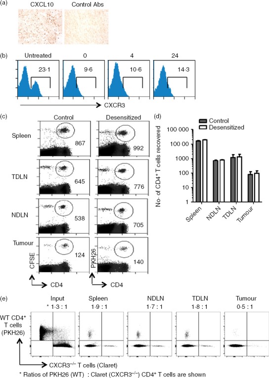
CXCR3 is not required for intra-tumoural recruitment of CD4+ T cells. (a) Methylcholanthrene-induced fibrosarcomas were stained by immunohistochemistry for detection of CXCL10 expression. (b). Desensitization of CXCR3 expression by CXCL10 treatment and CXCR3 expression assessed 0, 4 and 24 hr later. (c, d) Mice received 2 × 10⁷ splenic CD4+ T cells isolated from spleens of tumour-free Thy1.1 mice, half of which were treated with 500 nm CXCL10 and then labelled with PKH26 (desensitized fraction,). The other half was left untreated (control) and labelled with CFSE. After 24 hr, spleens, non-draining lymph nodes (NDLN), tumour-draining lymph nodes (TDLN) and tumours were harvested, stained and analysed by flow cytometry. (c) Representative plots showing numbers of cells recovered. (d) Absolute numbers of cells recovered from each organ. Results are representative of two independent experiments. (e). PKH26-labelled wild-type (WT) and Claret-labelled CXCR3−/− spleen cells were injected intravenously into a tumour-bearing mouse. Around 18 hr later, the spleen, TDLN, NDLN and tumour were harvested and single cell suspensions were stained with CD4-specific antibodies. Lymphocytes were gated using a live/dead marker and proportions of dye-labelled CD4+ cells in each of the compartments are shown.
