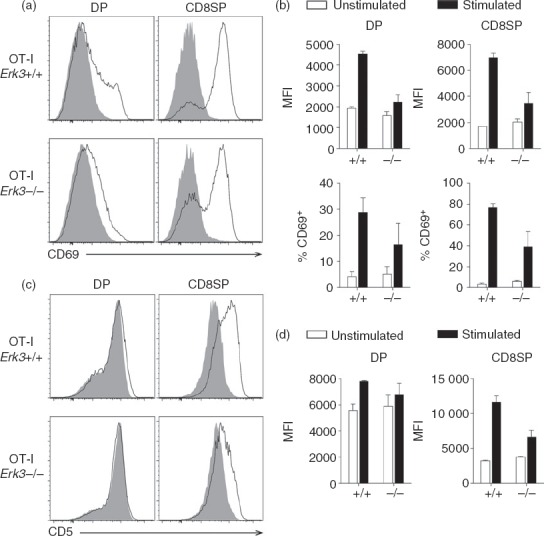
Figure 7.

Defective up-regulation of CD69 and CD5 expression in OT-I T-cell receptor (TCR) transgenic thymocytes lacking extracellular signal-regulated kinase 3 (ERK3) after TCR stimulation. OT-I thymocytes were incubated for 24 hr with (black line) or without (filled grey histogram) anti-CD3 antibodies for stimulation. The expression of CD69 (a) and CD5 (c) is shown for double-positive (DP) and CD8 single-positive (SP) thymocytes. (b) Compilation of the mean fluorescence intensity (MFI) of CD69 on CD69+ thymocytes (top panel) and percentage of CD69+ cells (bottom panel). (d) Compilation of the MFI of CD5 expression by unstimulated and stimulated cells is shown. The bar graphs represent the mean and the error bars are the standard error of the mean (SEM). The results of two independent experiments are shown.
