Abstract
Cleidocranial dysplasia (CCD) is an autosomal dominant disorder resulting in the skeletal and dental abnormalities due to the disturbance in ossification of the bones. Clavicle is the most commonly affected bone. The prevalence of CCD is one in millions of live births. In this report, we present a case of 10-years-old boy showing features of this condition.
Keywords: Clavicular hypoplasia, cleidocranial dysostosis, cleidocranial dysplasia
INTRODUCTION
Cleidocranial dysplasia (CCD) is a rare congenital disorder of autosomal dominant inheritance that leads to the disturbances in the growth of the bones of cranium, clavicle, and facial skeleton. The most important feature is clavicular hypoplasia which leads to the ability to approximate the shoulders anteriorly. Other clinical features includes frontal, parietal, occipital bossing, open fontanelles, wide pubic symphysis, and short stature.[1] Dental anomalies include multiple supernumerary teeth, retained deciduous dentition, delayed eruption or retention of the permanent dentition and high palate.[2,3] In this report, we present a 10-years-old male child with features of CCD.
CASE REPORT
A 10-years-old male child reported with his parents to our department with the chief complaints of disturbance in his dentition. General physical examination revealed a thin built, short stature, narrow thorax, and ability to approximate his shoulders anteriorly. He also had prominent forehead with frontal and occipital bossing, brachycephaly, mid-facial hypoplasia with depressed nasal bridge and hypertelorism [Figures 1 and 2]. Intraoral examination revealed a narrow high arched palate, unilateral anterior and posterior crossbite, multiple carious and retained deciduous teeth [Figure 3]. His clinical picture was suggestive of CCD.
Figure 1.
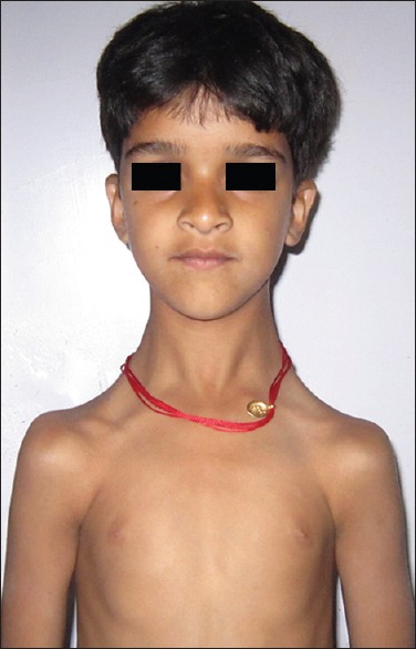
Frontal view of child showing prominent forehead, mid-facial hypoplasia with depressed nasal bridge and hypertelorism
Figure 2.
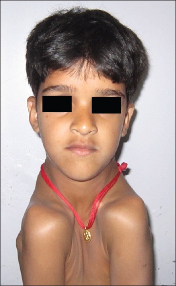
The photograph showing his ability to approximate shoulders anteriorly
Figure 3.
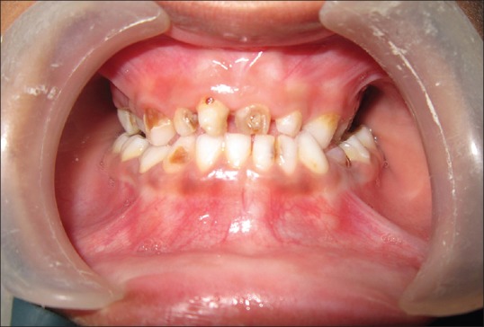
Intraoral photograph showing multiple carious and retained deciduous teeth, unilateral anterior and posterior crossbite
Radiological investigations included orthopantomogram (OPG) and chest radiograph. The OPG revealed multiple impacted permanent and supernumerary teeth [Figure 4]. Chest radiograph revealed narrow thorax with tapered width superiorly, severe hypoplastic clavicle over bilateral region [Figure 5]. Biochemical investigations showed normal serum calcium and phosphate level, but decreased serum alkaline phosphatase level. All investigations and radiograph were confirmatory for the diagnosis of CCD. The patient was then sent for evaluation and treatment of dental abnormalities, but he lost to regular follow-up.
Figure 4.
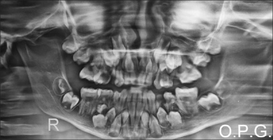
The orthopantomogram showing multiple impacted permanent and supernumerary teeth
Figure 5.
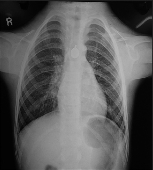
Chest radiograph showing narrow thorax and severe hypoplastic clavicle over bilateral region
DISCUSSION
Cleidocranial dysplasia is a rare autosomal dominant condition leading to disturbance in ossification of the bones of cranial and facial skeleton. The prevalence of CCD is one in millions of live births. This condition is associated with the mutation in the RUNX2 gene (also called Cbfa1 gene), which is located on the short arm of chromosome 6p21, encodes a protein necessary for the correct functioning of osteoblasts.[4] The Cbfa1 (RUNX2) gene controls normal development and growth of the human skeleton through the progression of intramembranous and endochondral ossification, and may be related to delayed ossification of the skull, teeth, pelvis, and clavicles.[5,6] It also results in delayed ossification of midline structures of the body, particularly membranous bone.
This condition is associated with various skeletal and dental deformities. The main skeletal deformity is hypoplasia of clavicle bone, affecting mostly lateral portion. In 10% of cases, clavicle is totally absent. This allows hypermobility of the shoulders resulting in the ability to touch them in front of the chest.[7]
The skull base is dysplastic with reduced growth, resulting in increased skull width and leading to brachycephaly and hypertelorism that are usually associated with frontal and bi-parietal bone bossing.[3] Other associated skeletal deformities are mid-face deficiency due to naso-orbito maxillary hypoplasia, prognathic mandible leading to class III skeletal malcclusion, depressed nasal bridge. Wide alar base, open skull fontanelle, deformities of the thoracic region, pelvic and pubic bones, short stature, short tapered fingers and anomalies of the phalangeal, tarsal, metatarsal, carpal and metacarpal bones.[1,8,9,10] Incidence of scoliosis, spina bifida and syringomelia have also been described. On rare occasions, brachial plexus irritation may occur.[11]
Dental abnormalities are typical main features of CCD, and they occur in 93.5% of affected patients.[12] The primary dentition usually develops relatively normally,[13] while the permanent dentition is severely disturbed. Supernumerary teeth, prolonged retention of the primary dentition, failed eruption of the permanent teeth, multiple crown and root abnormalities, crypt formation around impacted teeth, and ectopic locations of teeth are the more common dentition disturbances.[14] There is complete absence of cellular cementum and an increase in the amount of acellular cementum of the roots of the affected teeth.[15,16] A highly arched palate is a less common oral manifestation of CCD. In our patient, most of the above said that dental conditions were found.
Diagnosis of CCD is based on clinical and radiographic correlation. Management of such patient requires a multidisciplinary approach, including medical, dental and genetic specialties. Parental counseling and identifying the familial inheritance is very important.
Footnotes
Source of Support: Nil.
Conflict of Interest: None declared.
REFERENCES
- 1.Tanaka JL, Ono E, Filho EM, Castilho JC, Moraes LC, Moraes ME. Cleidocranial dysplasia: Importance of radiographic images in diagnosis of the condition. J Oral Sci. 2006;48:161–6. doi: 10.2334/josnusd.48.161. [DOI] [PubMed] [Google Scholar]
- 2.Daskalogiannakis J, Piedade L, Lindholm TC, Sándor GK, Carmichael RP. Cleidocranial dysplasia: 2 generations of management. J Can Dent Assoc. 2006;72:337–42. [PubMed] [Google Scholar]
- 3.Golan I, Baumert U, Hrala BP, Müssig D. Dentomaxillofacial variability of cleidocranial dysplasia: Clinicoradiological presentation and systematic review. Dentomaxillofac Radiol. 2003;32:347–54. doi: 10.1259/dmfr/63490079. [DOI] [PubMed] [Google Scholar]
- 4.Hemalatha R, Balasubramaniam MR. Cleidocranial dysplasia: A case report. J Indian Soc Pedod Prev Dent. 2008;26:40–3. doi: 10.4103/0970-4388.40322. [DOI] [PubMed] [Google Scholar]
- 5.Takeda S. Central control of bone remodeling. Biochem Biophys Res Commun. 2005;328:697–9. doi: 10.1016/j.bbrc.2004.11.071. [DOI] [PubMed] [Google Scholar]
- 6.Mundlos S. Cleidocranial dysplasia: Clinical and molecular genetics. J Med Genet. 1999;36:177–82. [PMC free article] [PubMed] [Google Scholar]
- 7.Juhl J. 7th ed. Philadelphia: Lippincott-Raven Publishers; 1998. Paul and Juhl's Essentials of Radiologic Imaging. [Google Scholar]
- 8.Zou SJ, D’Souza RN, Ahlberg T, Bronckers AL. Tooth eruption and cementum formation in the Run x2/Cbfa1 heterozygous mouse. Arch Oral Biol. 2003;48:673–7. doi: 10.1016/s0003-9969(03)00135-3. [DOI] [PubMed] [Google Scholar]
- 9.Garg RK, Agrawal P. Clinical spectrum of cleidocranial dysplasia: A case report. Cases J. 2008;1:377. doi: 10.1186/1757-1626-1-377. [DOI] [PMC free article] [PubMed] [Google Scholar]
- 10.Mohan RP, Suma GN, Vashishth S, Goel S. Cleidocranial dysplasia: Clinico-radiological illustration of a rare case. J Oral Sci. 2010;52:161–6. doi: 10.2334/josnusd.52.161. [DOI] [PubMed] [Google Scholar]
- 11.Weinstein S, Buckwalter J. 6th ed. Philadelphia: Lippincott Williams and Wilkins; 2005. Turek's Orthopaedics: Principles and their Application; pp. 251–2. [Google Scholar]
- 12.McNamara CM, O’Riordan BC, Blake M, Sandy JR. Cleidocranial dysplasia: Radiological appearances on dental panoramic radiography. Dentomaxillofac Radiol. 1999;28:89–97. doi: 10.1038/sj/dmfr/4600417. [DOI] [PubMed] [Google Scholar]
- 13.Jensen BL, Kreiborg S. Development of the dentition in cleidocranial dysplasia. J Oral Pathol Med. 1990;19:89–93. doi: 10.1111/j.1600-0714.1990.tb00803.x. [DOI] [PubMed] [Google Scholar]
- 14.Suba Z, Balaton G, Gyulai-Gaál S, Balaton P, Barabás J, Tarján I. Cleidocranial dysplasia: Diagnostic criteria and combined treatment. J Craniofac Surg. 2005;16:1122–6. doi: 10.1097/01.scs.0000179747.75918.58. [DOI] [PubMed] [Google Scholar]
- 15.Counts AL, Rohrer MD, Prasad H, Bolen P. An assessment of root cementum in cleidocranial dysplasia. Angle Orthod. 2001;71:293–8. doi: 10.1043/0003-3219(2001)071<0293:AAORCI>2.0.CO;2. [DOI] [PubMed] [Google Scholar]
- 16.Fukuta Y, Totsuka M, Fukuta Y, Takeda Y, Yoshida Y, Niitsu J, et al. Histological and analytical studies of a tooth in a patient with cleidocranial dysostosis. J Oral Sci. 2001;43:85–9. doi: 10.2334/josnusd.43.85. [DOI] [PubMed] [Google Scholar]


