

Abstract
There has always been a debate whether to remove or leave asymptomatic mesioangular impacted lower third molars. The present short communication discusses the merit of prophylactic removal to avoid clinical complications in future and the consequences to second molars.
Keywords: Lower wisdom teeth, mesioangular, third molars
INTRODUCTION
Impacted third molars are the most common developmental conditions that affect humans. The impactions are defined in relation to the geometric angle of impaction such as mesioangular, distoangular, and horizontal, and these can be partial or completely buried.
The NICE guidance on wisdom teeth describes the complications that may arise from the surgical removal of wisdom teeth but does not describe its benefits.[1] Clinical decision-making should be based on the relative benefits and harms for the patient. The important question is what the risk of retained wisdom teeth is in future for development of any pathology if left untreated.[2]
This short communication presents some of the practical difficulties frequently encountered in decision making for removal of mesioangular impacted lower third molars that are in contact with second molar.
CLINICAL EVIDENCE
Mesioangular-impacted lower third molars, which are completely covered with mucosa, create a deficient gingival collar that is not fluid-tight along distal cementoenamel junction of second molar.[3,4] This makes the second and third molars vulnerable to caries and rarely resorption of second molar if left untreated in long run.
Case 1
A 60-year-old man was advised to leave lower left mesioangular third molar that was asymptomatic and clinically covered with mucosa. On subsequent visit, he had occlusal decay with left lower second molar and was treated with gold crown by his dentist 15 years back. On recent visit, his orthopantomogram (OPG) showed mesioangular impaction with deep decay and recurrent decay with his left second molar under the crown distally and was referred for removal of lower third molar [Figure 1]. The lower left third molar was removed under local anesthesia, and the patient was referred back to dentist to treat recurrent decay with second molar [Figure 2].
Figure 1.
Orthopantomogram showing decayed mesioangular left lower third molar and recurrent decay with second molar
Figure 2.

Orthopantomogram postoperative after removal of wisdom tooth
Case 2
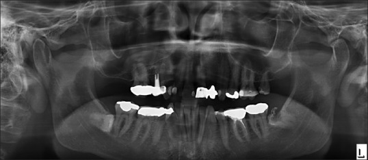
A 24-year-old man was referred for removal of wisdom teeth. Clinically, all his wisdom teeth were impacted and were asymptomatic. OPG showed lower third molars were mesioangular [Figure 3]. He was treated under general anesthetics in day surgery, and all his wisdom teeth were removed. His post-operative OPG showed distal resorption of right second molar which is rare but can be seen [Figure 4].
Figure 3.

Orthopantomogram showing mesioangular impacted third molars
Figure 4.

Orthopantomogram postoperative showing distal resorption of right second molar
DISCUSSION
This is well-known that mesioangular lower third molar are the potential cause of decay, and rarely resorption is associated with second molar. This raises another question that routine postoperative OPG is a must post-operatively after surgical removal of wisdom teeth to substantiate the findings of caries or resorption with second molars.
There is sufficient literature, which has been published from time to time, to reinstate these finding but NICE guidelines has remained the same.[4] There are no stringent guidelines to suggest that these situations should be dealt with prophylactic removal of third molars to avoid complications in future. Current UK clinical Guidelines for management of lower third molars are against the prophylactic removal of asymptomatic healthy impacted teeth.[3]
NICE guidelines were first published in 2000; since then the surgical skills, armamentarium, and tools for surgical pre assessment have improved considerably in avoiding surgical risk. It is time for NICE to review these guidelines to improve management of these situations.[2,5]
Footnotes
Source of Support: Nil.
Conflict of Interest: None declared.
REFERENCES
- 1.London: National Institute for Clinical Excellence; 2000. [Last accessed on 2014 Feb 9]. NHS National Institute for Clinical Excellence. NICE technology appraisal guidance number 1. Guidance on the extraction of wisdom teeth. Available from: http://www.nice.org.uk/nicemedia/pdf/wisdomteethguidance.pdf . [Google Scholar]
- 2.McArdle LW, McDonald F, Jones J. Distal cervical caries in mandibular second molars: An indication for the prophylactic removal of third molar teeth. Update? Br J Oral Maxillofac Surg. 2014;52:185–9. doi: 10.1016/j.bjoms.2013.11.007. [DOI] [PubMed] [Google Scholar]
- 3.Current clinical practice and parameters of care: The management of patients with third molar teeth. Faculty of Dental Surgery of the Royal College of Surgeons of England, 1997. [Last accessed on 2014 Feb 9]. Available from: http://www.rcseng.ac.uk/fds/publications.clinical.guidelines/clinical.guidelines/documents/3rdmolar.pdf .
- 4.McArdle LW, Renton T. The effects of NICE guidelines on the management of third molar teeth. Br Dent J. 2012;213:E8. doi: 10.1038/sj.bdj.2012.780. [DOI] [PubMed] [Google Scholar]
- 5.Allen RT, Witherow H, Collyer J, Roper-Hall R, Nazir MA, Mathew G. The mesioangular third molar- to extract or not to extract? Analysis of 776 consecutive third molars. Br Dent J. 2009;206:E 23. doi: 10.1038/sj.bdj.2009.517. [DOI] [PubMed] [Google Scholar]