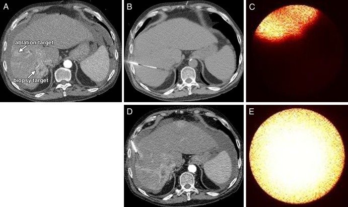
Figure 5:
A, CT scans in a 62-year-old man (patient 3) with HCV and alcoholic cirrhosis who underwent thermal ablation of an HCC in which biopsy was previously performed (ablation target), as well as percutaneous biopsy of a second, centrally necrotic focal hepatic lesion (biopsy target) in a single procedure setting. Contrast-enhanced CT guidance was used for performance of, B, percutaneous biopsy and, D, thermal ablation; optical molecular images show that both, C, biopsy target and, E, ablation target demonstrate avid ICG enhancement. Pathologic results for the biopsy lesion were consistent with a high-grade necrotic HCC.

