Abstract
Chemosynthetic Epsilonproteobacteria from deep-sea hydrothermal vents colonize substrates exposed to steep thermal and redox gradients. In many bacteria, substrate attachment, biofilm formation, expression of virulence genes and host colonization are partly controlled via a cell density-dependent mechanism involving signal molecules, known as quorum sensing. Within the Epsilonproteobacteria, quorum sensing has been investigated only in human pathogens that use the luxS/autoinducer-2 (AI-2) mechanism to control the expression of some of these functions. In this study we showed that luxS is conserved in Epsilonproteobacteria and that pathogenic and mesophilic members of this class inherited this gene from a thermophilic ancestor. Furthermore, we provide evidence that the luxS gene is expressed—and a quorum-sensing signal is produced—during growth of Sulfurovum lithotrophicum and Caminibacter mediatlanticus, two Epsilonproteobacteria from deep-sea hydrothermal vents. Finally, we detected luxS transcripts in Epsilonproteobacteria-dominated biofilm communities collected from deep-sea hydrothermal vents. Taken together, our findings indicate that the epsiloproteobacterial lineage of the LuxS enzyme originated in high-temperature geothermal environments and that, in vent Epsilonproteobacteria, luxS expression is linked to the production of AI-2 signals, which are likely produced in situ at deep-sea vents. We conclude that the luxS gene is part of the ancestral epsilonproteobacterial genome and represents an evolutionary link that connects thermophiles to human pathogens.
Introduction
Epsilonproteobacteria have been recognized as an ecologically significant group of bacteria in deep-sea geothermal environments as they are highly represented in moderately warm (25–35 °C) and oxidized as well as in relatively hot (40–70 °C) and anoxic vents (Longnecker and Reysenbach, 2001; Nakagawa et al., 2005; Campbell et al., 2006; Moussard et al., 2006; Huber et al., 2007; Sievert and Vetriani, 2012). At deep-sea hydrothermal vents, chemosynthetic Epsilonproteobacteria mediate the transfer of carbon and energy from the geothermal source to higher trophic levels, functioning as primary producers. The metabolic versatility observed in pure cultures of vent Epsilonproteobacteria (Campbell et al., 2006; Sievert and Vetriani, 2012), along with their high growth rates (Campbell et al., 2006) and ability to attach to substrates and form biofilms (Alain et al., 2004), is likely to contribute to the ecological success of these bacteria in environments characterized by high turbulence and steep thermal and redox gradients. Epsilonproteobacteria also include human pathogens (for example, Helicobacter and Campylobacter spp., Supplementary Figure S1) (Nakagawa et al., 2007), whose ability to colonize their hosts is related to high growth rates, attachment and biofilm formation (Stark et al., 1999; Joshua et al., 2006).
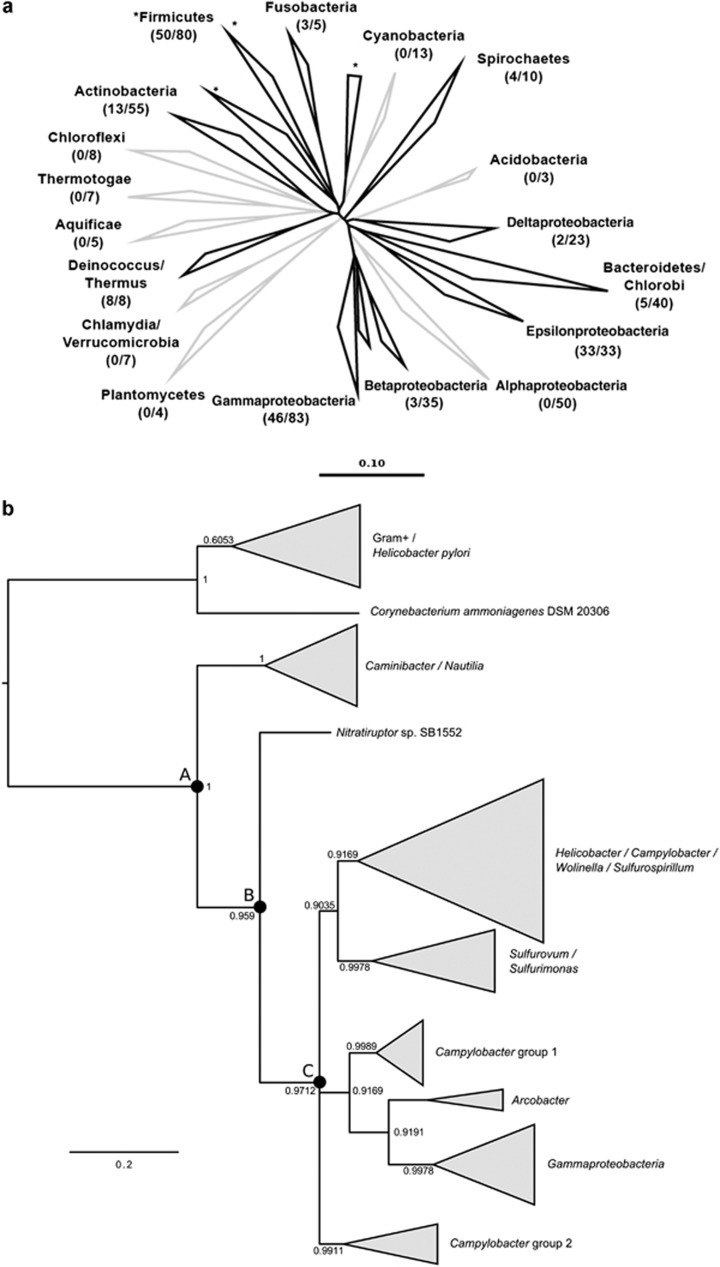
Quorum sensing (QS), a cell-to-cell communication mechanism that relies on cell density and the production of extracellular signaling molecules, is used by bacteria to control coordinated gene expression, including virulence genes and functions associated with biofilm formation and host colonization (Bassler et al., 1994; Keller and Surette, 2006). One QS system that appears to be widespread across the bacterial domain (Figure 1a), and that has been proposed to function as the universal language for interspecies communication, is based on a furanone derivative, known as autoinducer-2 (AI-2), synthesized by the LuxS enzyme (Kaper et al., 1999; Rader et al., 2007). The LuxS enzyme is an integral component of the activated methyl cycle (AMC), the major methyl donor in the cell (Vendeville et al., 2005; Turovskiy et al., 2007). In addition to its role as AI-2 synthase, the main function of the LuxS enzyme in the AMC is the conversion of S-ribosylhomocysteine to homocysteine, with the production of 4,5-dihydroxyl-2,3-pentanedione (DPD) as a by-product of this reaction (Chen et al., 2002; Winzer et al., 2003). Through processes of dehydration and cyclization, DPD is rearranged into a mixture of chemically related molecules that function as AI-2 chemical signals in QS. These molecules include 4-hydroxy-5-methyl-3 (2H) furanone (MHF), (2R, 4S)-2-methyl-2,3,3,4-tetrahydroxy-tetrhydrofuran (R-THMF) and furanosyl borate diester (S-THMF borate; (Schauder et al., 2001; Winzer et al., 2002; Chen et al., 2002; Winzer et al., 2003; Vendeville et al., 2005; Rader et al., 2007; Shen et al., 2010).
Figure 1.
(a) Distribution of the luxS gene among phyla of the domain Bacteria. The tree was constructed using the neighbor-joining method from the 16S ribosomal RNA genes of 469 representative bacterial species. The luxS homologs were indentified in 167 Beta-, Delta-, Gamma- and Epsilonproteobacteria, Firmicutes, Fusobacteria, Actinobacteria, Deinococcus/Thermus, Spirochaetes and Bacteroidetes/Chlorobi (branches shown in bold face). The number of luxS genes identified in each phylum is indicated in parenthesis, followed by the total number of genomes surveyed. The bar represents 0.10 base substitutions per nucleotide. The asterisks indicate branches that include Firmicutes. (b) Phylogenetic tree of LuxS amino acid sequences of Epsilonproteobacteria. The tree was constructed with Bayesian inference and maximum likelihood methods. The genera of the organisms within each collapsed branch are shown. Supplementary Table S6 lists all the organisms used in this analysis. The ancestral nodes A, B and C are shown as solid circles. Posterior probability values that support each topological element are shown at the branch nodes. Bar, 20% estimated substitutions.
The importance of luxS/AI-2 for the coordinated expression of virulence factors, biofilm formation and bioluminescence has been confirmed in a variety of microorganisms (Bassler et al., 1994; Forsyth and Cover, 2000; Wright et al., 2000; Elvers and Park, 2002; Shimizu et al., 2002; Stevenson and Babb, 2002; Winzer et al., 2002; McNab et al., 2003; Rhee et al., 2003). In Epsilonproteobacteria, the role of AI-2-mediated QS has been investigated exclusively in the human pathogens, Helicobacter and Campylobacter spp., where AI-2 secretion into the extracellular environment has been shown to occur by a LuxS-dependent mechanism (Forsyth and Cover, 2000; Joyce et al., 2000; Elvers and Park, 2002; Rader et al., 2007). In Helicobacter pylori, AI-2 modulates flagellar morphogenesis and gene expression in a dose-dependent manner (Rader et al., 2007; Shen et al., 2010; Rader et al., 2011). Similarly, in Campylobacter jejuni, luxS mutants showed reduced motility and attachment during host colonization (Plummer et al., 2012). LuxS-dependent motility/positioning patterns have been shown to be important virulence strategies required for colonization and dispersal of both Helicobacter and Campylobacter spp. in several animal models (Eaton et al., 1996; O'Toole et al., 2000; Ottemann and Lowenthal, 2002; Blaser and Atherton, 2004; Rader et al., 2007; Plummer et al., 2012).
As LuxS has been linked to the regulation of motility/colonization functions in pathogenic Epsilonproteobacteria (Eaton et al., 1996; Forsyth and Cover, 2000; Joyce et al., 2000; O'Toole et al., 2000; Wright et al., 2000; Elvers and Park, 2002; Rader et al., 2007; Shen et al., 2010; Plummer et al., 2012), we investigated the distribution of the luxS gene in Epsilonproteobacteria and we reconstructed the evolutionary history of the epsilonproteobacterial LuxS lineage. Furthermore, we used Sulfurovum lithotrophicum and Caminibacter mediatlanticus as model strains for mesophilic and thermophilic vent Epsilonproteobacteria, respectively, to investigate the expression of the luxS gene and the production of a QS signal during growth. Finally, we identified luxS transcripts in Epsilonproteobacteria-dominated chemosynthetic biofilms collected from active deep-sea hydrothermal vents.
Materials and methods
Organisms and growth conditions
S. lithotrophicum (DSM 23290T; Inagaki et al., 2004), a microaerobic, facultative anaerobic thiosulfate-oxidizing mesophile, was obtained from the German Collection of Microorganisms and Cell Cultures (Braunschweig, Germany). Caminibacter mediatlanticus strain TB-2 (DSM 16658T), a strictly anaerobic, hydrogen-oxidizing thermophile was isolated and described previously in our laboratory (Voordeckers et al., 2005). The two reporter strains, Vibrio harveyi strains BB170 and BB120 (Bassler et al., 1993), were kindly provided to us by Dr Chikindas (Rutgers University, New Brunswick, NJ, USA) and Dr Bassler (Princeton University, Princeton, NJ, USA). S. lithotrophicum was grown anaerobically (N2/CO2, 80%: 20%) at 30 °C in 356 MJ basal medium (Inagaki et al., 2004) supplemented with 10 mM Na2S2O3•5H2O and 14 mM KNO3 as electron donor and acceptor, respectively. C. mediatlanticus was grown at 55 °C in modified SME medium (Stetter et al., 1983) supplemented with 14 mM KNO3 under a gas phase of H2/CO2 (20%: 80%). Strains of V. harveyi were grown in modified Autoinducer Bioassay (AB) medium (Turovskiy and Chikindas, 2006). Time course experiments for S. lithotrophicum and C. mediatlanticus were performed in 1 liter of culture medium in triplicate.
Autoinducer-2 bioassay
The AI-2 detection assay utilizes the luxN-null mutant, V. harveyi BB170, as a reporter strain that produces bioluminescence only in response to AI-2 QS signals (Bassler et al., 1994). The assay was initiated by growing V. harveyi BB170 cultures for 16 h in modified Autoinducer-2 Bioassay (AB) medium (Turovskiy and Chikindas, 2006) with aeration at 30 °C. The cultures were diluted 1:5000 in fresh AB medium to obtain ∼105 CFUs per ml. Cell-free supernatant (CFS) preparations of S. lithotrophicum and C. mediatlanticus were added to the diluted V. harveyi to a 10% v/v final concentration, and these cultures were shaken (140 r.p.m.) at 30 °C for 5 and 6 h, respectively. Bioluminescence measurements were taken every 30 min with a Luminoscan TL plus luminometer (ThermoFisher Scientific, Waltham, MA, USA). Uninoculated S. lithotrophicum and C. mediatlanticus media were used as negative controls, whereas the CFS from the wild-type V. harveyi strain BB120 was used as a positive control. All bioluminescence induction values obtained from the three independent growth experiments for each bacterium are averages of duplicate AI-2 bioassays.
The activity of AI-2 is expressed in relative bioluminescence units. The relative bioluminescence units were normalized by subtracting the light intensity values of the negative control from the light intensity values of all samples (including the positive controls) in the assays. The relative bioluminescence unit values were expressed for both S. lithotrophicum and C. mediatlanticus as percentages of bioluminescence induction, where each measured time point was divided by the maximum bioluminescence induction value obtained from each bacterium, including the positive controls.
The CFSs of S. lithotrophicum and C. mediatlanticus were prepared from 10 ml of cell suspensions at different stages during the time course growth experiments. The CFS of V. harveyi BB120 (positive control) was prepared from cell suspensions of approximately OD600 of 0.5–0.6. All cell suspensions were filtered through a 0.2 μm-pore-size filter, centrifuged at 6000 × g for 30 min (Nalgene Nunc International, Rochester, NY, USA) and transferred into new tubes. Samples were stored at −20 °C until further processing.
Microscopy and cell counts
Cells were routinely stained with 0.1% acridine orange and visualized with an Olympus BX 60 microscope (Center Valley, PA, USA) with an oil immersion objective (UPlanF1 100/1.3). Samples for direct cell counts were collected to monitor cell growth in the time course experiments and at different time points during the AI-2 bioassay to determine the number of V. harveyi BB170 cells per ml (Supplementary Table S2).
RNA extraction and quantitative reverse transcription PCR (qRT-PCR) analyses of luxS transcripts in model organisms
Total RNA was isolated from S. lithotrophicum and C. mediatlanticus cultures at different phases during growth. Then, 75 ml of S. lithotrophicum cultures and 50 ml of C. mediatlanticus cultures were preserved in a 1:1 volume of RNAlater (Ambion, ThermoFisher Scientific) and stored at −80 °C until further processing. RNA was extracted from cell pellets using the RNAeasy Mini kit (Qiagen, Valencia, CA, USA) and treated with TURBO DNase (Ambion), as recommended by the manufacturer. The quantity and quality of the RNA obtained was evaluated both spectrophotometrically on a NanoDrop ND-1000 spectrophotometer (ThermoFisher Scientific) and by agarose gel electrophoresis.
Absolute quantification of luxS and 16S rRNA transcripts was carried out by qRT-PCR using the iScript One-Step RT-PCR kit with SYBR Green (Bio-Rad, Hercules, CA, USA) and the real-time detection system iCycler iQ (Bio-Rad). Reactions, each in triplicate, were carried out according to the manufacturer's instructions, using 0.15 μg of total RNA for S. lithotrophicum and 0.3 μg of total RNA for C. mediatlanticus in 15 μl reactions. Primers were manually designed and analyzed using the Oligo Analyzer 3.1 from IDT (Integrated DNA Technologies, Coralville, IA, USA) to have a composition suitable for use in qRT-PCR (Supplementary Table S3). Specificity of each pair of primers was confirmed by PCR amplification using the genomic DNA of the model organisms followed by sequencing. The efficiency of the PCR amplifications was determined from the slopes of the dilution curves of the target RNA.
Standard curves for the luxS and 16S rRNA genes were constructed using dilutions (range of standards: 103–107 copies per ml for S. lithotrophicum and C. mediatlanticus in a 15 μl reaction) of the corresponding PCR amplicon (Chini et al., 2007; Cuebas et al., 2011) using the iQ SYBR Green Supermix (Bio-Rad). The cycle threshold (Ct) values obtained were used to calculate the absolute number of transcript copies per ng of RNA for both strains (Supplementary Tables S4 and S5). The number of 16S rRNA transcripts was monitored as the internal standard to ensure that equal amounts of RNA were used in each reaction and to normalize the levels of luxS transcripts across samples (Supplementary Figure S2). Values for 16S rRNA and luxS transcript copies per ng presented here are an average of triplicate qRT-PCR experiments for the three independent growth experiments carried out for each model organism. Negative controls omitting the templates were included.
Sampling of natural biofilms from deep-sea hydrothermal vents
Samples of microbial biofilms were collected during R/V Atlantis cruises AT 15-15 (January–February 2007) and AT 15–28 (December 2007–January 2008) using the Deep-Submergence Vehicle Alvin. Both biofilms used in this study were collected using experimental microbial colonization devices made of stainless steel mesh. The first biofilm (designated as low T biofilm) was collected from a diffuse flow vent with no associated megafauna (‘Mk 33', fluid temperature: 10–28 °C, depth 2506 m, East Pacific Rise, 9° 50 N, 104° 18 W). The second biofilm (designated as high T biofilm) was collected from the wall of a sulfide structure colonized by Alvinella pompejana tubeworms venting 20–50 °C fluids (‘Jumeaux' site, depth 2621 m, East Pacific Rise, 12° 48 N, 103° 56 W).
Vent natural biofilms: RNA extraction, RT-PCR detection of luxS transcripts and library construction
Samples of the low T and high T biofilms were resuspended in RNAlater (Ambion) and stored at −80 °C. Total RNA was extracted from the biofilm samples using the RNAeasy kit (Qiagen) and the DNase-treated RNA (0.5 μg) was reversed transcribed in 25 μl reactions using SuperScript III One-Step RT-PCR System with Platinum Taq DNA Polymerase (Invitrogen, Carlsbad, CA, USA). Two sets of primers, the first based on the luxS genes of S. lithotrophicum and Sulfurimons denitrificans (SL/SD luxS primers; Supplementary Table S3) and the second based on the luxS genes of C. mediatlanticus and Nautilia profundicola (CM/NP luxS primers: Supplementary Table S3), were designed and used in RT-PCR reactions to amplify the luxS gene transcripts from the low T and high T biofilms. Both primer sets were used to amplify luxS transcripts in the biofilm samples.
Touchdown PCR conditions for all luxS amplifications were as follow: an initial denaturation for 5 min at 94 °C, followed by 10 cycles of denaturation at 94 °C for 30 s, annealing at 44 °C for 1 min, extension at 72 °C for 1 min and 30 s, followed by 30 cycles of denaturation at 94 °C for 30 s, annealing at 40 °C for 1 min, extension at 72 °C for 1 min and 30 s and a final extension of 5 min at 72 °C.
The amplified luxS transcripts were gel-purified using the MinElute Gel Extraction Kit (Qiagen), cloned into the pCR4-TOPO plasmid vector and the ligation products were transformed into Escherichia coli One Shot TOP10 Competent Cell using the TOPO TA Cloning Kit for Sequencing (Invitrogen). In all, 52 and 46 randomly chosen clones from the two luxS environmental libraries were analyzed, respectively, for insert-containing plasmids by direct PCR followed by gel electrophoresis of the amplified products. The resulting PCR fragments were screened by restriction fragment length polymorphism analysis using the RsaI restriction endonuclease (Boehringer Mannheim, Mannheim, Germany). The restriction fragment length polymorphism reactions were analyzed on a 3% MetaPhor Agarose (Lonza, Rockland, ME, USA) gel. Representative luxS genes with unique restriction fragment length polymorphism patterns were purified using the MiniPrep Plasmid Purification Kit (Qiagen) and sequenced.
PCR amplification of the luxS gene from reference strains
Genomic DNA extracted from 10 ml cultures of Caminibacter sp. strain TB1 (Voordeckers et al., 2005), Nautilia spp. strains MT3 and MT4 (Voordeckers et al., 2008), Nautilia nitratireducens strain MB-1 (Perez-Rodriguez et al., 2010) and S. lithotrophicum (Inagaki et al., 2004) was used for the amplification of partial luxS genes using the luxS primers CM/NP and SL/SD (Caminibacter/Nautilia and Sulfurovum/Sulfurimonas spp., respectively; Supplementary Table S3) and the touchdown PCR conditions described above. Long-term stocks of these reference strains, prepared by adding 50 μl of dimethyl sulfoxide to 1 ml of liquid cultures, are stored at −80 °C. Live cultures are available upon request.
Phylogenetic analyses
The distribution of the luxS gene within the Bacteria was investigated in 465 representative taxa within the bacterial domain by genome search or PCR amplification in reference strains. A phylogenetic tree based on the 16S ribosomal RNA genes of the taxa surveyed was then constructed using the ARB neighbor-joining method (http://www.arb-home.de/home.html; Figure 1a).
The phylogenetic tree in Figure 1b was constructed by aligning trimmed LuxS amino acid sequences from GenBank, using Muscle (Edgar, 2004) and Gblocks (Castresana, 2000). Trees were reconstructed with Bayesian inference and maximum likelihood methods. The maximum likelihood phylogeny was inferred from the alignment using PHYML (Guindon and Gascuel, 2003) with the CpREV substitution model and 1000 bootstrap resamplings. The substitution model was selected based on AIC values using ProTest3 (Darriba et al., 2011). Bayesian phylogeny was inferred using MrBayes (Ronquist and Huelsenbeck, 2003) performing 500 000 generations with two parallel searches with the CpREV amino acid matrix model (selected using forward selection among all possible substitution models) and a burn-in of 125 000 generations. The tree was rooted using the mid-point convergence method and manually using LuxS from Gram-positive bacteria as the outgroup. Both rooting methodologies gave the same results. The organisms used in the phylogenetic tree reconstruction shown in Figure 1b are listed in Supplementary Table S6. Ancestral state reconstruction of the LuxS sequence was performed using the MEGA package (Tamura et al., 2013) using the maximum likelihood option with the CpREV amino acid matrix model of substitution.
For the construction of the phylogenetic tree shown in Figure 3, the luxS sequences obtained by PCR from reference strains and natural vent biofilm communities were translated into amino acid sequences using EMBOSS Transeq (http://www.ebi.ac.uk/emboss/transeq/), aligned using ClustalX v 1.8 (Thompson et al., 1997) and manually adjusted using Seaview (Galtier et al., 1996). Phylogenetic distances were calculated using the CpREV substitution model and the maximum likelihood method to evaluate tree topologies and their robustness was tested by bootstrap analysis with 100 resamplings.
Nucleotide sequence accession numbers
The luxS sequences from this study are available through GenBank under the following accession numbers: KF876791-KF876794 for luxS genes in Caminibacter sp. strain TB1, Nautilia sp. strain MT3, Nautilia sp. strain MT4 and N. nitratireducens strain MB1; KF876760-KF876777 for luxS transcripts obtained from the low T biofilm; and KF876778-KF876790 for luxS transcripts obtained from high T biofilm.
Results
Distribution of the LuxS enzyme in the bacterial domain and reconstruction of the evolutionary history of the epsilonproteobacterial LuxS lineage
In this study we investigated the distribution of the luxS gene by searching representative genomes within the bacterial domain and we identified luxS gene homologs in 167 out of 469 bacterial taxa (Supplementary Tables S7 and S8). Representative species coding for luxS were found within the Beta- (5%), Delta- (9%), Gamma- (55%) and Epsilonproteobacteria (100%), Firmicutes (63%), Fusobacteria (60%), Actinobacteria (24%), Deinococcus/Thermus (100%), Spirochaetes (40%) and Bacteroidetes/Chlorobi (13%) (Figure 1a and Supplementary Table S7).
Bayesian phylogenetic analyses of the LuxS enzyme of Epsilonproteobacteria indicated that the lineage found in pathogenic, commensal and mesophilic Epsilonproteobacteria shared a common ancestor with members of the order Nautiliales (node A in Figure 1b; 100% posterior probability) and with Nitratiruptor sp. (node B in Figure 1b). All members of the Nautiliales and Nitratiruptor sp. are deep-sea vent thermophilic chemolithoautotrophs (Campbell et al., 2006; Sievert and Vetriani, 2012) (Figure 1b and Supplementary Table S6). A reconstruction of the amino acid sequence of the LuxS ancestral state indicated that the sequence corresponding to node A (Figure 1b) was 83.1% and 72.3% identical to the LuxS of C. mediatlanticus and Campylobacter fetus, respectively.
All the mesophilic Epsilonproteobacteria, which include environmental species of the genera Sulfurovum, Sulfurimonas, Arcobacter and Sulfurospirillum, the commensal organism Wolinella succinogenes and the pathogenic species, Campylobacter and Helicobacter, shared a common ancestor (node C in Figure 1b) nested within the thermophilic lineage. However, the topology of this section of the LuxS tree showed a polytomy; hence the evolutionary relationships between mesophilic, commensal and pathogenic Epsilonproteobacteria could not be resolved. Interestingly, a group of LuxS sequences found in members of the Gammaproteobacteria (including species of the genera Vibrio, Photobacterium, Salmonella, Escherichia and Enterobacter) clustered with the Arcobacter lineage and nested within a group of Campylobacter sequences (Campylobacter group 1 in Figure 1b). Finally, our analysis showed that the LuxS enzyme of H. pylori was related to the Gram+ lineage (Figure 1b).
Quantitative detection of luxS transcripts and AI-2 activity in S. lithotrophicum and C. mediatlanticus
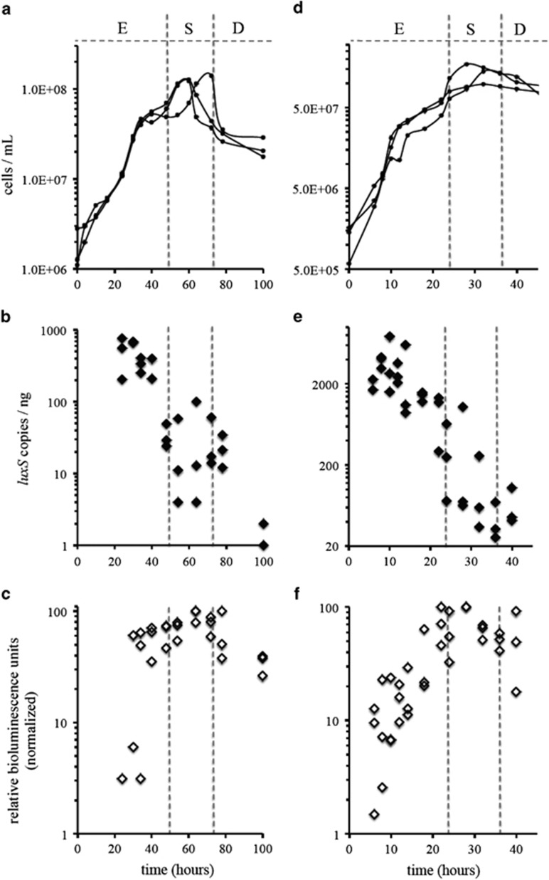
We carried out a time course experiment and quantified luxS transcripts at different phases during the growth cycle of these bacteria (Figures 2a and d). We found that in both S. lithotrophicum and C. mediatlanticus, the expression of the luxS gene was highest during the mid-exponential phase of growth and gradually dropped to baseline levels during the stationary phase (Figures 2b and e). The drop in luxS transcripts was ∼336-fold in S. lithotrophicum from 30–100 h of growth and ∼59-fold in C. mediatlanticus from 8 to 40 h of growth (Figures 2b and e).
Figure 2.
Growth curves for S. lithotrophicum (a) and C. mediatlanticus (d) cultured under nitrate-reducing conditions. Expression of the luxS gene (filled symbols) and induction of bioluminescence (open symbols) in S. lithotrophicum (b, c) and C. mediatlanticus (e, f). Exponential (E), stationary (S) and death (D) growth phases are labeled within each graph. All experiments were carried out in triplicates.
To establish whether S. lithotrophicum and C. mediatlanticus produced an AI-2-like QS signal during the time course experiment, we carried out an AI-2 detection assay using V. harveyi strain BB170, a reporter strain that produces bioluminescence in response to AI-2 QS signals (Bassler et al., 1994; Turovskiy et al., 2007). Indeed, supplementing cultures of V. harveyi BB170 with CFSs of S. lithotrophicum and C. mediatlanticus induced a bioluminescence response (Figures 2c and f). In both microorganisms, the observed bioluminescence induction increased with cell densities during growth (Figures 2c and f). Maximum bioluminescence induction (expressed in relative bioluminescence units) was observed during the stationary phase of growth, at 68.7±8.1 h for S. lithotrophicum and at 26±3.5 h for C. mediatlanticus (Supplementary Table S1). In fact, bioluminescence induction in V. harveyi BB170 was significantly correlated to cell density in both S. lithotrophicum (r=0.60, P<0.01, n=25) and C. mediatlanticus (r=0.63, P<0.01, n=33). Bioluminescence induction using the CFSs of V. harveyi strain BB120 (wild type) as a positive control was 2.5- and 2.2-fold higher than that measured when the CFSs from S. lithotrophicum and C. mediatlanticus were used, respectively (Supplementary Table S2). The net positive bioluminescence induction measured in V. harveyi BB170 in response to the CFSs of S. lithotrophicum and C. mediatlanticus indicated that growth of these organisms was necessary for the synthesis of AI-2-like molecules and to elicit a bioluminescence response in the reporter strain.
When changes in luxS transcription and bioluminescence induction at different times during growth were compared, a negative correlation was established between the two variables. Expression of the luxS gene (Figures 2b and e) in both S. lithotrophicum (r (24)=−0.68, P<0.01) and C. mediatlanticus (r (31)=−0.54, P<0.01) is inversely correlated to bioluminescence induction in V. harveyi (Figures 2c and f).
Detection of luxS transcripts in biofilm communities from deep-sea hydrothermal vents
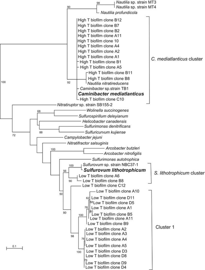
Following our finding that laboratory strains of vent Epsilonproteobacteria express the luxS gene during growth and elicit a QS response in a V. harveyi reporter strain, we hypothesized that the luxS gene was expressed in situ at deep-sea vents during the formation of microbial biofilms. To this end, we collected recently established biofilms (3 to 7 days old) from two vents located on the East Pacific Rise at 9° and 13° North, respectively, that were characterized by different thermal and biological regimes. Both the low T and high T biofilms were dominated by Epsilonproteobacteria (93.1% and 86.6%, respectively; data not shown). The luxS transcripts related to Epsilonproteobacteria were amplified by RT-PCR from RNA extracted from the biofilm communities. However, luxS transcripts from the low T biofilm were detected only when the SL/SD primers were used, whereas luxS transcripts from the high T biofilm were obtained only with the CM/NP primers. Phylogenetic analysis of the amino acid sequences of the LuxS enzyme deduced from RT-PCR-amplified transcripts showed that the high T biofilm expressed enzymes related to C. mediatlanticus, Caminibacter sp. strain TB-1 and N. nitratireducens (Figure 3; 98–99% amino acid identity to LuxS of C. mediatlanticus). In contrast, the LuxS retrieved from the low T biofilm was distributed in three clusters. The majority of the sequences (cluster 1 in Figure 3) were grouped in three subclusters with 82–83% amino acid identity to the LuxS of S. lithotrophicum. Two sequences were related to the enzyme of S. lithotrophicum and Sulfurovum sp. strain NBC37-1 (low T biofilm A6 and B8; 88% and 87% identity to the enzyme of S. lithotrophicum, respectively), whereas a single sequence (low T biofilm C12) was placed in a discrete cluster (83% identity to the LuxS of S. lithotrophicum).
Figure 3.
Maximum likelihood phylogenetic tree inferred from the amino acid sequence deduced from a fragment of the luxS gene transcripts obtained from the high and low T biofilm communities collected from deep-sea hydrothermal vents on the East Pacific Rise. The tree shows the position of the biofilm-derived LuxS sequences relative to that of pure cultures of Epsilonproteobacteria. The model organisms, Caminibacter mediatlanticus and Sulfurovum lithotrophicum, are shown in bold face. Percentages >50% of 100 bootstrap resampling that support each topological element are shown at the branch nodes. Bar, 10% estimated substitutions.
Discussion
Thermophilic ancestry of the LuxS lineage and luxS gene flow in Epsilonproteobacteria
Bayesian reconstruction of the LuxS evolutionary history indicated a thermophilic origin of the epsilonproteobacterial lineage (Figure 1b). This finding is supported by the observation that the LuxS sequences from mesophilic, commensal and pathogenic Epsilonproteobacteria are nested within the thermophilic clades and that the ancestral state reconstruction of the LuxS sequence (corresponding to node A in Figure 1b) showed higher amino acid identity to the enzymes found in modern thermophiles than those found in modern mesophiles. As all members of the Nautiliales and Nitratiruptor sp. are thermophiles, the most parsimonious interpretation of the data is that the ancestral LuxS proteins represented by nodes A and B were in thermophilic Epsilonproteobacteria, and that a more recent ancestor was transferred to mesophilic Epsilonproteobacteria between nodes B and C (Figure 1b). This interpretation is in line with phylogenomic and 16S rRNA gene-based reconstructions of the epsilonproteobacterial tree that showed the Nautiliales as the first line of divergence (Zhang and Sievert, 2014). Overall, our data suggest that the epsilonproteobacterial luxS lineage originated in geothermal environments. However, the direction of the luxS gene flow among mesophilic Epsilonproteobacteria (for example, from environmental strains to commensals/pathogens of warm-blooded animals or vice versa) was less defined and remains unresolved (Figure 1b).
Our phylogenetic analysis revealed two events of luxS horizontal gene transfer within the Epsilonproteobacteria. H. pylori appears to have acquired the gene from Enterococcus faecium, a Gram-positive bacterium that, like H. pylori, inhabits the human gastrointestinal tract (Figure 1b). This finding is in line with previous studies that showed that the LuxS from H. pylori was related to the enzyme found in members of the Firmicutes (Lerat and Moran, 2004; Doherty et al., 2010). Moreover, marine Gammaproteobacteria of the genera Photobacterium and Vibrio, along with E. coli and other human-associated bacteria, appear to share a common luxS ancestor with Arcobacter nitrofigilis and A. butzleri, a marine organism and a human pathogen, respectively (Figure 1b and Supplementary Table S6). These findings indicate that bacteria inhabiting similar ecological niches, regardless of their taxonomic distance, share closely related forms of the luxS gene. Because habitat and niche play an important role in horizontal gene transfer (Polz et al., 2013), we suggest that interspecies QS within the same habitat is an important driver in the evolution of luxS.
luxS expression and AI-2 activity in laboratory cultures of vent Epsilonproteobacteria
Here we present one of the few studies on how the expression of the luxS gene relates to AI-2 activity. In Salmonella typhimurium, the constitutive expression of luxS suggests that AI-2 production is regulated by the availability of the LuxS substrate, S-ribosylhomocysteine (Beeston and Surette, 2002). In contrast, the expression of luxS in E. coli and Edwardsiella spp. is under the control of catabolite repression, and it is correlated with growth and with AI-2 activity levels (Wang et al., 2005; Zhang et al., 2008; Zhang and Sun, 2012). In comparison, the number of luxS transcripts and AI-2 extracellular activity levels in S. lithotrophicum and C. mediatlanticus showed highest luxS expression during the mid and late exponential phases of growth, whereas maximum AI-2 activity levels occurred during the stationary phase (Figures 2b–f). This temporal uncoupling between luxS expression and AI-2 activity levels is conserved in both vent strains and may be indicative of a mechanism of feedback inhibition of luxS expression, perhaps controlled by the concentration of AI-2.
LuxS function in vent Epsilonproteobacteria: QS vs AMC
The dual function of LuxS as AI-2 synthase and in recycling methionine via the homocysteine precursor in the AMC cannot be ignored when investigating its role in new species. However, in geothermal environments, where reduced sulfur is not limiting, the de novo synthesis of methionine is thought to be energetically inexpensive; hence, the function of LuxS in recycling methionine may not be as critical as in other habitats (Winzer et al., 2003). This consideration suggests that luxS expression in vent Epsilonproteobacteria may be more directly involved in AI-2 production rather than in recycling methionine. Moreover, the ability of H. pylori to synthesize cysteine under an incomplete AMC via the reverse transsulfuration pathway further suggests the importance of LuxS in AI-2 production in Epsilonproteobacteria (Doherty et al., 2010; Shen et al., 2010).
Although we have not identified the specific furanone derivative responsible for the signaling activity that we measured in our experiments, previous studies showed that the precursor of AI-2, DPD, spontaneously cyclizes into different isomers, including S-THMF borate with AI-2 activity in V. harveyi, R-THMF with AI-2 activity in S. typhimurium and the less active metabolic by-product, MHF (Feather, 1981; Nedvidek et al., 1992). In particular, MHF has been shown to exhibit at least a 10-fold reduction in activity compared with S-THMF borate when tested as a QS-inducing molecule in V. harveyi reporter strains (Feather, 1981; Nedvidek et al., 1992; Schauder et al., 2001; Winzer et al., 2003). The bioluminescence induced by S. lithotrophicum and C. mediatlanticus was very similar (2.5- and 2.2-fold lower, respectively) to that induced by the CFSs of the wild-type V. harveyi strain BB120, suggesting that the CFSs of the vent strains contained an active form of AI-2-like molecule with a much higher activity than MHF. Furthermore, as bioluminescence induction by S. lithotrophicum and C. mediatlanticus was measured using similar concentration of V. harveyi cells in all the assays (Supplementary Table S2), our results indicate that the gradual increase in bioluminescence induction during growth (Figures 2a and d) reflected the increasing production of AI-2-like molecules in the CFS of the two strains (Figures 2c and f). We argue that the significant correlation between AI-2 activity levels and cell density in our model organisms could, in principle, provide a measure of population density.
As no AI-2 receptors have yet been identified in Epsilonproteobacteria, the complete luxS/AI-2 QS circuit remains unclear. Two main classes of AI-2 receptors have been characterized: the LuxP family receptors, found in Vibrio spp. (Chen et al., 2002), and the LrsB family, found in S. typhimurium and other enteric bacteria (Miller et al., 2004). In addition, the AI-2-binding capability has been demonstrated in RbsB-like receptors of the ribose binding protein family (Armbruster et al., 2011). However, no homologs to these receptors have been found in Epsilonproteobacteria. Hence, other receptors must exist, as responses to AI-2 have been reported in bacteria lacking LuxP, LsrB and RbsB receptors. For instance, the established role of luxS/AI-2 in cell–cell signaling in H. pylori in the absence of LuxP, LsrB or RbsB homologs suggests the existence of yet unidentified receptors (Doherty et al., 2010). This is further supported by the small sequence similarity between LuxP, LrsB and RbsB (Pereira et al., 2009) that likely evolved independently while converging toward the same function.
Vent Epsilonproteobacteria express the luxS gene in situ during the formation of biofilms
We demonstrated that the luxS gene was expressed in situ in two biofilms from active vents characterized by different thermal regimes. The luxS gene transcripts that we identified in these biofilms reflected the distribution of specific taxa of Epsilonproteobacteria along the thermal gradient. Transcripts related to Sulfurovum and Sulfurimonas spp., two sulfur-oxidizing mesophiles, were detected in the low T biofilm, whereas transcripts related to Caminibacter and Nautilia spp., two hydrogen-oxidizing thermophiles, were identified in the high T biofilm (Figure 3). In light of our finding that in S. lithotrophicum and C. mediatlanticus the expression the luxS gene is correlated to AI-2-like activity and to a QS response in V. harveyi, the detection of epsilonproteobacterial luxS transcripts in vent microbial biofilms implies that AI-2 signals are produced in situ. It is reasonable to hypothesize that, similar to what has been reported in H. pylori and C. jejuni, AI-2 activity in vent Epsilonproteobacteria might be involved in controlling the genes involved in flagellar motility, adhesion and other functions associated with the formation of biofilms.
Concluding remarks
Epsilonproteobacteria are a versatile group of bacteria that include chemolithoautotrophic thermophiles and mesophiles inhabiting marine geothermal environments, symbionts of invertebrates, commensals of mammals and human pathogens (Dubilier et al., 2008; Ruehland and Dubilier, 2010). Identifying the genes encoded by the core—or ancestral—genome shared by Epsilonproteobacteria may reveal how these microorganisms evolved to colonize very diverse habitats. The luxS gene is part of the epsilonproteobacterial core genome and represents an evolutionary link that connects vent thermophiles to human pathogens. Why is luxS/AI-2, rather than other QS mechanisms, conserved in Epsilonproteobacteria? These bacteria live in environments, such as the deep-sea vents, the rumen of cows and the human stomach, characterized by harsh physicochemical conditions. DPD, the precursor of AI-2, is very stable over a broad pH range, whereas other QS signaling molecules, such as acylhomoserine lactones and autoinducing peptides, appear to have inherent susceptibility to hydrolytic degradation (Globisch et al., 2012). In line with this hypothesis, a study where the vent hyperthermophiles Thermotoga maritima and Pyrococcus furiosus were found to produce AI-2 via a luxS-independent biotic/abiotic reaction pathway also evidenced the stability of AI-2 at elevated temperature (Nichols et al., 2009). Hence, it is possible that the chemical stability of DPD has played a role in the selection of AI-2 as a chemical signal in Epsilonproteobacteria.
Although it is known that luxS/AI-2 regulates functions associated with biofilm formation in pathogenic Epsilonproteobacteria (Eaton et al., 1996; O'Toole et al., 2000; Ottemann and Lowenthal, 2002; Blaser and Atherton, 2004; Rader et al., 2007; Plummer et al., 2012), its involvement in attachment and colonization of substrates in geothermal environments remains unknown. Future efforts leading to the development of a genetic system in vent Epsilonproteobacteria will provide the necessary tools to further understand the luxS/AI-2 QS system in geothermal environments.
Acknowledgments
We express our appreciation to Dr Michael Chikindas and Dr Bonnie Bassler for making reporter strains and laboratory equipment for the AI-2 assays available to us. We thank Dr Stefan Sievert for inviting us on cruise AT 15-28 and Dr Donato Giovannelli for his assistance with phylogenetic analyses. We also thank the crew of R/V Atlantis and the crew and pilots of the DSV Alvin for their skilled operations at sea. This work was supported by NSF Grants MCB 04-56676, MCB 08-43678 and OCE 11-36451 to CV and by a PEO scholar award and a Carnegie Postdoctoral Fellowship to IP-R. This paper is C-DEBI contribution 232.
The authors declare no conflict of interest.
Footnotes
Supplementary Information accompanies this paper on The ISME Journal website (http://www.nature.com/ismej)
Supplementary Material
References
- Alain K, Zbinden M, Le Bris N, Lesongeur F, Querellou J, Gaill F, et al. Early steps in microbial colonization processes at deep-sea hydrothermal vents. Environ Microbiol. 2004;6:227–241. doi: 10.1111/j.1462-2920.2003.00557.x. [DOI] [PubMed] [Google Scholar]
- Armbruster CE, Pang B, Murrah K, Juneau RA, Perez AC, Weimer KED, et al. RbsB (NTHI_0632) mediates quorum signal uptake in nontypeable Haemophilus influenzae strain 86-028NP. Mol Microbiol. 2011;82:836–850. doi: 10.1111/j.1365-2958.2011.07831.x. [DOI] [PMC free article] [PubMed] [Google Scholar]
- Bassler BL, Wright M, Showalter RE, Silverman MR. Intercellular signalling in Vibrio harveyi: sequence and function of genes regulating expression of luminescence. Mol Microbiol. 1993;9:773–786. doi: 10.1111/j.1365-2958.1993.tb01737.x. [DOI] [PubMed] [Google Scholar]
- Bassler BL, Wright M, Silverman MR. Multiple signalling systems controlling expression of luminescence in Vibrio harveyi: sequence and function of genes encoding a second sensory pathway. Mol Microbiol. 1994;13:273–286. doi: 10.1111/j.1365-2958.1994.tb00422.x. [DOI] [PubMed] [Google Scholar]
- Beeston AL, Surette MG. pfs-dependent regulation of autoinducer 2 production in Salmonella enterica serovar Typhimurium. J Bacteriol. 2002;184:3450–3456. doi: 10.1128/JB.184.13.3450-3456.2002. [DOI] [PMC free article] [PubMed] [Google Scholar]
- Blaser MJ, Atherton JC. Helicobacter pylori persistence: biology and disease. J Clin Invest. 2004;113:321–333. doi: 10.1172/JCI20925. [DOI] [PMC free article] [PubMed] [Google Scholar]
- Campbell BJ, Engel AS, Porter ML, Takai K. The versatile epsilon-proteobacteria: key players in sulphidic habitats. Nat Rev Microbiol. 2006;4:458–468. doi: 10.1038/nrmicro1414. [DOI] [PubMed] [Google Scholar]
- Castresana J. Selection of conserved blocks from multiple alignments for their use in phylogenetic analysis. Mol Biol Evol. 2000;17:540–552. doi: 10.1093/oxfordjournals.molbev.a026334. [DOI] [PubMed] [Google Scholar]
- Chen X, Schauder S, Potier N, Van Dorsselaer A, Pelczer I, Bassler BL, et al. Structural identification of a bacterial quorum-sensing signal containing boron. Nature. 2002;415:545–549. doi: 10.1038/415545a. [DOI] [PubMed] [Google Scholar]
- Chini V, Foka A, Dimitracopoulos G, Spiliopoulou I. Absolute and relative real-time PCR in the quantification of tst gene expression among methicillin-resistant Staphylococcus aureus: evaluation by two mathematical models. Lett Appl Microbiol. 2007;45:479–484. doi: 10.1111/j.1472-765X.2007.02208.x. [DOI] [PubMed] [Google Scholar]
- Cuebas M, Villafane A, McBride M, Yee N, Bini E. Arsenate reduction and expression of multiple chromosomal ars operons in Geobacillus kaustophilus A1. Microbiology. 2011;157:2004–2011. doi: 10.1099/mic.0.048678-0. [DOI] [PubMed] [Google Scholar]
- Darriba D, Taboada GL, Doallo R, Posada D. ProtTest 3: fast selection of best-fit models of protein evolution. Bioinformatics. 2011;27:1164–1165. doi: 10.1093/bioinformatics/btr088. [DOI] [PMC free article] [PubMed] [Google Scholar]
- Doherty NC, Shen F, Halliday NM, Barrett DA, Hardie KR, Winzer K, et al. In Helicobacter pylori, LuxS is a key enzyme in cysteine provision through a reverse transsulfuration pathway. J Bacteriol. 2010;192:1184–1192. doi: 10.1128/JB.01372-09. [DOI] [PMC free article] [PubMed] [Google Scholar]
- Dubilier N, Bergin C, Lott C. Symbiotic diversity in marine animals: the art of harnessing chemosynthesis. Nat Rev Microbiol. 2008;6:725–740. doi: 10.1038/nrmicro1992. [DOI] [PubMed] [Google Scholar]
- Eaton KA, Suerbaum S, Josenhans C, Krakowka S. Colonization of gnotobiotic piglets by Helicobacter pylori deficient in two flagellin genes. Infect Immun. 1996;64:2445–2448. doi: 10.1128/iai.64.7.2445-2448.1996. [DOI] [PMC free article] [PubMed] [Google Scholar]
- Edgar RC. MUSCLE: multiple sequence alignment with high accuracy and high throughput. Nucleic Acids Res. 2004;32:1792–1797. doi: 10.1093/nar/gkh340. [DOI] [PMC free article] [PubMed] [Google Scholar]
- Elvers KT, Park SF. Quorum sensing in Campylobacter jejuni: detection of a luxS encoded signalling molecule. Microbiology. 2002;148:1475–1481. doi: 10.1099/00221287-148-5-1475. [DOI] [PubMed] [Google Scholar]
- Feather MS. Amine-assisted sugar dehydration reactions. Prog Food Nutr Sci. 1981;5:37–45. [Google Scholar]
- Forsyth MH, Cover TL. Intercellular communication in Helicobacter pylori: luxS is essential for the production of an extracellular signaling molecule. Infect Immun. 2000;68:3193–3199. doi: 10.1128/iai.68.6.3193-3199.2000. [DOI] [PMC free article] [PubMed] [Google Scholar]
- Galtier N, Gouy M, Gautier C. SEAVIEW and PHYLO_WIN: two graphic tools for sequence alignment and molecular phylogeny. Comput Appl Biosci. 1996;12:543–548. doi: 10.1093/bioinformatics/12.6.543. [DOI] [PubMed] [Google Scholar]
- Globisch D, Lowery CA, McCague KC, Janda K. Uncharacterized 4,5-dihydroxy-2,3-pentanedione (DPD) molecules revealed through NMR spectroscopy: implications for a greater signaling diversity in bacterial species. Angew Chem Int Ed. 2012;51:4204–4208. doi: 10.1002/anie.201109149. [DOI] [PMC free article] [PubMed] [Google Scholar]
- Guindon S, Gascuel O. A simple, fast, and accurate algorithm to estimate large phylogenies by maximum likelihood. Syst Biol. 2003;52:696–704. doi: 10.1080/10635150390235520. [DOI] [PubMed] [Google Scholar]
- Huber JA, Welch DB, Morrison HG, Huse SM, Neal PR, Butterfield DA, et al. Microbial population structures in the deep marine biosphere. Science. 2007;318:97–100. doi: 10.1126/science.1146689. [DOI] [PubMed] [Google Scholar]
- Inagaki F, Takai K, Nealson KH, Horikoshi K. Sulfurovum lithotrophicum gen. nov., sp. nov., a novel sulfur-oxidizing chemolithoautotroph within the epsilon-Proteobacteria isolated from Okinawa Trough hydrothermal sediments. Int J Syst Evol Microbiol. 2004;54:1477–1482. doi: 10.1099/ijs.0.03042-0. [DOI] [PubMed] [Google Scholar]
- Joshua GW, Guthrie-Irons C, Karlyshev AV, Wren BW. Biofilm formation in Campylobacter jejuni. Microbiology. 2006;152:387–396. doi: 10.1099/mic.0.28358-0. [DOI] [PubMed] [Google Scholar]
- Joyce EA, Bassler BL, Wright A. Evidence for a signaling system in Helicobacter pylori: detection of a luxS-Encoded autoinducer. J Bacteriol. 2000;182:3638–3643. doi: 10.1128/jb.182.13.3638-3643.2000. [DOI] [PMC free article] [PubMed] [Google Scholar]
- Kaper JB, Sperandio V, Mellies JL, Nguyen W, Shin S. Quorum sensing controls expression of the type III secretion gene transcription and protein secretion in enterohemorrhagic and enteropathogenic Escherichia coli. Proc Natl Acad Sci USA. 1999;96:15196–15201. doi: 10.1073/pnas.96.26.15196. [DOI] [PMC free article] [PubMed] [Google Scholar]
- Keller L, Surette MG. Communication in bacteria: an ecological and evolutionary perspective. Nat Rev Microbiol. 2006;4:249–258. doi: 10.1038/nrmicro1383. [DOI] [PubMed] [Google Scholar]
- Lerat E, Moran NA. The evolutionary history of quorum-sensing systems in bacteria. Mol Biol Evol. 2004;21:903–913. doi: 10.1093/molbev/msh097. [DOI] [PubMed] [Google Scholar]
- Longnecker K, Reysenbach AL. Expansion of the geographic distribution of a novel lineage of epsilon-Proteobacteria to a hydrothermal vent site on the Southern East Pacific Rise. FEMS Microbiol Ecol. 2001;35:287–293. doi: 10.1111/j.1574-6941.2001.tb00814.x. [DOI] [PubMed] [Google Scholar]
- McNab R, Ford SK, El-Sabaeny A, Barbieri B, Cook GS, Lamont RJ. LuxS-based signaling in Streptococcus gordonii: autoinducer 2 controls carbohydrate metabolism and biofilm formation with Porphyromonas gingivalis. J Bacteriol. 2003;185:274–284. doi: 10.1128/JB.185.1.274-284.2003. [DOI] [PMC free article] [PubMed] [Google Scholar]
- Miller ST, Xavier KB, Campagna SR, Taga ME, Semmelhack MF, Bassler BL, et al. Salmonella typhimurium recognizes a chemically distinct form of the bacterial quorum-sensing signal Al-2. Mol Cell. 2004;15:677–687. doi: 10.1016/j.molcel.2004.07.020. [DOI] [PubMed] [Google Scholar]
- Moussard H, Corre E, Cambon-Bonavita MA, Fouquet Y, Jeanthon C. Novel uncultured Epsilonproteobacteria dominate filamentous sulphur mat from the 13°N hydrothermal vent field, East Pacific Rise. FEMS Microbiol Ecol. 2006;58:449–463. doi: 10.1111/j.1574-6941.2006.00192.x. [DOI] [PubMed] [Google Scholar]
- Nakagawa S, Takai K, Inagaki F, Hirayama H, Nunoura T, Horikoshi K, et al. Distribution, phylogenetic diversity and physiological characteristics of epsilon-Proteobacteria in a deep-sea hydrothermal field. Environ Microbiol. 2005;7:1619–1632. doi: 10.1111/j.1462-2920.2005.00856.x. [DOI] [PubMed] [Google Scholar]
- Nakagawa S, Takaki Y, Shimamura S, Reysenbach AL, Takai K, Horikoshi K. Deep-sea vent ɛ-proteobacterial genomes provide insights into emergence of pathogens. Proc Natl Acad Sci USA. 2007;104:12146–12150. doi: 10.1073/pnas.0700687104. [DOI] [PMC free article] [PubMed] [Google Scholar]
- Nedvidek W, Ledl F, Fischer P. Detection of 5-hydroxymethyl-2-methyl-3(2H)-furanone and of α-dicarbonyl compounds in reaction mixtures of hexoses and pentoses with different amines. Z Lebensm Unters Forsch. 1992;194:222–228. [Google Scholar]
- Nichols JD, Johnson MR, Chou CJ, Kelly RM. Temperature, not LuxS, mediates AI-2 formation in hydrothermal habitats. FEMS Microbiol Ecol. 2009;68:173–181. doi: 10.1111/j.1574-6941.2009.00662.x. [DOI] [PubMed] [Google Scholar]
- O'Toole PW, Lane MC, Porwollik S. Helicobacter pylori motility. Microbes Infect. 2000;2:1207–1214. doi: 10.1016/s1286-4579(00)01274-0. [DOI] [PubMed] [Google Scholar]
- Ottemann KM, Lowenthal AC. Helicobacter pylori uses motility for initial colonization and to attain robust infection. Infect Immun. 2002;70:1984–1990. doi: 10.1128/IAI.70.4.1984-1990.2002. [DOI] [PMC free article] [PubMed] [Google Scholar]
- Pereira CS, de Regt AK, Brito PH, Miller ST, Xavier KB. Identification of functional LsrB-like autoinducer-2 receptors. J Bacteriol. 2009;191:6975–6987. doi: 10.1128/JB.00976-09. [DOI] [PMC free article] [PubMed] [Google Scholar]
- Perez-Rodriguez I, Ricci J, Voordeckers JW, Starovoytov V, Vetriani C. Nautilia nitratireducens sp nov., a thermophilic, anaerobic, chemosynthetic, nitrate-ammonifying bacterium isolated from a deep-sea hydrothermal vent. Int J Syst Evol Microbiol. 2010;60:1182–1186. doi: 10.1099/ijs.0.013904-0. [DOI] [PubMed] [Google Scholar]
- Plummer P, Sahin O, Burrough E, Sippy R, Mou K, Rabenold J, et al. Critical role of LuxS in the virulence of Campylobacter jejuni in a guinea pig model of abortion. Infect Immun. 2012;80:585–593. doi: 10.1128/IAI.05766-11. [DOI] [PMC free article] [PubMed] [Google Scholar]
- Polz MF, Alm EJ, Hanage WP. Horizontal gene transfer and the evolution of bacterial and archaeal population structure. Trends Genet. 2013;29:170–175. doi: 10.1016/j.tig.2012.12.006. [DOI] [PMC free article] [PubMed] [Google Scholar]
- Rader BA, Campagna SR, Semmelhack MF, Bassler BL, Guillemin K. The quorum-sensing molecule autoinducer 2 regulates motility and flagellar morphogenesis in Helicobacter pylori. J Bacteriol. 2007;189:6109–6117. doi: 10.1128/JB.00246-07. [DOI] [PMC free article] [PubMed] [Google Scholar]
- Rader BA, Wreden C, Hicks KG, Sweeney EG, Ottemann KM, Guillemin K. Helicobacter pylori perceives the quorum-sensing molecule AI-2 as a chemorepellent via the chemoreceptor TlpB. Microbiology. 2011;157:2445–2455. doi: 10.1099/mic.0.049353-0. [DOI] [PMC free article] [PubMed] [Google Scholar]
- Rhee JH, Kim SY, Lee SE, Kim YR, Kim CM, Ryu PY, et al. Regulation of Vibrio vulnificus virulence by the LuxS quorum-sensing system. Mol Microbiol. 2003;48:1647–1664. doi: 10.1046/j.1365-2958.2003.03536.x. [DOI] [PubMed] [Google Scholar]
- Ronquist F, Huelsenbeck JP. MrBayes 3: Bayesian phylogenetic inference under mixed models. Bioinformatics. 2003;19:1572–1574. doi: 10.1093/bioinformatics/btg180. [DOI] [PubMed] [Google Scholar]
- Ruehland C, Dubilier N. Gamma- and epsilonproteobacterial ectosymbionts of a shallow-water marine worm are related to deep-sea hydrothermal vent ectosymbionts. Environ Microbiol. 2010;12:2312–2326. doi: 10.1111/j.1462-2920.2010.02256.x. [DOI] [PubMed] [Google Scholar]
- Schauder S, Shokat K, Surette MG, Bassler BL. The LuxS family of bacterial autoinducers: biosynthesis of a novel quorum-sensing signal molecule. Mol Microbiol. 2001;41:463–476. doi: 10.1046/j.1365-2958.2001.02532.x. [DOI] [PubMed] [Google Scholar]
- Shen F, Hobley L, Doherty N, Loh JT, Cover TL, Sockett RE, et al. In Helicobacter pylori auto-inducer-2, but not LuxS/MccAB catalysed reverse transsulphuration, regulates motility through modulation of flagellar gene transcription. BMC Microbiol. 2010;10:210. doi: 10.1186/1471-2180-10-210. [DOI] [PMC free article] [PubMed] [Google Scholar]
- Shimizu T, Ohtani K, Hayashi H. The luxS gene is involved in cell-cell signalling for toxin production in Clostridium perfringens. Mol Microbiol. 2002;44:171–179. doi: 10.1046/j.1365-2958.2002.02863.x. [DOI] [PubMed] [Google Scholar]
- Sievert SM, Vetriani C. Chemoautotrophy at deep-sea vents: past, present, and future. Oceanography. 2012;25:218–233. [Google Scholar]
- Stark RM, Gerwig GJ, Pitman RS, Potts LF, Williams NA, Greenman J, et al. Biofilm formation by Helicobacter pylori. Lett Appl Microbiol. 1999;28:121–126. doi: 10.1046/j.1365-2672.1999.00481.x. [DOI] [PubMed] [Google Scholar]
- Stetter KO, König H, Stackebrandt E. Pyrodictium, a new genus of submarine disc-shaped sulfur reducing archaebacteria growing optimally at 105°C. Syst Appl Microbiol. 1983;4:535–551. doi: 10.1016/S0723-2020(83)80011-3. [DOI] [PubMed] [Google Scholar]
- Stevenson B, Babb K. LuxS-mediated quorum sensing in Borrelia burgdorferi, the Lyme disease spirochete. Infect Immun. 2002;70:4099–4105. doi: 10.1128/IAI.70.8.4099-4105.2002. [DOI] [PMC free article] [PubMed] [Google Scholar]
- Tamura K, Stecher G, Peterson D, Filipski A, Kumar S. MEGA6: molecular evolutionary genetics analysis version 6.0. MolBiol Evol. 2013;30:2725–2729. doi: 10.1093/molbev/mst197. [DOI] [PMC free article] [PubMed] [Google Scholar]
- Thompson JD, Gibson TJ, Plewniak F, Jeanmougin F, Higgins DG. The CLUSTAL_X windows interface: flexible strategies for multiple sequence alignment aided by quality analysis tools. Nucleic Acids Res. 1997;25:4876–4882. doi: 10.1093/nar/25.24.4876. [DOI] [PMC free article] [PubMed] [Google Scholar]
- Turovskiy Y, Chikindas ML. Autoinducer 2 bioassay is a qualitative, not quantitative method influenced by glucose. J Microbiol Methods. 2006;66:497–503. doi: 10.1016/j.mimet.2006.02.001. [DOI] [PubMed] [Google Scholar]
- Turovskiy Y, Kashtanov D, Paskhover B, Chikindas ML. Quorum sensing: fact, fiction, and everything in between. Adv Appl Microbiol. 2007;62:191–234. doi: 10.1016/S0065-2164(07)62007-3. [DOI] [PMC free article] [PubMed] [Google Scholar]
- Vendeville A, Winzer K, Heurlier K, Tang CM, Hardie KR. Making ‘sense' of metabolism: autoinducer-2, LuxS and pathogenic bacteria. Nat Rev Microbiol. 2005;3:383–396. doi: 10.1038/nrmicro1146. [DOI] [PubMed] [Google Scholar]
- Voordeckers J, Starovoytov V, Vetriani C. Caminibacter mediatlanticus sp. nov., a thermophilic, chemolithoautotrophic, nitrate ammonifying bacterium isolated from a deep-sea hydrothermal vent on the Mid-Atlantic Ridge. Int J Syst Evol Microbiol. 2005;55:773–779. doi: 10.1099/ijs.0.63430-0. [DOI] [PubMed] [Google Scholar]
- Voordeckers JW, Do M, Hügler M, Ko V, Sievert SM, Vetriani C. Culture dependent and independent analyses of 16S rRNA and ATP citrate lyase genes: a comparison of microbial communities from different black smoker chimneys on the Mid-Atlantic Ridge. Extremophiles. 2008;12:627–640. doi: 10.1007/s00792-008-0167-5. [DOI] [PubMed] [Google Scholar]
- Wang L, Li J, March JC, Valdes JJ, Bentley WE. luxS-Dependent gene regulation in Escherichia coli K-12 revealed by genomic expression profiling. J Bacteriol. 2005;187:8350–8360. doi: 10.1128/JB.187.24.8350-8360.2005. [DOI] [PMC free article] [PubMed] [Google Scholar]
- Winzer K, Hardie KR, Williams P. LuxS and autoinducer-2: their contribution to quorum sensing and metabolism in bacteria. Adv Appl Microbiol. 2003;53:291–396. doi: 10.1016/s0065-2164(03)53009-x. [DOI] [PubMed] [Google Scholar]
- Winzer K, Sun Y, Green A, Delory M, Blackley D, Hardie KR, et al. Role of Neisseria meningitidis luxS in cell to cell signaling and bacteremic infection. Infect Immun. 2002;70:2245–2248. doi: 10.1128/IAI.70.4.2245-2248.2002. [DOI] [PMC free article] [PubMed] [Google Scholar]
- Wright A, Joyce EA, Bassler BL. Evidence for a signaling system in Helicobacter pylori: detection of a luxS-Encoded autoinducer. J Bacteriol. 2000;182:3638–3643. doi: 10.1128/jb.182.13.3638-3643.2000. [DOI] [PMC free article] [PubMed] [Google Scholar]
- Zhang M, Sun K, Sun L. Regulation of autoinducer 2 production and luxS expression in a pathogenic Edwardsiella tarda strain. Microbiology. 2008;154:2060–2069. doi: 10.1099/mic.0.2008/017343-0. [DOI] [PubMed] [Google Scholar]
- Zhang M, Sun L. Edwardsiella ictaluri LuxS: activity, expression, and involvement in pathogenicity. Pol J Microbiol. 2012;61:263–271. [PubMed] [Google Scholar]
- Zhang Y, Sievert SM. Pan-genome analyses identify lineage- and niche-specific markers of evolution and adaptation in Epsilonproteobacteria. Front Microbiol. 2014;5:110. doi: 10.3389/fmicb.2014.00110. [DOI] [PMC free article] [PubMed] [Google Scholar]
Associated Data
This section collects any data citations, data availability statements, or supplementary materials included in this article.


