Abstract
Introduction:
Lipomas are among the most common tumors of the human body. However, they are uncommon in the oral cavity and are observed as slow growing, painless, and asymptomatic yellowish submucosal masses. Surgical excision is the treatment of choice and recurrence is not expected.
Case Report:
The case of a 30-year-old woman with a huge lipoma on the tip of her tongue since 3 years, is presented. She had difficulty with speech and mastication because the tongue tumor was filling the oral cavity. Clinical examination revealed a yellowish lesion, measuring 8 cm in maximum diameter, protruding from the lingual surface. The tumor was surgically excised with restoration of normal tongue function and histopathological examination of the tumor confirmed that it was a lipoma.
Conclusion:
Tongue lipoma is rarely seen and can be a cause of macroglossia. Surgical excision for lipoma is indicated for symptomatic relief and exclusion of associated malignancy.
Key Words: Lipoma, Tongue, Tumor
Introduction
Lipomas are the most common benign soft tissue mesenchymal neoplasm; however, they are not common in the oral cavity (1,2) and are usually observed as slow-growing, painless, and asymptomatic masses. It is known that, with continued growth, their size may interfere with speech and mastication (3,4). Oral lipomas can occur in various anatomic sites including the major salivary glands, buccal mucosa, lip, tongue, palate, vestibule, and the floor of the mouth. Various case reports have described lipomas and its variants in several locations (5-7). The buccal mucosa and the tongue are the predominant sites in adults. Some studies show a male preference while other studies show no gender differences (8-10). The tumors are either encapsulated, non-encapsulated, or present in an infiltrating manner. Oral lipoma usually occurs as a solitary lesion. The color is often yellow in tone, depending on the thickness of the overlying mucosa.
Case Report
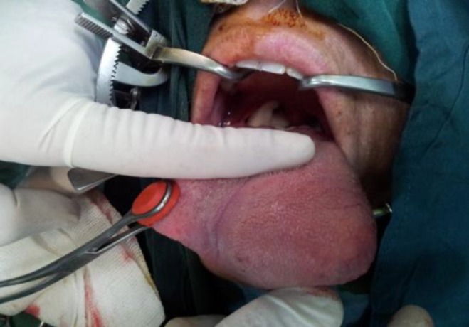
A 30-year-old female was presented to the head and neck clinic with a slow-growing mass on the tip of her tongue, which had been present for 3 years. Her speech was not clear due to the bulkiness of the mass and she also had difficulties swallowing. Clinical examination revealed a yellowish lesion measuring 8 cm in diameter protruding from the lingual surface and covered by a mucosa that was rich in vessels. During palpation, the lesion was observed to be rubbery and compressible (Fig.1).
Fig1.
Mass in antrolateral part of the tongue; during palpation, the lesion was observed to be rubbery and compressible.
Although the tongue movement was limited, there was no ankyloglossia. Taste and somatic sensation were intact.
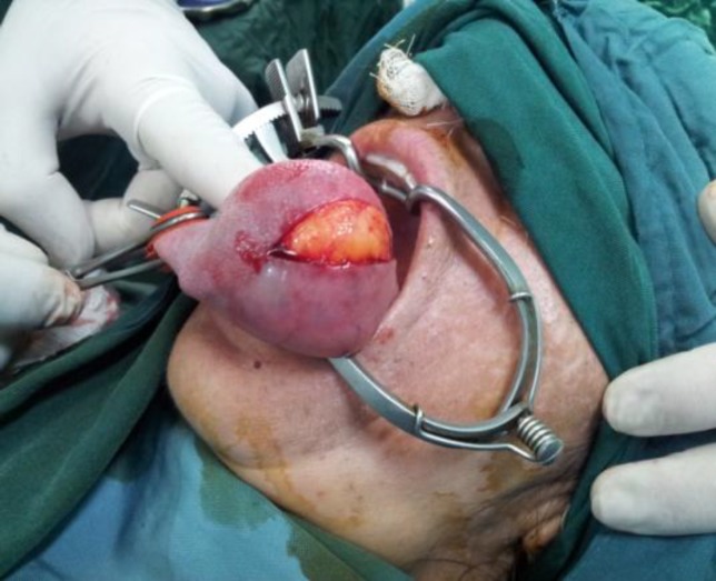
Under general anesthesia, through a longitudinal incision along the edge of the tongue, the tumor was removed (Fig. 2).
Fig 2.
Under general anesthesia, through a longitudinal incision along the edge of the tongue, the tumor was removed
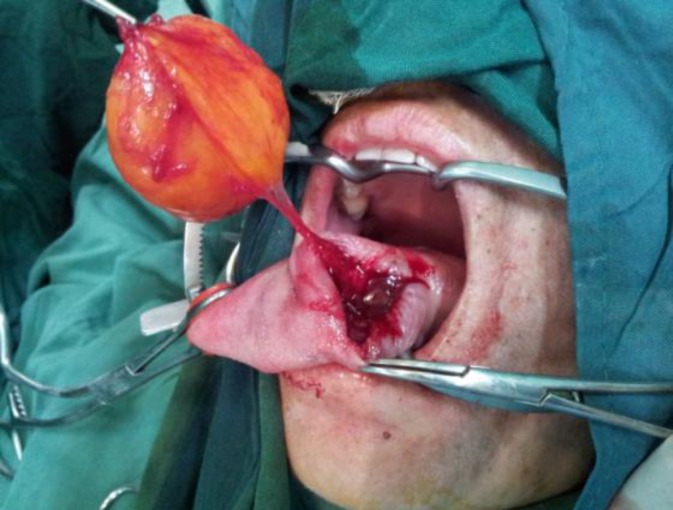
The tumor was yellowish in color and well-encapsulated (Fig.3). The mucosal layers were close together with absorbable sutures obliterating the dead space. The musculature volume of the left side of tongue was decreased.
Fig 3.
The tumor, which was yellowish in color, 8 cm in diameter, and well-encapsulated, was completely removed
Histological examination revealed mature adipocytes without cellular atypia. Lipoblast, which is pathognomonic for malignant liposarcoma, was not present.
Discussion
The majority of tongue tumors are malignant in nature. Lingual lipoma, which accounts for 0.3% of tongue neoplasms, is a benign condition. Similarly, the occurrence in the oral cavity is rare and reported as 2% to 4% of all lipomas (11,12). It is typically described as well- circumscribed, submucosal, with less than 1 cm swelling, and located on the lateral edge of the anterior two-thirds of the tongue surface (13). Microscopically, it is composed of mature adipocytes; however, in 20% of cases, it demonstrates histological variants that include spindle cell lipoma, pleomorphic lipoma, angiolipoma, fibrolipoma, myxoidlipoma, and atypical lipoma.
In this study the patient was a 30-year-old female with a slow growing mass present since the last 3 years, which measured 8 cm in diameter. The mass was painless but she had difficulties swallowing and tongue movement was impaired; however, taste and somatic sensation was intact.
In other studies: Chunkitchung reported a 62-year-old man with a 6cm mass in his tongue that was slow growing for 2 years. He had difficulties swallowing large food items. Moreover, his speech was not very clear due to the bulkiness of the mass (14).
Magadum reported a 60-year-old man with a 3cm mass in his tongue, which he had first noticed about 10 years earlier. Because of the absence of pain and bleeding, he was not initially alarmed, but later he complained of masticatory problems (15).
Chidzonga reported a 58-year-old female with an 11cm mass that had been present for 3 years. She had a large “anterior open bite” and slurred speech with the tumor bobbing up and down and in and out of the mouth when speaking. Despite the feeding and breathing difficulties, she was well nourished and not in any particular distress (16). Chandak also reported a 75-year-old man with a mass on the anterior border of the tongue, which he had first noticed 16 years earlier. He had difficulty in mastication and swallowing, and frequently used to wake up from sleep because of obstruction in his airway (17).
Finally Colella reported a 75-year-old man with a 10 cm mass in his tongue from 30 years ago. His speech was not very clear due to the bulkiness of the mass and he had difficulties swallowing (18).
These studies are generally without gender predilection (4,9,10,19-21); however, some studies have shown a male preponderance (13). Lipomas may be observed as solitary or multiple lesions, such as Gardner’s or Bournville’s syndrome (19,22), or as macroglossia (22-26) or lipomatosis (27).
Their clinical course is usually asymptomatic until they grow to large sizes (19,22). In the present case, the large size interfered with speech and mastication, similarly to a case reported by Gray and Baker (22). Large tumors have been shown to cause dentofacial deformities and anterior open bite (9,10). On rare occasions, the infiltration is so extensive that it can cause muscle dysfunction or sensory changes due to pressure on nerve trunks. Pain is rarely severe (28,29). The average duration of the lipoma before excision is 3.2 years with a range of 6 weeks to 15 years (21). The usual range in size is 0.5 to 8 centimeters (21). The present case was 8 centimeters in diameter.
The differential diagnosis includes well-differentiated liposarcoma, ranula, dermoid cyst, thyroglossal duct cyst, ectopic thyroid tissue, pleomorphic adenoma, and mucoepidemoid carcinoma angiolipoma, fibrolipoma, and malignant lymphoma (19,23-26). The definitive diagnosis is by microscopic examination, which shows adult fat tissue cells embedded in a stroma of connective tissue and surrounded by a fibrous capsule (26). Lipoma has a characteristic radiographic appearance. On CT scan it shows a high density from 83 to 143 Hamsfield units with well or poorly defined margins depending on the capsule (19). Ultrasonography shows a lesion, which is round or elliptical in shape with an intact or mostly intact capsule (30).
Surgical excision is the most common form of treatment (19,21). Recurrence is reduced by wide surgical excision while preserving the surrounding structures. Well-encapsulated lipomas, as the present case, easily shell out with no possibility of recurrence or damage to the surrounding structures. It is still advisable to excise them with a little cuff of surrounding normal tissue to prevent recurrence while still conserving surrounding structures (11).
Conclusion
Tongue lipoma is rarely seen and can be a cause of macroglossia. Surgical excision for lipoma is indicated for symptomatic relief and exclusion of associated malignancy
References
- 1.Fletcher CDM, Unni KK, Mertens F. Pathology and genetics: tumours of soft tissue and bone. World Health Organization classification of tumours: IARC Press; 2000. Adipocytic tumors; pp. 9–46. [Google Scholar]
- 2.de Freitas MA, Freitas VS, de Lima AA, Pereira FB Jr, dos Santos JN. Intraorallipomas: a study of 26 cases in a Brazilian population. Quintessence Int. 2009;40(1):79–85. [PubMed] [Google Scholar]
- 3.Dattilo DJ, Ige J T, Nwana EJC. Intraoral lipoma of the tongue and submandibular space. J Oral MaxillofacSurg. 1996;54(7):915–7. doi: 10.1016/s0278-2391(96)90549-2. [DOI] [PubMed] [Google Scholar]
- 4.Keskin G, Ustundag E, Ercin C. Multiple infiltrating lipomas of the tongue. J LaryngolOtol. 2002;116(05):395–7. doi: 10.1258/0022215021910906. [DOI] [PubMed] [Google Scholar]
- 5.Taira Y, Yasukawa K, Yamamori I, Lino M. Oral lipoma extending superiorly from mandibular gingivobuccal fold to gingival: a case report and analysis of 207 patients with oral lipoma in Japan. Odontology j. 2012;100(1):104–8. doi: 10.1007/s10266-011-0027-0. [DOI] [PubMed] [Google Scholar]
- 6.Rafieiyan N, Hamian M, Anbari F, Abdolsamadi HR. Lipoma of the tongue: a case report. DJH . 2011;2(1):59–61. [Google Scholar]
- 7.Lee SH, Yoon HJ. Bilateral asymmetric tongue classic lipomas. Oral Surg Oral Med Oral Pathol Oral RadiolEndod . 2012;114(1):e15–8. doi: 10.1016/j.tripleo.2011.07.045. [DOI] [PubMed] [Google Scholar]
- 8.Epivatianos A, Markopoulos AK, Papanayonou P. Benign tumors of adipose tissue of the oral cavity: a clinicopathologic study of 13 cases. J Oral MaxillofacSurg. 2000;58(10):1113–17. doi: 10.1053/joms.2000.9568. [DOI] [PubMed] [Google Scholar]
- 9.Fregnani ER, Pires FR, Falzoni R, Lopes MA, Vargas PA. Lipomas of the oral cavity: clinical findings, Histological classification and proliferative activity of 46 cases. Int J Oral MaxillofacSurg. 2003;32:49–53. doi: 10.1054/ijom.2002.0317. [DOI] [PubMed] [Google Scholar]
- 10.Lawoyin JO, Akande OO, Kolude B, Agbaje JO. Lipoma of the oral cavity: clinicopathological review of seven cases form Ibadan. Niger J Med. 2001;10(4):189–91. [PubMed] [Google Scholar]
- 11.Moore PL, Goede A, Phillips DE, Carr RP. Atypical lipoma of the tongue. J LaryngolOtol. 2001;115(10):859–61. doi: 10.1258/0022215011909198. [DOI] [PubMed] [Google Scholar]
- 12.Thomas S, Varghese BT, Sebastian P, Koshy CM, Mathews A, Abraham EK. Intramuscular lipomatosis of tongue. Postgrad Med J. 2002;78(919):295–7. doi: 10.1136/pmj.78.919.295. [DOI] [PMC free article] [PubMed] [Google Scholar]
- 13.Guillou L, Dehon A, Charlin B, Madarnas P. Pleomorphic lipoma of the tongue: case report and literature review. J Otolaryngol. 1986;15(5):313–6. [PubMed] [Google Scholar]
- 14.Chun-Kit Chung J, Wai-Man Ng R. A huge tongue lipoma. Otolaryngology–Head and Neck Surgery. 2007;137:830–1. doi: 10.1016/j.otohns.2007.07.014. [DOI] [PubMed] [Google Scholar]
- 15.Magadum D, Sanadi A, Manish Agrawal J, Suresh Agrawal M. Classic tongue lipoma: a common tumour at a rare site. BMJ Case Rep.[internet] 2013 doi: 10.1136/bcr-2012-007987. doi:10.1136/bcr-2012-007987. [DOI] [PMC free article] [PubMed] [Google Scholar]
- 16.Chidzonga MM, Mahomva L, Marimo C. Gigantic tongue lipoma: A case report. Med Oral Patol Oral Cir Bucal. 2006;11:437–9. [PubMed] [Google Scholar]
- 17.Chandak S, Pandilwar PK, Chandak T, Mundhada R. Huge lipoma of tongue. Contemporary Clinical Dentistry. 2012;3(4):507–9. doi: 10.4103/0976-237X.107457. [DOI] [PMC free article] [PubMed] [Google Scholar]
- 18.Colella G, Biondi P, Caltabiano R, Vecchio GM, Amico P, Magro G. Giant intramuscular lipoma of the tongue: a case report and literature review. Cases Journal. 2009;2 doi: 10.4076/1757-1626-2-7906. [DOI] [PMC free article] [PubMed] [Google Scholar]
- 19.Del Castillo- Pando De Vera JL, Cebrian– Carretero JL, Gomez-Garan E. Chronic lingual ulceration cased by lipoma of the oral cavity. Med Oral. 2004;9(2):166–7. [PubMed] [Google Scholar]
- 20.Enzenger FM. Soft tissue tumors. St Louis: Mosby; 1983. pp. 223–7. [Google Scholar]
- 21.Furlong MA, Fanburg-Smith JC, Childers EL. Lipoma of the oral and Maxillofacial region: Site and subclassifications of 125 cases. Oral Surg Oral Med Oral Pathol Oral Rad Endod . 2004;98:441–50. doi: 10.1016/j.tripleo.2004.02.071. [DOI] [PubMed] [Google Scholar]
- 22.Gray A R, Barker G R. Sublingual lipoma. Report of an unusually large lesion. J Oral MaxillofacSurg. 1991;49:747–50. doi: 10.1016/s0278-2391(10)80241-1. [DOI] [PubMed] [Google Scholar]
- 23.Capodiferro S, Scully C, Maiorano E, Lo Muzio L, Favia G. Liposarcomacircumscriptum (lipoma-like) of the tongue: report of a case. Oral Dis. 2004;10(6):398–400. doi: 10.1111/j.1601-0825.2004.01040.x. [DOI] [PubMed] [Google Scholar]
- 24.Favia G, Maiorano E, Orsini G, Piatelli A. Myxoidliposarcoma of the oral cavity with involvement of the periodontal tissues. J ClinPeriodontol. 2004;28(2):109–12. doi: 10.1034/j.1600-051x.2001.028002109.x. [DOI] [PubMed] [Google Scholar]
- 25.Nunes FD, Loducca SVL, de Oliveira EMF, de Araujo VC. Well-differentiated liposarcoma of the tongue. Oral Oncol. 2002;38(1):117–19. doi: 10.1016/s1368-8375(01)00030-6. [DOI] [PubMed] [Google Scholar]
- 26.Piattelli A, Rubies C, Fioroni M, Steches G. Spindle-cell lipoma of the cheek: a case report. Oral Oncol. 2000;36(5):495–6. doi: 10.1016/s1368-8375(00)00035-x. [DOI] [PubMed] [Google Scholar]
- 27.Katou F, Shirai N, Moategi K, Satoh R, Satoh S. Symmetrical lipomatosis of the tongue presenting as macroglossia. Report of two cases. J Craniomaxillofac. 1993;21(7):298–301. doi: 10.1016/s1010-5182(05)80351-8. [DOI] [PubMed] [Google Scholar]
- 28.Pelissier A, Sawaf MH, Shabana AH. Infiltrating (intramuscular) benign lipoma of the head and neck. J Oral MaxillofacSurg. 1991;49(11):1231–6. doi: 10.1016/0278-2391(91)90425-l. [DOI] [PubMed] [Google Scholar]
- 29.Piattelli A, Fioroni M, Rubini C. Intramuscular lipoma of the cheek: a case report. J Oral MaxillofacSurg. 2000;58(7):817–19. doi: 10.1053/joms.2000.7281. [DOI] [PubMed] [Google Scholar]
- 30.Zhong LP, Zhao SF, Chen GF, Ping FY. Ultrasonographic appearance of lipoma in the oral and maxillofacial region. Oral Surg Oral Med Oral Pathol Oral RadiolEndod. 2004;98(6):738–40. doi: 10.1016/j.tripleo.2004.04.022. [DOI] [PubMed] [Google Scholar]


