Abstract
A 90-year-old patient presented with a large cutaneous horn (cornu cutaneum) of nine-year duration arising at her right cheek. The lesion was removed by surgery. Histology was reported as cornu cutaneum with a well-differentiated squamous cell carcinoma at its base. Cutaneous horn is morphological designation for protuberant mass of keratin that resembles the horn of an animal. Such lesions appear on sun-exposed skin areas like upper parts of the face and ears in elderly patients. Large cutaneous horns (> 1 cm) tend to be more commonly associated with squamous cell carcinoma compared to smaller cutaneous horns, particularly when present on the face.
Keywords: actinic keratosis, aged, cornu cutaneum, cutaneous horn, precancerous conditions, skin cancer
Case
Cutaneous horn is a relatively uncommon lesion, also known by the Latin name "cornu cutaneum". Although there are multiple reports of cutaneous horns, giant cutaneous horns are much rarer. Cutaneous horns resemble animal horns at first glance. Most of them develop on face and scalp, but any part of the body can be involved.
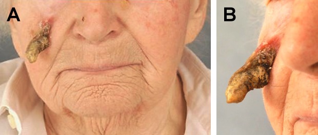
A 90-year-old woman presented with a nine-year history of a large cutaneous horn arising at her right cheek. No history of trauma could be elicited. The patient had a history of long-term sun exposure due to farm activities and had solar keratosis on face and extremities. Clinical examination revealed actinic keratosis on left forehead and firm keratinized conical lesion with a height of 5 cm [Fig. 1] on right cheek. There was no regional lymphadenopathy. Excision of the lesion with an elliptical incision and primary closure of defect was done. Histopathological examination revealed a completely excised cornu cutaneum with a well-differentiated squamous cell carcinoma at its base [Fig. 2].
Figure 1.

A: cornu cutaneum on the right cheek and actinic keratosis on left forehead, B: cornu cutaneum on right cheek, detail.
Figure 2.
A: Histologic examination revealed a highly differentiated squamous cell carcinoma at the base of the cutaneous horn (haematoxylin and eosin, original magnification x40). B: Dermal invasion of the carcinoma cells (original magnification ×200).
Discussion
Cutaneous horn (cornu cutaneum), though uncommon, has already been described as early as 1588 in a woman in London.[1] During the sixteenth and seventeenth century cutaneous horns were believed to be a natural anomaly. Data on the incidence or prevalence of cutaneous horns are not available. Patients usually present with an irregular mass, ranging in size from a few millimeters to several centimeters. Specimens have been reported up to 25 cm in length and 35 cm in circumference. They are mainly composed of compact keratin and differ from animal horns by the absence of central bone. Cutaneous horns can be variable in size and shape, such as cylindrical, conical, pointed, transversely or longitudinally corrugated, or curved like a ram's horn. Although cutaneous horns may arise from any site of the body, approximately 30% are found on the upper face and scalp.[1] Other common locations include sun-exposed areas of the body such as chest, neck, shoulders and dorsum of the hand. Cutaneous horns have also been described in non-sun-exposed areas such as lower lip mucosa, penis and nasal vestibule.[2]
Large or giant cutaneous horns are very rare and more commonly occur on a malignant base, but a predisposing size has not be established, although horns with a larger width-to-height ratio at the base have a greater risk of malignancy.[3] The primary lesions associated with cutaneous horns may be benign, premalignant, or malignant. These conditions include seborrheic keratosis, viral warts, molluscum contagiosum, trichilemmoma, haemangioma, sebaceous adenoma, basal cell carcinoma, actinic keratosis, Bowen's disease, keratoacanthoma, Kaposi's sarcoma, squamous cell carcinoma and sebaceous carcinoma.[3] According to Yu et al.[3], benign, premalignant, or malignant pathology can be expected in 61.1%, 23.2%, and 15.7% of cases. Of the malignant lesions, squamous cell carcinoma is the most common underlying entity.[4] Cutaneous horns may also arise in the presence of concomitant and distant malignancies such as renal cell carcinoma.[5]
Although the exact pathogenesis of cutaneous horns is unknown, certain epidemiologic patterns and associations regarding the condition have been defined. Individuals aged >50 years tend to be most affected, but cutaneous horns can also occur in young patients even on photoprotected areas.[6]
Surgery is the initial treatment, primarily involving excisional biopsy of the lesion. The final pathologic diagnosis determines the definitive therapy.
Conclusion
The primary lesions associated with cutaneous horns may be benign, premalignant, or malignant. Up to 40% of cutaneous horns have been shown to have an underlying premalignant or malignant lesion. Squamous cell carcinoma should always be included in the differential diagnosis as a common cause of this entity, particularly when present on the face.
References
- Bondeson J. Everard Home, John, Hunter, and cutaneous horns: a historical review. Am J Dermatopathol. 2001;23:362–369. doi: 10.1097/00000372-200108000-00014. [DOI] [PubMed] [Google Scholar]
- Fernandes NF, Sinha S, Lambert WC, Schwartz RA. Cutaneous horn: a potentially malignant entity. Acta Dermatovenerol Alp Pannonica Adriat. 2009;18:189–193. [PubMed] [Google Scholar]
- Yu RC, Pryce DW, Macfarlane AW, Stewart TW. A histopathological study of 643 cutaneous horns. Br J Dermatol. 1991;124:124–452. doi: 10.1111/j.1365-2133.1991.tb00624.x. [DOI] [PubMed] [Google Scholar]
- Solivan GA, Smith KJ, James WD. Cutaneous horn of the penis: Its association with squamous cell carcinoma and HPV 16 infections. J Am Acad Dermatol. 1990;23:969–972. doi: 10.1016/0190-9622(90)70315-9. [DOI] [PubMed] [Google Scholar]
- Ostürk S, Cil Y, Sengezer M, Vigit T, Eski M, Oscan A. Squamous cell carcinoma arising in the giant cutaneous horns accompanied with renal carcinoma. Eur J Plast Surg. 2006;28:483–485. [Google Scholar]
- Solanki LS, Dhingra M, Raghubanshi G, Thami GP. An innocent giant. Indian J Dermatol. 2014;59:633. doi: 10.4103/0019-5154.143582. [DOI] [PMC free article] [PubMed] [Google Scholar]


