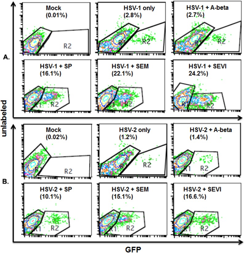
Figure 3.
Flow cytometry to detect viral infection rate. (A) HSV-1 infection (1 mL per well) in HEK 293T cells. HEK 293T cells were cultured in a 6-well plate. When the cells were 80% confluent, the cells were mock-infected or infected with HSV-1 at an MOI of 0.1. Virus was pre-incubated with 10 μg/mL of A-beta, SEM amyloids, SEVI, or SP (1:1000 dilution) for 1 h at 37 °C before infection. The cells were fixed at 8 h post-infection, stained with anti-gD conjugated with FITC, and measured by flow cytometry to detect the percentage of FITC-positive (infected) cells. The percentage of infected cells is shown in the lower right-hand corner; (B) HSV-2 infection in HEK 293T cells. The same experiments as described in A, but using HSV-2 instead of HSV-1. Mock-infected cells in the absence or presence of 10 μg/mL of SEM or 10 μg/mL of SEVI were used to control for autofluorescence. Results are representative of one of three experiments.

