Abstract
The entry of sodium and calcium play a key effect on myocyte subjected to cardiac arrest by hyperkalemia. They cause cell swelling, acidosis, consumption of adenosine triphosphate and trigger programmed cell death. Cardiac arrest caused by hypocalcemia maintains intracellular adenosine triphosphate levels, improves diastolic performance and reduces oxygen consumption, which can be translated into better protection to myocyte injury induced by cardiac arrest.
Keywords: Heart Arrest, Induced; Myocardial Ischemia; Hyperkalemia; Hypocalcemia
Abstract
A entrada de sódio e cálcio desempenham efeito chave no miócito submetido à parada cardíaca por hiperpotassemia. Eles provocam edema celular, acidose, consumo de trifosfato de adenosina e desencadeiam processo de morte celular programada. A parada cardíaca provocada por hipocalcemia mantém os níveis intracelulares de trifosfato de adenosina, melhora o rendimento diastólico e reduz o consumo de oxigênio, o que pode ser traduzido em melhor proteção do miócito às lesões provocadas pela parada cardíaca induzida.
| Abbreviations, acronyms & symbols | |
|---|---|
| ATP | Adenosine triphosphate |
| CPB | Cardiopulmonary bypass |
| HTK | Histidine-tryptophan-ketoglutarate |
| Em | Resting potential |
| CPK | Creatine phosphokinase |
INTRODUCTION
The first cardiac surgeries were performed on a beating heart, being limited to the correction of minor illnesses, such as suturing of cardiac wounds, pericardial drainage, and closing of the arterial channel. At that time, complications were related to myocardial depression[1,2]. In 1953, John Gibbon performed the first open heart surgery, making it possible to approach intracardiac diseases[3] by using substances that trigger controlled cardiac arrest, commonly known as cardioplegic agents[4].
Although several myocardial protection methods produce satisfactory results, none can be considered ideal. The perfect cardioplegic agent must meet the following requirements[5]:
Cardiac arrest: fast and efficient induction, with relaxed myocardium and minimum ATP consumption;
Myocardial protection: protective effects to delay irreversible cell injury caused by global ischemia as well as to limit the extent of the reperfusion injury;
Reversibility: immediate reversal of cardiac arrest with heart rate and force of contraction, allowing for early weaning from cardiopulmonary bypass (CPB);
Low toxicity: short half-life without toxic effects to other systems after termination of CPB;
Allowing good visualization of the operative field.
In this context, the aim of this study is to discuss a few biochemical and physiological considerations related to hyperkalemic and hypocalcemic cardioplegia.
Hyperkalemic Cardioplegia
In 1955, Melrose et al.[6] introduced myocardial perfusion using hypothermic crystalloid solution containing 2.5% potassium citrate, but soon gave up employing this technique due to myocardial necrosis and death of several patients. Gay & Ebert[7] reintroduced this technique with lower concentrations of potassium and they were able to provide metabolic and functional benefits to the myocardium without any apparent structural change. Based on these results, the use of potassium-rich, hypothermic crystalloid solution for myocardial protection became widespread. In 1975, Hearse et al.[8] introduced the St. Thomas solution, containing 10-30 mM potassium chloride. With these solutions, stone heart was no longer a problem and solutions containing adequate concentrations of potassium to induce cardiac arrest became the gold standard in myocardial protection[9,10].
However, Cohen et al.[11]later found a correlation between the use of these solutions and postoperative myocardial dysfunction due to failure to fully protect the myocardium from ischemia and reperfusion injury, which led to the search for safer alternatives for patients.
Metabolic pathways of hyperkalemic cardioplegia
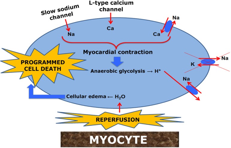
Elevated extracellular potassium concentration (10-40mM) alters resting potential (Em) for myocyte, from -85mV to between -65mV and -40mV, leading to fast sodium channels inactivation. The new Em blocks conduction of myocardial action potential, thereby inducing depolarized arrest. However, slow sodium channels are not fully inactivated (sodium window), causing a slow increase in its intracellular concentration (Figure 1)[5,10,12].
Fig. 1.
Schematic of the metabolic pathways of hyperkalemic cardioplegia harmful to myocytes
In addition, the L-type calcium channel (dihydropyridine), which is activated when Em is between -20mV and -30mV, allows calcium to enter the cytosol[13], a phenomenon known as calcium window[5]. The Na+/Ca2 + exchanger is then activated on reverse mode, removing intracellular sodium and allowing Ca2 + influx. H+ ions (resulting from ischemia) leave the cell via Na+/H+ antiporter, contributing to even higher sodium influx (Figure 1)[5,10].
The Na+/K+ pump is inhibited by the increased concentration of extracellular potassium, acidosis, and hypothermia, allowing intracellular Na levels to continue elevated and, consequently, the Na+/Ca2 + exchanger to continue working on reverse mode. The elevated intracellular calcium causes myocyte contraction, without action potential being triggered and with energy consumption. Elevated sodium levels lead to cellular edema and cytolysis during reperfusion[5]. Thus, hyperkalemic cardioplegia can neither inhibit calcium influx in myocardial cells nor avoid its negative effects (Figure 1)[12,13].
Calcium physiology in cardiac fibers
Calcium functions as an effector in cardiac fibers, connecting the ventricular contraction phase to the excitation phase through action potential. Such mechanism is known as excitation-contraction coupling[14] and it is also present in skeletal striated muscle cells; however, there are a few differences in cardiac fibers with important implications to their contraction[15].
Similar to skeletal muscle, when action potential travels through the myocardial membrane, it also propagates to the interior of cardiac muscle fibers along the t-tubule membranes. Action potential in t-tubules, in turn, cause the instant release of calcium ions from the sarcoplasmic reticulum into the sarcoplasm. Then, in a few thousandths of a second, calcium ions diffuse into myofibrils and catalyze chemical reactions that promote sliding of actin and myosin filaments, which in turn produces muscle contraction. So far, the excitation-contraction coupling mechanism is the same as that of skeletal muscle; however, from this point on, a fundamental difference starts to emerge. Besides the calcium ions released into the sarcoplasm from the sarcoplasmic reticulum, great quantities of calcium are also diffused into the sarcoplasm during t-tubule action potential[15].
In fact, without the extra calcium in the t-tubules, cardiac muscle contraction strength would be considerably reduced since cardiac muscle cisternae are less developed than in skeletal muscle and thus are unable to store enough calcium to produce effective muscle contraction. On the other hand, cardiac muscle t-tubules have a diameter five times greater than that of skeletal t-tubules. Within cardiac t-tubules, there are large quantities of negatively charged mucopolysaccharides that bind and store calcium ions, making them always available to be diffused into cardiac muscle fiber when action potential occurs in the t-tubule. The extra supply of calcium from t-tubules is at least one of the factors that contribute to longer action potential in cardiac muscle and its contraction for 0.3 seconds as opposed to 0.1 second in skeletal muscle[15].
At the end of the action potential plateau, influx of calcium ions into muscle fibers is suddenly interrupted, and the calcium ions in the sarcoplasm are rapidly pumped back into the sarcoplasmic reticulum and t-tubules, thereby ending contraction until the next action potential[15].
Of particular interest is the fact that the strength of cardiac muscle contraction depends on the concentration of calcium ions in the extracellular fluids, which is usually not true of striated muscle. This is likely because the amount of calcium ions in t-tubules is directly proportional to its concentration in extracellular fluids. Therefore, the availability of calcium ions to cause cardiac contraction is directly dependent on the extracellular fluid calcium ion concentration[15].
Hypocalcemic cardioplegia
In the absence of extracellular calcium ions, there is no calcium influx, neither via cell membrane nor via sarcoplasmic reticulum, preventing myofilament contraction. Even if there is action potential, there is no myocardial contraction[5].
By investigating the effects of hypocalcemic cardioplegic solution in immature sheep hearts subjected to ischemia and hypothermia, Aoki et al.[16] noted that in the presence of physiological calcium concentration there was worsening of left ventricular and diastolic functions as well as increase in oxygen consumption and in coronary vascular resistance when compared to hypocalcemia, indicating that low levels of calcium protected the heart from the deleterious effects of hypothermia and ischemia.
Baker et al.[17], when comparing the effects of several concentrations of calcium in St. Thomas solution in immature rabbit hearts, found that the lower the concentration of calcium, the lower the concentration of creatine phosphokinase (CPK). However, they also found that recovery of aortic flow did not follow the same distribution. It showed parabolic behavior with its zenith at 0.3 mmol/L, when the heart obtained the highest aortic flow values.
Comparing hypocalcemic and normocalcemic solutions in blood cardioplegia in immature pig hearts, with or without ischemia, under CPB, Bolling et al.[18] observed that the nonischemic group benefited from both types of cardioplegia. On the other hand, in the ischemic group, myocardial function and ATP levels worsened and coronary vascular resistance increased in those undergoing normocalcemic cardioplegia whereas for those undergoing hypocalcemia, values were the same as the ones for the nonischemic group, indicating that hypocalcemia protected the heart from ischemia.
In assessing oxygen consumption in rat hearts subjected to hypocalcemic or hyperkalemic cardioplegia, Burkhoff et al.[19] ascertained that the hypocalcemic group consumed less oxygen compared to the group that used a potassium-rich solution.
Kronon et al.[20] concluded that a cardioplegic solution containing physiological calcium concentration is deleterious to immature pig hearts, except when magnesium was added to the solution (10 mEq/L), which protected the myocardium from the deleterious effects of calcium. In another study, the same authors found that when magnesium (either 5-6 mEq/L or 10-12 mEq/L) was added to the hypocalcemic solution there was significant improvement in myocardial contraction and reduction in diastolic rigidity[21].
In a study of rabbit hearts subjected to cardioplegia solutions either with low calcium concentration (0.5 mmol/L) or without calcium, Mori et al.[22] reported that hearts subjected to the low calcium cardioplegic solution showed better recovery from left ventricular developed pressure (dP/dt) and coronary flow, lower CPK values, and less cellular edema.
Robinson et al.[23], using lower concentrations of calcium in St. Thomas solution (0.6 mmol/L), found there was 64% improvement in recovery of aortic flow, 84% reduction in CPK as well as reduction in postischemic reperfusion arrhythmia and faster return to normal sinus rhythm when compared to the traditional solution.
Zweng et al.[24] reported improved recovery of aortic flow, developed pressure, and dP/dt in immature rabbit hearts with calcium concentrations between 0.6 and 1.2 mmol/L, subjected to an hour of normothermic ischemia.
Several authors have shown that cardioplegia without calcium is associated with worse recovery of cardiac function[22,24-26] and of coronary[22] and aortic[24] flow as well as increase in creatine phosphokinase[22,25] and cellular edema[22]. These changes are due to a phenomenon known as the "calcium paradox", which does not happen when small quantities of calcium are added to the solution[14,27].
Bretschneider solution is an example of a hypocalcemic solution. In addition to traces of calcium, it also incorporates the benefits of histidine, tryptophan, ketoglutarate, and mannitol.
Histidine (amino acid) acts as a temperature-dependent buffer system. When placed under 22oC, histidine keeps pH at 7.02-7.20 whereas when the temperature is 4oC, pH value is about 7.40. This means that the optimal temperature of the solution is 4oC, since it maintains physiological pH. This amino acid also inhibits matrix metalloproteinases, which are responsible for the development of tissue damage from cold storage. Furthermore, histidine regulates cell membrane permeability, enhancing the osmotic effect of mannitol[28].
Tryptophan contributes to maintaining the integrity of cell membranes[28]. Hachida et al.[29] reported that the absence of tryptophan in the solution caused significant decrease in maximum developed pressure and coronary blood flow.
In addition, Hachida et al.[29] also assessed the absence of ketoglutarate in the solution, which led to significant decrease in maximum developed pressure and significant increase in creatine phosphokinase MB fraction.
CONCLUSION
Hypocalcemic cardioplegia provides safe protection of myocytes, both in immature and older hearts, since it prevents depletion of ATP and acidosis and it is associated with good recovery of ventricular function. Hypocalcemic cardioplegic solutions, such as histidine-tryptophan-ketoglutarate, have been commonly used in cardiac surgery worldwide, but it is still not the ideal solution. New studies are needed to find cardioplegia that is fast-acting and atoxic, metabolizes rapidly, promotes good visualization of the operative field, and adequately protects the myocyte.
| Authors' roles & responsibilities | |
|---|---|
| MABO | Analysis and/or interpretation of data; statistical analysis; final approval of the manuscript; conception and design of the study; carrying out of operations and/or experiments; drafting of the manuscript or critical review |
| ACB | Drafting of the manuscript or critical review |
| CAS | Drafting of the manuscript or critical review |
| PHHB | Drafting of the manuscript or critical review |
| JLLC | Drafting of the manuscript or critical review |
| DMB | Final approval of the manuscript; conception and design of the study |
Footnotes
No financial support.
Work carried out at São José do Rio Preto Medical School (FAMERP), São José do Rio Preto, SP, Brazil.
REFERENCES
- 1.Rosky LP, Rodman T. Medical aspects of open-heart surgery. N Engl J Med. 1966;274(16):886–893. doi: 10.1056/NEJM196604212741606. [DOI] [PubMed] [Google Scholar]
- 2.Prates PR. Pequena história da cirurgia cardíaca: e tudo aconteceu diante de nossos olhos... Rev Bras Cir Cardiovasc. 1999;14(3):177–184. [Google Scholar]
- 3.Miller BJ, Gibbon JH, Jr, Greco VF, Smith BA, Cohn CH, Allbritten FF., Jr The production and repair of interatrial septal defects under direct vision with the assistance of an extracorporeal pump-oxygenator circuit. J Thorac Surg. 1953;26(6):598–616. [PubMed] [Google Scholar]
- 4.Godoy MF, Braile DM. Cardioplegia: exegesis. Arq Bras Cardiol. 1994;62(4):277–278. [PubMed] [Google Scholar]
- 5.Fallouh HB, Kentish JC, Chambers DJ. Targeting for cardioplegia: arresting agents and their safety. Curr Opin Pharmacol. 2009;9(2):220–226. doi: 10.1016/j.coph.2008.11.012. [DOI] [PubMed] [Google Scholar]
- 6.Melrose D, Dreyer B, Bentall H, Baker JB. Elective cardiac arrest. Lancet. 1955;266(6879):21–22. doi: 10.1016/s0140-6736(55)93381-x. [DOI] [PubMed] [Google Scholar]
- 7.Gay WA, Jr, Ebert PA. Functional, metabolic, and morphologic effects of potassium-induced cardioplegia. Surgery. 1973;74(2):284–290. [PubMed] [Google Scholar]
- 8.Hearse DJ, Stewart DA, Braimbridge MV. Hypothermic arrest and potassium arrest: metabolic and myocardial protection during elective cardiac arrest. Circ Res. 1975;36(4):481–489. doi: 10.1161/01.res.36.4.481. [DOI] [PubMed] [Google Scholar]
- 9.Shiroishi MS. Myocardial protection: the rebirth of potassium-based cardioplegia. Tex Heart Inst J. 1999;26(1):71–86. [PMC free article] [PubMed] [Google Scholar]
- 10.Chambers DJ, Hearse DJ. Developments in cardioprotection: "polarized" arrest as an alternative to "depolarized" arrest. Ann Thorac Surg. 1999;68(5):1960–1966. doi: 10.1016/s0003-4975(99)01020-6. [DOI] [PubMed] [Google Scholar]
- 11.Cohen NM, Damiano RJ, Jr, Wechsler AS. Is there an alternative to potassium arrest? Ann Thorac Surg. 1995;60(3):858–863. doi: 10.1016/0003-4975(95)00210-C. [DOI] [PubMed] [Google Scholar]
- 12.Silveira LM, Filho, Petrucci O, Jr, Carmo MR, Oliveira PP, Vilarinho KA, Vieira RW, et al. Padronização de modelo de coração isolado "working heart" com circulação parabiótica. Rev Bras Cir Cardiovasc. 2008;23(1):14–22. doi: 10.1590/s0102-76382008000100004. [DOI] [PubMed] [Google Scholar]
- 13.Chen RH. The scientific basis for hypocalcemic cardioplegia and reperfusion in cardiac surgery. Ann Thorac Surg. 1996;62(3):910–914. doi: 10.1016/s0003-4975(96)00462-6. [DOI] [PubMed] [Google Scholar]
- 14.Oliveira MAB, Godoy MF, Braile DM, Lima-Oliveira APM. Solução cardioplégica polarizante: estado da arte. Rev Bras Cir Cardiovasc. 2005;20(1):69–74. [Google Scholar]
- 15.Guyton AC. Músculo cardíaco; o coração como bomba. In: Guyton AC, editor. Fisiologia humana e mecanismos das doenças. 6 ed. Rio de Janeiro: Guanabara Koogan; 2008. p. p.130. [Google Scholar]
- 16.Aoki M, Nomura F, Kawata H, Mayer JE., Jr Effect of calcium and preischemic hypothermia on recovery of myocardial function after cardioplegic ischemia in neonatal lambs. J Thorac Cardiovasc Surg. 1993;105(2):207–212. [PubMed] [Google Scholar]
- 17.Baker EJ, 5th, Olinger GN, Baker JE. Calcium content of St. Thomas' II cardioplegic solution damages ischemic immature myocardium. Ann Thorac Surg. 1991;52(4):993–999. doi: 10.1016/0003-4975(91)91266-x. [DOI] [PubMed] [Google Scholar]
- 18.Bolling K, Kronon M, Allen BS, Ramon S, Wang T, Hartz RS, et al. Myocardial protection in normal and hypoxically stressed neonatal hearts: the superiority of hypocalcemic versus normocalcemic blood cardioplegia. J Thorac Cardiovasc Surg. 1996;112(5):1193–1200. doi: 10.1016/S0022-5223(96)70132-0. [DOI] [PubMed] [Google Scholar]
- 19.Burkhoff D, Kalil-Filho R, Gerstenblith G. Oxygen consumption is less in rat hearts arrested by low calcium than by high potassium at fixed flow. Pt 2Am J Physiol. 1990;259(4):H1142–H1147. doi: 10.1152/ajpheart.1990.259.4.H1142. [DOI] [PubMed] [Google Scholar]
- 20.Kronon M, Bolling KS, Allen BS, Rahman S, Wang T, Halldorsson A, et al. The relationship between calcium and magnesium in pediatric myocardial protection. J Thorac Cardiovasc Surg. 1997;114(6):1010–1019. doi: 10.1016/S0022-5223(97)70015-1. [DOI] [PubMed] [Google Scholar]
- 21.Kronon MT, Allen BS, Hernan J, Halldorsson AO, Rahman S, Buckberg GD, et al. Superiority of magnesium cardioplegia in neonatal myocardial protection. Ann Thorac Surg. 1999;68(6):2285–2291. doi: 10.1016/s0003-4975(99)01142-x. [DOI] [PubMed] [Google Scholar]
- 22.Mori F, Suzuki K, Noda H, Kato T, Tsuboi H, Miyamoto M, et al. Evaluation of a new calcium containing cardioplegic solution in the isolated rabbit heart in comparison to a calcium-free, low sodium solution. Jpn J Surg. 1991;21(2):192–200. doi: 10.1007/BF02470908. [DOI] [PubMed] [Google Scholar]
- 23.Robinson LA. Calcium in neonatal cardioplegia. Ann Thorac Surg. 1991;51(6):1043–1044. doi: 10.1016/0003-4975(91)91050-6. [DOI] [PubMed] [Google Scholar]
- 24.Zweng TN, Iannettoni MD, Bove EL, Pridjian AK, Fox MH, Bolling SF, et al. The concentration of calcium in neonatal cardioplegia. Ann Thorac Surg. 1990;50(2):262–267. doi: 10.1016/0003-4975(90)90746-s. [DOI] [PubMed] [Google Scholar]
- 25.Hendriks FF, Jonas J, van der Laarse A, Huysmans HÁ, van Rijk-Zwikker GL, Schipperheyn JJ. Cold ischemic arrest: comparison of calcium-free and calcium-containing solutions. Ann Thorac Surg. 1985;39(4):312–317. doi: 10.1016/s0003-4975(10)62620-3. [DOI] [PubMed] [Google Scholar]
- 26.Pearl JM, Laks H, Drinkwater DC, Meneshian A, Sun B, Gates RN, et al. Normocalcemic blood or crystalloid cardioplegia provides better neonatal myocardial protection than does low-calcium cardioplegia. J Thorac Cardiovasc Surg. 1993;105(2):201–206. [PubMed] [Google Scholar]
- 27.Rebeyka IM, Axford-Gatley RA, Bush BG, del Nido PJ, Mickle DA, Romaschin AD, et al. Calcium paradox in an in vivo model of multidose cardioplegia and moderate hypothermia. Prevention with diltiazem or trace calcium levels. J Thorac Cardiovasc Surg. 1990;99(3):475–483. [PubMed] [Google Scholar]
- 28.Antunovic M, Aleksic D. Preparation and testing of solutions for organ perfusion and preservation in transplantation. Vojnosanit Pregl. 2008;65:596–600. doi: 10.2298/vsp0808596a. [DOI] [PubMed] [Google Scholar]
- 29.Hachida M, Ookado A, Nonoyama M, Koyanagi H. Effect of HTK solution for myocardial preservation. J Cardiovasc Surg (Torino) 1996;37(3):269–274. [PubMed] [Google Scholar]


