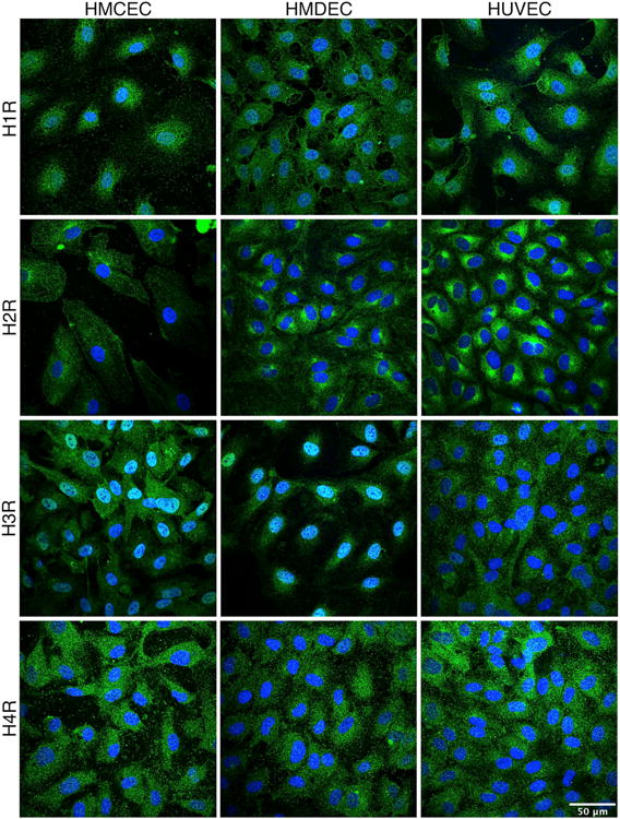
Fig. 3. Identification the Histamine receptor on EC using immunocytochemistry.

HUVEC, HCMEC, and HDMEC were immunolabeled to visualize localization of the H1R (A), H2R (B), H3R (C) and H4R (D), all labeled in green. Nuclei are labeled in blue. Images are projections of confocal z-stacks, and are representative of three separate experiments for each EC type. Images of the negative controls for labeling are provided in the supplemental data (Suppl. Fig. 1).
