



Abstract
Peptidoglycan is a potent immune adjuvant derived from bacterial cell walls. Previous investigations suggest that intestinal epithelium may absorb peptidoglycan from the lumen. Nonetheless, how peptidoglycan is taken up and crosses intestinal epithelium remains largely unclear. Here, we first characterized peptidoglycan transport in vitro using IEC-18 and HT29-CL19A cells, which represent less mature epithelial cells in intestinal crypts. With fluorescent microscopy, we visualized internalization of dual-labeled peptidoglycan by enterocytes. Engulfed peptidoglycan was found to form a complex with peptidoglycan recognition protein-3, which may facilitate delivering peptidoglycan in vivo. Utilizing electronic microscopy, we revealed that uptake of apical peptidoglycan across intestinal epithelial monolayers was involved in phagocytosis, multivesicular body formation, and exosome secretion. We also studied transport of peptidoglycan using the transwell system. Our data indicated that apically loaded peptidoglycan was exocytosed to the basolateral compartment with exosomes by HT29-CL19A cells. The peptidoglycan-contained basolateral exosome extracts induced macrophage activation. Through gavaging mice with labeled peptidoglycan, we found that luminal peptidoglycan was taken up by columnar epithelial cells in crypts of the small intestine. Furthermore, we showed that pre-confluent immature but not post-confluent mature C2BBe1 cells engulfed peptidoglycan via a toll-like receptor 2-dependent manner. Together, our findings suggest that (1) crypt-based immature intestinal epithelial cells play an important role in transport of luminal peptidoglycan over the intestinal epithelium; and (2) luminal peptidoglycan is transcytosed across intestinal epithelia via a toll-like receptor 2-meciated phagocytosis-multivesicular body-exosome pathway. The absorbed peptidoglycan and its derivatives may facilitate maintenance of intestinal immune homeostasis.
Keywords: enterocytes, transcellular transport, mucosal immunophysiology, bacterial cell wall
INTRODUCTION
The intestinal epithelium serves as a vast interface between the body and luminal commensal microorganisms. It has been demonstrated that approximately 1014 commensal bacteria live in the lumen of an adult human intestine (Ley et al., 2006). The host-enteric microbe interactions occur constantly at the intestinal mucosa. Advances in previous studies have suggested that the cross-talk between commensal bacteria and intestinal epithelial cells provides nutrients to the host, regulates intestinal structure, protects against incoming pathogenic microbes, and promotes immune homeostasis (Tappenden and Deutsch, 2007). Several investigators have revealed the translocation of commensal bacterial derivatives across the intestinal barrier in normal circumstance (Gewirtz et al., 2001; Lichtman et al., 1991; Martin et al., 1984). These findings have suggested a novel mechanism through which commensal bacteria in the intestinal lumen communicate with immune cells in the lamina propria and submucosa.
Peptidoglycan (PGN) is a macromolecule in the bacterial cell wall. Numerous in vivo studies have demonstrated biological properties of PGN and its derivatives such as adjuvant activity, macrophage activation, and induction of slow-wave sleep (Boneca, 2005; Martin et al., 1984). In cell culture models, early reports also identified the immunomodulatory properties of PGN towards various immune cells (Boneca, 2005; McCurdy et al., 2003). In the intestinal lumen, PGN-contained cell wall fragments can be released from commensal bacteria after digestion with Paneth cell derived lysozyme. It has been suggested that absorption of PGN may take place at the intestinal mucosa (Lichtman et al., 1991; Martin et al., 1984). However, the characteristics of transporting PGN across intestinal epithelial barrier have not precisely been defined.
Recently, we showed that enteral administration of cell wall components derived from probiotics results in enhancing antimicrobial immunity (Bu et al., 2006). We also found that bacterial cell wall components cross the mucosal barrier and enter the intestinal lamina propria in normal mice (Bu et al., 2006). In this study, we further examined whether and how PGN crosses intestinal epithelial barrier. We revealed that luminal PGN is taken up by epithelial cells in the intestinal crypts. Using a model of crypt-based intestinal epithelial monolayers, we demonstrated that immature intestinal epithelial cells transport apical PGN to basolateral compartment via a mechanism involved in toll-like receptor 2- (TLR2) dependent phagocytosis, multivesicular bodies (MVB) formation, and exosome secretion.
MATERIAL AND METHODS
Intestinal epithelial cell lines and cell culture
Intestinal epithelial cells including IEC-18, HT29-CL19A, and C2BBe1 lines were used. IEC-18 cells are a nontransformed small intestinal epithelial cell line derived from undifferentiated crypt epithelial cells in normal rats (Quaroni et al., 1979), whereas HT29-CL19A and C2BBe1 cells are human intestinal epithelial cell lines derived from colonic adenocarcinoma (Zweibaum et al., 1991; Peterson and Mooseker, 1992). Confluent monolayers of these cells form paracellular junctional barrier. The cell lines of IEC-18 and C2BBe1 were obtained from American Type Culture Collection (ATCC, Rockville, MD). HT29-CL19A cell line (Jilling and Kirk, 1996; Claud et al., 2002) was kindly provided by Dr. Tamas Jilling (Evanston Northwestern Healthcare Research Institute, Evanston, IL). Cultures of IEC-18 cells were performed as previously described (Bu et al., 2007; Tan et al., 2000). C2BBe1 cells were cultured in a water-saturated atmosphere with 5% CO2 at 37°C in Minimum Essential Medium (Eagle) supplemented with 2 mM L-glutamine, 1.5 g/L sodium bicarbonate, 0.1 mM non-essential amino acids, and 1.0 mM sodium pyruvate, 50 U/ml penicillin, 50 μg/ml streptomycin, and 10% fetal bovine serum. HT29-CL19A cells were grown in Dulbecco’s Modified Eagle’s Minimum Essential Medium containing 50 U/ml penicillin, 50 μg/ml streptomycin, and 10% fetal bovine serum at 37° C in a CO2-humidified incubator. Cells were maintained in T-75 tissue culture flasks. In experiments, cells were grown on 35-mm culture dishes or chamber slides to confluence prior PGN exposure. In some experiments, HT29-CL19A and C2BBe1 cells were cultured using transwell systems. Briefly, cells were seeded on transwell inserts (3.0 μm pore polycarbonate membrane insert) and the culture media were changed every 24 – 48 h. Electrical resistance of cell monolayers was measured after 2–3 weeks using EVOM-G (World Precision Instruments, Sarasota, FL). The cell cultures were considered to constitute a polarized epithelial monolayer when resistances were ≥170 ohms.cm2 and stable.
Labeling of PGN with BacLight-Green Stain and biotin
The BacLight-Green Stain (Invitrogen Molecular Probes, Carlsbad, CA) is a non-nucleic acid fluorescent labeling reagent. It has a high affinity to the bacterial cell wall components such as PGN. The reagent is non-fluorescent when not associated with bacteria or cell wall components. The protocol suggested by the manufacture was followed. Briefly, PGN (Sigma-Aldrich, St. Louis, MO) was suspended in PBS at concentration of 1 mg/ml. Then, 1 μl of working dye solution was added into 1 ml of PGN suspension. The sample was incubated at room temperature for 15 min. At the end of the reaction, the labeled PGN was pelleted by centrifugation at 10,000g for five min, washed with PBS three times, and suspended in PBS at concentration of 1 mg/ml. In some experiments, BacLight-Green Stain-labeled PGN was biotinylated with EZ-Link biotin-hydrazide (Pierce, Rockford, IL) using a protocol provided by the manufacture. To this end, BacLight-Green Stain-labeled PGN was suspended in MES Buffer containing 0.1 M 2-N-morpholino ethanesulfonic acid (MES, pH 4.7 – 5.5) at 5 mg/ml. Thereafter, 25 μl of biotin hydrazide solution (50 mM biotin hydrazide reagent) and 12.5 μl of 1-ethyl-3-[3-dimethylaminopropy]carbodiimide hydrochloride (100 mg/ml) solution per 1 ml of PGN suspension were sequentially added and mixed thoroughly. The biotinylating reaction was carried out for 2 h. At the end of the reaction, the labeled PGN was sequentially washed with MES buffer and PBS, and solubilized by sonication.
Determination of PGN internalization in vitro by fluorescent microscopy
The protocol was modified from a previously described method (Neal et al., 2006). BacLight-Green Stain and biotin dual-labeled PGN was prepared as described above. Intestinal epithelial cells were grown on chamber slides. To determine PGN internalization, chamber slides with confluent cell monolayers were washed with cold PBS, added fresh culture medium, and set on ice for 10 min. Then, dual-labeled PGN (100 μg/ml) was added to medium. Chamber slides were set on ice for additional 30 min to allow PGN adhering on the cell surface. Thereafter, cells were cultured in a water-saturated atmosphere with 5% CO2 at 37°C for 2 h. At the end of incubation, cells were washed three times with ice-cold PBS, fixed with 4% paraformaldehyde (pH 7.2) for 10 min, and washed with PBS three times. To labeled extracellular biotinylated PGN with an additional fluorescent dye, fixed-cells were incubated with strepavidin-Alex 633 (1:500 dilution, Invitrogen Molecular Probes, Carlsbad, CA) for 30 min at room temperature followed by washing with PBS three times. Finally, slides were examined under an upright fluorescence microscope (model MD R, Leica, Wetzlar, Germany) using BacLight-Green stain, Alexa Fluor 633, or DAPI filter sets. Under the fluorescence microscope, fluorescent particles identified with the BacLight-Green stain filter set were considered as internalized or external PGN, whereas fluorescent particles visualized with the Alexa Fluor 633 filter set were considered as extracellular PGN. In addition, cells were examined using DIC optics under the upright light microscope in order to obtain the sketch of cells. Images were acquired with a digital camera (model C4742-95, Hamamatsu, Hamamatsu City, Japan). For each area examined, images of BacLight-Green stain, Alexa Fluor 633 and DIC observation were collected. The time of measurements, image capturing, and image intensity gain at all wavelengths were optimally adjusted and kept constant. Images were transferred to a G5 Macintosh computer (Apple Computer), analyzed by image-analysis software (Openlab®), and assembled with Adobe Photoshop 8.0® software.
Determination of PGN phagocytosis in vitro with electron microscopy (EM)
Intestinal epithelial cells were exposed to unlabelled PGN for up to 24 h. In some experiments, cells were pretreated with murine mAb against human TLR2 (clone T2.5, 20 μg/ml, eBioscience Inc., San Diego, CA) or isotype control IgG (20 μg/ml) for 30 min before PGN exposure. At the end of treatments, cells were extensively washed with cold PBS, fixed in 0.1 M sodium phosphate-buffered (pH 7.2) 2.5% glutaraldehyde solution at 4°C for 30 min, and postfixed in 0.1 M sodium phosphate-buffered (pH 7.2) 1% OsO4 solution at 4°C for 2 h. After dehydration in an ethanol gradient (50–100% for 10 min each), samples were embedded in EPON812 at 60°C for two days. Ultrathin sections (70 nm) were stained using uranyl acetate and lead citrate. Sections were examined using an electron microscope (Zeiss-900) at 50 kV.
Immunofluorescent staining
Cells were fixed with 4% paraformaldehyde (pH 7.2) for 10 min. A standard protocol of our laboratory (Bu et al., 2007) for immunofluorescent staining was followed. Murine monoclonal antibody against human peptidoglycan recognition protein 3 (PGLYRP-3, Imgenex Corp., San Diego, CA) and a rabbit anti mouse IgG antibody labeled with Alexa Fluor 633 (Invitrogen Molecular Probes, Carlsbad, CA) were respectively used as primary and secondary antibodies. The nuclei were counterstained with 4′,6-diamidino-2-phenylindole (DAPI, Vector Laboratory, Burlingame, CA). The stained cells were examined under an upright fluorescence microscope as mentioned above.
Animals
C57BL/6J mice (male, 6 – 9 wks) were obtained from Jackson Laboratory (Bar Harbor, Maine). They were housed in a specific pathogen-free animal facility at Children’s Memorial Research Center (CMRC). All animal experiments were conducted in accordance with NIH guidelines under protocols approved by the Institutional Animal Care and Use Committee of CMRC.
Detection of absorption of PGN in vivo
Mice were fasted overnight with free access to water. In the morning, mice were gavaged with 100 μl 1mg/ml BacLight-Green Stain-labeled PGN solution. After 90 min, they were sacrificed with CO2 inhalation. Then ileum was collected, extensively flushed with saline, and processed for cryosections (5 μm). Sections were fixed with 4% paraformaldehyde (pH 7.2) for 10 min, counterstained with DAPI, and examined under an upright fluorescence microscope as described above.
Isolation of exosome (van Niel et al., 2001)
Briefly, HT-29C19A cells were seeded at a density of 8 × 10⁵ cells/cm2 on polyethylene terephtalate cell culture inserts with a pore diameter of 3 mm and a surface area of 44 cm2 (Corning Cat# 3420). We routinely used 10 plates because of the small production of exosomes in intestinal epithelial cells. Cells formed confluent monolayers with a stable electronical resistance ≥170 ohms.cm2 after three weeks. To avoid contamination by exosomes present in the serum itself, the medium was changed to a serum-free medium that contains bovine serum albumin (4 g/L). Then, cells were processed for experimental treatments as described in experimental designs. Thereafter, culture media in contact with the basolateral side of cells were collected. Exosomes recovered from the medium through several steps of centrifugations including a first centrifugation at 300g for 10 min at 4°C to remove dead cells or cellular debris, second (2000g for 20 min) and third (10,000g for 30 min) centrifugations to eliminate all remaining membrane fragments from the conditioned-media. The final supernatant was ultracentrifuged and washed three times in PBS at 100,000g for 1 h at 4° C using a 70 Ti rotor (Beckman Instruments, Inc., Fullerton, CA). Pellets were finally resuspended in 100 μl PBS and stored at −80° C. The specificity of intestinal epithelial exosome extracts was confirmed by western blot using an antibody against A33 antigen as described before (van Niel et al., 2001; Mallegol et al., 2007).
SLP-HS assay
SLP (Silkworm larvae plasma) reagent contains a pro-phenol-oxidase cascade system and enables to rapidly quantify the small amount of PGN by the absorbance change caused by the color change of the cascade system (Kobayashi et al., 2000). The SLP-HS assay kit was obtained from Wako Pure Chemical Industries, Ltd. (Richmond, VA). The protocol suggested by the manufacture was modified. Briefly, 50 μl of samples were added to a 96-well plate. Then, an equal volume of SLP-diluent reconstituted SLP regent was added to each well. Afterwards, the plate was incubated for 30 min at 30°C. The optical density of the plate was read at 690nm. The amount of PGN in the sample was calculated according to a standard curve.
Isolation of peritoneal residential macrophages
A standard protocol of our laboratory was followed (Bu et al., 2006). Briefly, mice were sacrificed with inhalation of CO2. The peritoneum was lavaged with cold serum-free RPMI-1640 several times. The exudate cells were washed and plated at 1 × 10⁶ cells/ml in cell culture plates. After incubation for 2 h at 37°C in a humidified air atmosphere containing 5% CO2, non-adherent cells were removed by washing with Hanks balanced salt solution (HBSS) buffered with 10 mM HEPES (pH 7.2). The macrophage specificity was assessed by determining cell surface F4/80 antigen (a murine macrophage specific marker) with flow cytometric analysis as described before (Wang et al., 2009). Cells were then cultured with RPMI 1640 medium with 10% FBS overnight and used for various experiments.
Measurement of macrophage activation
Two procedures were applied to determine macrophage activation in response to stimulations. First, peritoneal macrophages were processed for treatment as experimental designs. Six hours after treatments, cells were examined under an inverted microscope to determine morphological changes of cells. In addition, Raw 264.2 macrophages (obtained from ATCC) were seeded in a 96-well plate for overnight. Then, cells were treated according to experimental designs. Overnight after treatments, conditioned-media from Raw 264.2 cell culture was collected, and IL-6 was detected using murine IL-6 Quantikine colorimetric sandwich ELISA kit (R & D System, Inc, Minneapolis, MN). The protocol provided by the manufacturer was followed. Standard curves were generated for IL-6 using the standard provided in the kit, and the concentration of IL-6 in the cell supernatant was determined by interpreting from the appropriate standard curve.
Protein isolation and western blotting
The standard protocol of our laboratory for protein extraction was followed (Tan et al., 2000; Zhu and Tan, 2005; Zhu et al., 2003). For western blotting, total cellular proteins or exosome extracts were resolved on 4–20% SDS-PAGE gel, and transferred onto a nitrocellulose membrane. The membranes containing sample proteins were used for determining PGLYRP-3 or A33 proteins. Specifically, blots preincubated with PBS containing 5% nonfat dry milk were reacted with a mouse mAb against human PGLYRP-3 (1:500, Imgenex Corp., San Diego, CA) or rabbit polyclonal antibody against human A33 (1:200, Santa Cruz Biotechnology Inc, Santa Cruz, CA) for 1 h at room temperature. After incubation, the blot was washed four times with PBS containing 0.05% Tween 20 (PBS-T), and then incubated with PBS-T containing 1:10,000 diluted HRP-conjugated appropriate secondary antibodies for 1 h at room temperature. After additional washing with PBS-T, immune complexes on the blot were visualized by the ECL system.
Statistics and data analysis
For data analysis, we used analysis of variance and one-way analysis of variance (ANOVA) followed by Fisher’s LSD test least significant difference post-hoc test to assess the significant of differences. P< 0.05 was considered significant.
RESULTS
1. Internalization of PGN by crypt-based intestinal epithelial cells
IEC-18 and HT29-CL19A cells were used in this experiment. These two cell lines have characteristics of less mature epithelial cells in intestinal crypts (Morris et al., 1994; Jilling and Kirk, 1996; Augeron and Laboisse, 1984; Quaroni et al., 1979). To determine whether PGN uptake into intestinal epithelial cells occurs, confluent IEC-18 cells were incubated with dual-labeled PGN. The internalized PGN was determined using a method that was modified from a previously described protocol (Neal et al., 2006). It was revealed that PGN aggregates were present inside IEC-18 cells 2 h after PGN exposure (Figure 1). In addition, HT29-CL19A cells were also revealed to engulf PGN (data not shown). Together, the data suggested that crypt-based intestinal epithelial cells are able to engulf PGN in vitro.
Figure 1. Internalization of PGN by IEC-18 cells.

IEC-18 cells were treated with PGN that was dual labeled with BacLight-Green Stain and biotin. After 2 h, the cells were washed, fixed, and stained with strepavidin-Alex633 as described in the Methods. Under a fluorescent microscope, internalized PGN particles (marked with circles) were visualized with BacLight-Green not Alex633 optics, whereas extracellular PGN particles (marked with arrows) were visualized with both BacLight-Green and Alex633 optics. The phase contrast appearance and merged image of the green and red signal is shown in the merged panel. Control panel shows the merged image of cells which were treated with unlabeled PGN. Original magnification, X20. Data are representative of three separate experiments.
2. Internalized PGN is associated with PGLYRP-3 in intestinal epithelial cells
Peptidoglycan recognition proteins (PGRP or PGLYRP) are a group of innate immunity molecules conserved from insects to mammals (Royet and Dziarski, 2007). Previous studies have demonstrated that human intestinal epithelial cells express PGLYRP-3 (Lu et al., 2006). With western blot, we confirmed that HT29-CL19A and undifferentiated C2BBe1 cells expressed PGLYRP-3 (Figure 2A). Furthermore, the cytoplasm of undifferentiated C2BBe1 cells showed a strong punctiform PGLYRP-3 immunofluorescent staining (Figure 2B). Because PGLYRP-3 has high affinity to PGN, we hypothesized that internalized PGN is bound onto PGLYRP-3 in intestinal epithelial cells. To test the hypothesis, undifferentiated C2BBe1 cells were exposed to BacLight-Green Stain-labeled PGN for 4 h. Staining the cells with a monoclonal antibody against human PGLYRP-3 revealed that engulfed PGN was colocalized with PGLYRP-3 in the cytoplasm (Figure 3), suggesting that internalization of PGN resulted in recruitment of PGLYRP-3 to PGN.
Figure 2. PGLYRP-3 is expressed in intestinal epithelial cells.

(A) PGLYRP-3 is constitutively expressed in human intestinal epithelial cells. Total cellular proteins were isolated from HT29-CL19A and undifferentiated C2BBe1 cells. Thirty micrograms of protein were subjected to SDS-PAGE, transferred to membranes, and analyzed using Western blot analysis with anti-PGLYRP-3 mAb. Lane 1, positive control provided by Imgenex Corp; Lane 2, protein extracted from HT29-CL19A cells; Lane 3, protein extracted from undifferentiated C2BBe1 cells. (B) Cellular localization of PGLYRP-3 protein in undifferentiated C2BBe1 cells. The cells were stained with immunofluorescence using mAb against human PGLYRP-3 followed by counterstaining the nuclei with DAPI as described in the Methods. Their staining profiles were merged. Original magnification, X63. Data in each panel are representative of two separate experiments.
Figure 3. Internalized PGN is colocalized with PGLYRP-3 in intestinal epithelial cells.

Pre-confluent C2BBe1 cells were exposed to BacLight-Green Stain labeled-PGN. After 4 h, the cells were washed, fixed, and stained with immunofluorescence using murine mAb against human PGLYRP-3 as a primary antibody and Alex633-labeled anti-mouse IgG as the secondary antibody. Nuclei were counterstained with DAPI. The cells were examined under a fluorescent microscope. (A) Internalized PGN appeared under BacLight-Green optic. (B) PGYLRP3 was visualized under Alex633 optic. (C) Nucleus was visualized using DAPI filter set. (D) The images of panels A – C were merged using Photoshop software. Original magnification, X63. Data in each panel are representative of three separate experiments.
3. Internalized PGN is distributed into multiple subcellular compartments in crypt-based intestinal epithelial cells
To further characterize internalization of PGN by crypt-based intestinal epithelial cells, HT29-CL19A cells were cultured in transwell systems for three weeks. Then, the monolayers were exposed to unmodified PGN from the apical side for up to 24 h followed by electron microscopic examination. Under the EM, PGN particles were found in a series of compartments in HT29-CL19A cells. They attached and fused with the membrane of the cells, underwent internalization, and existed in the cytoplasma (Figure 4A). Some engulfed PGN particles were localized in phagosomes and phagolysosomes (Figure 4B), whereas others in phagosomes mixed together with MVB (Figure 4C). Overnight after exposure to PGN, the amount of MVB was found to increase in cytoplasm (Figure 4D). In some cells, MVB were projected from the cell membrane suggesting the secretion of exosomes (Figure 4E). Taken together, the data suggested that PGN is internalized by crypt-based intestinal epithelial cells from apical sides, processed in phagosomes, phagolysosomes, and MVB, and exocytosed with exosomes.
Figure 4. Internalization and cellular fate of PGN within intestinal epithelial cells.

HT29-CL19A cell monolayers were exposed to PGN for 2 (Panels A – B), 6 (Panel C) and 24 h (Panels D – E) from apical sides. At the end of the treatments, the cells were washed, fixed, and processed for EM. Original magnification, X12,000 (A – B), X20,000 (C – D), and X25,000 (E). P, PGN. White arrows indicate phagosomes. Black arrows indicate phagolysosomes. Asterisks indicate MVB. Data in each panel are representative of three separate experiments.
4. Uptake of PGN by intestinal crypt columnar epithelial cells in vivo
To examine whether PGN engulfment by intestinal epithelial cells occurs in vivo, C57BL/6 mice were subjected to feed with BacLight-Green Stain-labeled PGN. As the control, a group of mice was gavaged with PBS containing working dye solution of BacLight-Green Stain (1 μl/ml PBS). Ninety minutes after PGN exposure, mice were sacrificed. Segments of proximal and distal ileum were collected for examination under an upright fluorescent microscope. In the mice fed with BacLight-Green Stain-labeled PGN, strong fluorescent signals were present in the intestinal crypt epithelial cells and in the lamina propria but not in enterocytes in the upper villi region (Figure 5A). In contrast, only weak autofluorescent signals were detected in the intestinal tissues of vehicle-fed mice (Figure 5B). The data suggested that luminal PGN is absorbed by crypt cells in the intestine under the normal physiological state.
Figure 5. Absorption of PGN occurs in crypts of the small intestines.
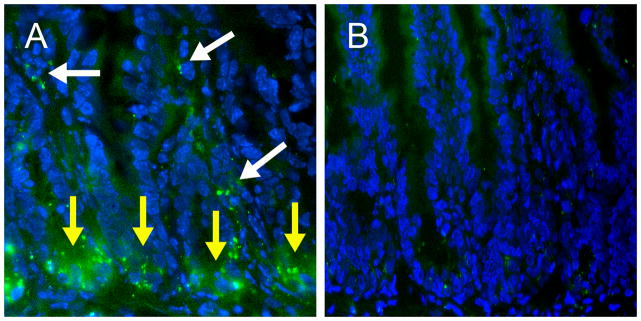
Mice (n=3) were gavaged with BacLight-Green stain labeled PGN (Panel A) or the vehicle (Panel B). After 90 min, they were sacrificed. The small intestines were processed from cryosections, counterstaining with DAPI (blue), and examination under a fluorescent microscope. Yellow arrows indicate labeled PGN (green) in the crypt area and white arrows indicate labeled PGN (green) in the lamina propria. X10. Merged images are shown. Data in each panel are representative of two separate experiments.
5. Matured enterocytes are unable to engulf PGN
Because intestinal epithelial cells in the crypts are less mature than the cells in villus regions, we tested the hypothesis that immature but not mature enterocytes are able to engulf PGN in a series of experiments. First, we examined uptake of PGN using pre- and post-confluent C2BBe1 cells. After exposure to PGN for 2 h, the cytoplasm of pre-confluent C2BBe1 cells contained a large amount of PGN (Figure 6A), whereas post-confluent C2BBe1 cells did not engulf PGN (Figure 6B). The data suggested that the function of phagocytosis of PGN was diminished when intestinal epithelial cells are mature. The pre-confluent C2BBe1 cells lack polarization, cell-cell contact, and organization of tight junctions. Therefore, we further confirmed this finding using HT29-CL19A cell monolayers, an in vitro model representing immature intestinal epithelial cells with the apico-basolateral polarization characteristic. Specifically, HT29-CL19A cells were cultured in transwell systems for three weeks to allow development of paracellular-tight junction complexes. PGN was delivered to the monolayers from apical side. The cells were processed for EM examination 2 h after PGN exposure. Under electronic microscopy, PGN aggregates were revealed to exist in the cytoplasm of HT29-CL19A cells (Figure 6C). Together, these data indicated that immature but not mature intestinal epithelial cells have the ability to engulf PGN in vitro.
Figure 6. PGN is engulfed by immature but not mature intestinal epithelial cells.

Cells were cultured in transwell systems. PGN was added to apical compartments of pre-confluent C2BBe1 cells (Panel A), post-confluent C2BBe1 cells (Panel B), and post-confluent HT29-CL19A cells (Panel C). After 2 h, the cells were washed, fixed, and processed for EM. Original magnification, X7,000 (A) and X12,000 (B, C). P, PGN. The arrow indicates engulfed-PGN underwent degradation in a phagolysosome. Data in each panel are representative of three separate experiments.
6. TLR2 mediates engulfment of PGN by undifferentiated C2BBe1 cells
TLR2 is a type 1 transmembrane protein involved in microbial pattern recognition (Takeda et al., 2003). It has a high affinity to PGN and synthetic PGN derivatives (Asong et al., 2009). Previously, Caco-2/C2BBe1 cells have been revealed to express TLR2 (Cario et al., 2000). Thus, we investigated whether neutralization of TLR2 attenuated uptake of PGN by immature C2BBe1 cells. To this end, undifferentiated C2BBe1 cells and the cells pretreated with either mAb against human TLR2 or control IgG were exposed to PGN for 2 h followed by EM examination. We found that undifferentiated C2BBe1 cells internalized PGN within 2 h (Figure 7A). Pretreatment with anti-TLR2 antibody markedly blocked PGN internalization in C2BBe1 cells (Figure 7B). However, the control IgG did not affect PGN uptake (Figure 7C). Together, the results suggest that TLR2 plays a role in the process.
Figure 7. TLR2 facilitates phagocytosis of PGN by immature intestinal epithelial cells.

Pre-confluent C2BBe1 cells were pretreated with medium alone (Panel A), anti-TLR2 antibody (20 μg/ml, Panel B), or isotype control IgG (20 μg/ml, Panel C) for 30 min. Then, cells were co-incubated with PGN for 2 h. At the end of treatments, cells were extensively washed with PBS and processed for EM examination as described in Methods. Original magnification, X12,000. P, PGN. Data in each panel are representative of two separate experiments.
7. Exosome is a carrier involving transcellular transport of PGN across intestinal epithelial monolayers
Confluent HT29-CL19A monolayers on transwell filters were used in this experiment. First, the transepithelial electrical resistance (TER) of HT29-CL19A monolayers was measured to ensure that there were no PGN-induced changes in transepithelial function. Similarly to a previous report (Wu et al., 2007), TER of HT29-CL19A monolayers was not affected by PGN treatment (Figure 8A), suggesting that the integrity of the cell monolayers. Then, we examined whether PGN is exocytosed from intestinal epithelial cells with exosomes because PGN particles were associated with MVB, a cell apparatus involving production of exosomes. To this end, confluent HT29-CL19A cells were exposed apically to PGN. Exosomes were recovered from the lower chamber conditioned-media 24 h later. We determined the significant amount of transcytosed PGN in the exosome extracts by SLP-HS assay (Figure 8B).
Figure 8. PGN is transcellular transported by intestinal epithelial cells via an exosome associated mechanism.
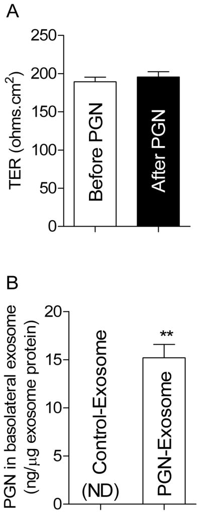
HT29-CL19A cells were cultured on transwell filters for three weeks. The cell monolayers with stable TER were treated from apical side with PGN. The TER of monolayers was measured prior and after PGN treatment (Panel A). Twenty-four hours after PGN exposure, exosomes were isolated from basolateral and then processed for quantifying PGN with SLP-HS assay as described in the Methods (Panel B). n=3. ND, Not detected. Data in each panel are representative of two separate experiments.
In addition, we measured PGN-like biological activities of basolateral exosome extracts from PGN-treated cells using macrophage activation assays. First, the extracts were boiled and then used for treatment of murine peritoneal macrophages in vitro. We found that macrophages altered their shapes within 6 hrs after the stimulation (Figure 9A). Furthermore, murine Raw 264.7 macrophages were stimulated with boiled exosome extracts isolated from PGN-treated HT29-CL19A cells followed by measurement of IL-6 in the culture supernatant. We revealed that the extracts induced IL-6 release in Raw 264.7 cells (Figure 9B). Together, the results suggested that (1) apically added PGN translocates across the HT29-CL19A monolayer using exosome as a carrier; and (2) PGN in exosome extracts has biological activity.
Figure 9. Macrophages are activated by exosomes isolated from basolateral medium of PGN-treated HT29-CL19A monolayer.

(A) PGN-contained exosomes induced pseudopod formation in macrophages. Peritoneal macrophages from C57BL/6J mice were treated for 6 h with culture medium alone, boiled-exosome extracts isolated from basolateral medium of controls HT29-CL19A monolayer cultures (Control-Exosome), or boiled-exosome extracts isolated from basolateral medium of PGN-treated HT29-CL19A monolayer cultures (PGN-Exosome). The cells were examined under an inverted microscope. X40. Data in each panel are representative of three separate experiments. (B) PGN-contained exosomes induced IL-6 production in macrophages. Raw 264.7 cells were subjected to treatment with medium alone, exosome isolated from control HT29-CL19A cells (Control-Exosome), or PGN-contained exosomes (PGN-Exosome). After 24 h, the culture supernatants were processed for measuring the IL-6 level as described in Materials and Methods. n = 4. ND, Not detected. Results are the means ± SEM. **, P < 0.01 compared with the control-exosome group.
DISCUSSION
A number of independent lines of evidence suggest that PGN does get through the intestinal epithelium (Lichtman et al., 1991; Martin et al., 1984; Bu et al., 2006). However, cellular mechanisms through which PGN is absorbed in the intestinal mucosa are unclear. The apicolaterally located tight junctions form a paracellular seal between the lateral membranes of adjacent cells and act as a paracellular barrier in the intestinal epithelium. Therefore, it is unlikely that PGN translocates across intestinal epithelial lining through paracellular pathways in normal circumstance. In the present study, we tested the hypothesis that phagocytosis and exocytosis are main mechanisms involved in transferring luminal PGN to lamina propria. We demonstrated that absorption of PGN occurs in the intestinal crypt region. Furthermore, we found that PGN was present in the lamina propria of both villus and crypt regions after gavaging mice. This data suggests that PGN is rapidly diffuse within the lamina propria after crossing epithelium in the crypt. With confluent IEC-18 cells, a crypt-derived rat intestinal epithelial cell line, we showed that PGN is engulfed by intestinal epithelial cells. The junctional integrity of IEC-18 has been shown to be looser than that of the human intestinal epithelial cell lines derived from colonic adenocarcinoma such as Caco-2, C2BBe1 and HT29-CL19A cells (Duizer et al., 1996). To rule out the contribution of the paracellular pathway to internalization of PGN by intestinal epithelial cells, we used monolayers of HT29-CL19A cells (a crypt-based human intestinal epithelial cell line) and confirmed that PGN is transported from the apical to basolateral compartment in intestinal epithelial cells. Furthermore, we characterized internalization of PGN by crypt-based intestinal epithelial cells using HT29-CL19A cell line under EM. Our results suggest that the apical to basolateral transport of PGN by immature intestinal epithelial cells is involved in PGN binding to apical membrane of intestinal epithelial cells, PGN engulfing, processing PGN in several membrane-bounded carriers such as phagosomes and MVB, and PGN releasing with exosomes to the basal side of the cells.
Enterocytes are derived from stem cells in the crypts. They exhibit morphological and functional heterogeneity along the crypt-villus axis in the small intestine. The cells undergo maturation as they migrate from the proliferation zone in the crypts to the absorptive area at the villus tips. It has been shown that functional characteristics of enterocytes in the crypts differ from the cells in the villi (Welsh et al., 1982). Furthermore, crypt-based immature columnar cells have been revealed to engulf cell debris (Cheng and Leblond, 1974). In the present study, we demonstrated that these cells engulf luminal PGN in vivo. To further determine whether crypt-based immature columnar cells but not mature enterocytes in villi are able to internalize PGN, we used two human intestinal epithelial cell lines including HT29-CL19A and C2BBe1 cells. Comparing to HT29-CL19A cells, C2BBe1 intestinal epithelial cells differentiate spontaneously after confluence. From the morphological view of points, the postconfluent HT29-CL19A cells are less mature than postconfluent C2BBe1 cells. For instance, postconfluent C2BBe1 cells have a typical enterocytic differentiation that is associated with formation of apical long microvilli (Peterson and Mooseker, 1992). This morphological feature is similar to one observed in mature enterocytes at the villi of intestines (Zweibaum et al., 1991; Cheng and Leblond, 1974), indicating that differentiated C2BBe1 cells are morphologically analogous to fully mature enterocyte-like cells. In contrast, HT29-CL19A cells remain a relatively immature feature although they exhibit epithelial differentiation and form a polarized monolayer with the presence of tight junctions. Furthermore, postconfluent HT29-CL19A cells have an immature brush border in their apical surface (Augeron and Laboisse, 1984). This morphological feature is similar to that observed in crypt-base columnar cells (e.g. progenitor cells and immature epithelial cells) in intestines (Cheng and Leblond, 1974). In addition, the electrophysiological features of HT29-CL19A cells are also similar to crypt cells in intestines (Morris et al., 1994; Jilling and Kirk, 1996). Therefore, we used post-confluent HT29-CL19A cells as a tissue culture model for immature intestinal epithelial cells in the crypts, whereas post-confluent C2BBe1 cells as mature intestinal epithelial cells in the villi of adults. We demonstrated that (1) post-confluent HT29-CL19A cells internalize PGN from the apical side; (2) pre-confluent C2BBe1 cells take up PGN but the cells lose their function of engulfing PGN after development of post-confluent maturation, and (3) PGN is transported across HT29-CL19A monolayers from apical to basolateral sides. Together, we proposed that crypt-based intestinal epithelial cells play an important role in transport of luminal PGN over the intestinal epithelium.
TLR2 is an important membrane protein involved in recognition of conserved microbial molecules such as PGN, lipoteichoic acid, and bacterial lipoproteins (Takeuchi et al., 1999; Schwandner et al., 1999; Takeuchi et al., 2000). In the intestinal epithelium, TLR2 is restrictively expressed in crypt enterocytes (Furrie et al., 2005; Cario et al., 2000). Although whether PGN is an activator for TLR2 is still under debate (Travassos et al., 2004; Dziarski and Gupta, 2005), it has clearly been shown that TLR2 directly binds synthetic PGN derivatives and PGN fragments derived from bacteria (Asong et al., 2009). In the present study, we revealed PGN absorption occurs in the crypt region, which is correlated with the pattern of TLR2 expression in intestines. Furthermore, we found that in vitro uptake of PGN by immature intestinal epithelial cells is attenuated by mAb against TLR2. These results in conjunction with previous findings suggest that TLR2 mediates engulfment of PGN by immature intestinal epithelial cells.
Exosomes are biologically active membrane vesicles released into the extracellular environment by different cell types including epithelial, hematopoietic, and some tumor cells (Kesimer et al., 2009). They are derived from the fusion of MVB with the plasma membrane (Fevrier and Raposo, 2004). Previous studies showed that intestinal epithelial cells secrete exosomes (van Niel et al., 2001). Exosomes released by intestinal epithelial cells contain MHC class I, MHC class II, CD63, CD26/dipeptidyl-peptidase IV, and A33 antigen, indicating the role of intestinal epithelial cell-derived exosomes in antigen presentation. A recent investigation demonstrated that intestinal epithelial exosomes mediate the transfer of luminal antigenic information, facilitate immune surveillance at mucosal surfaces, and potentiate peptide presentation to T cells (Mallegol et al., 2007). Their work suggests that these membrane vesicles constitute a powerful link between luminal antigens and local immune cells. Here, we revealed that epithelial exosomes appear to be an efficient intercellular carrier allowing luminal PGN being transmitted to basolateral side. The PGN-contained exosomes are able to induce macrophage activation. Therefore, the results of the present study and previous investigations put forward that basolaterally releasing exosomes contributes to transferring luminal antigens and commensal bacterial components from the intestinal lumen to immune cells in lamina propria, which in turn regulate homeostasis of immune cells in intestinal mucosa.
It is interesting to note that engulfment of PGN is colocalized with PGLYRP-3 in intestinal epithelial cells. PGLYRP-3, a secreted protein, is a member of mammalian peptidoglycan recognition protein family (Lu et al., 2006). Previous studies demonstrate that PGLYRP3 is expressed throughout epithelium in both villi and crypts in intestines but not Paneth cells (Lu et al., 2006; Mathur et al., 2004). Mammalian PGLYRP-3 has been revealed to have Zn2+-dependent bactericidal activity against both Gram-positive and Gram-negative bacteria (Wang et al., 2007). It has high affinity to peptidoglycan (Guan et al., 2004). In the present study, we found that PGLYRP-3 protein is present throughout the cytoplasm with a distinctive punctiform in intestinal epithelial cells, which could be in agreement with the fact that this molecule is normally secreted. Furthermore, we revealed that PGLYRP-3 is redistributed and colocalized to phagocytosed PGN in intestinal epithelial cells. Together, these findings lead to hypothesis that intestinal PGLYRP-3 functions as a carrier for PGN in addition to killing bacteria.
In addition, we used a commercial PGN in the present study. It has been reported that bacterial lipoproteins and lipoteichoic acid are present in commercially available preparations of PGN (Travassos et al., 2004; Hashimoto et al., 2006) because they covalently bind each other. Thus, it is highly possible that other bacterial components could also be engulfed by intestinal epithelial cells in our study. Indeed, teichoic acid and bacterial lipoproteins are expected to constantly cross-link with PGN in the intestinal lumen. Therefore, we hypothesize that teichoic acid and bacterial lipoproteins are simultaneously taken up when crypt-based epithelial cells engulf PGN. Previously, translocation of staphylococcal enterotoxins across epithelia was revealed (Hamad et al., 1997). In addition, Gewirtz and his associates found transcytosis of flagellin through intestinal cell monolayers (Gewirtz et al., 2001; Lyons et al., 2004). Together, our results, in conjunction with previous research, strongly suggest that transport of bacterial products across intestinal epithelial lining can take place not only in pathological conditions but also in normal physiological circumstance.
In conclusion, our study suggests that luminal PGN is taken up by immature intestinal epithelial cells in crypts of the small intestine. The PGN transcytosis is involved in TLR2-dependent phagocytosis, MVB formation, and exosome secretion in the cells. Engulfed PGN forms a complex with PGLYRP-3 in intestinal epithelial cells, which may facilitate delivering PGN in vivo. Together, we hypothesize that a phagosome-MVB-exosome pathway contributes to transport of luminal PGN over intestinal epithelium in crypts of the small intestine. Gram-positive bacteria contain a thick PGN cell wall, which can be degraded by lysozyme. They are the dominant microbial species in the small intestine (reviewed by (Sartor, 2008)). In addition, high concentration of lysozyme is expected near the crypt region in the small intestine because the enzyme is secreted by Paneth cells. Thus, uptake of PGN by immature intestinal epithelial cells at the crypt is physiologically meaningful. Together, absorbed PGN and its derivatives may contribute to maintenance of intestinal immune homeostasis.
Supplementary Material
Acknowledgments
This work was supported in part by the Grant R01DK064240 (to X.-D.T.) from National Institutes of Health, the Excellence in Academic Medicine Award from Illinois Department of Public Aid (to X.-D.T.), and Eloise and Warren Batts Investigator Chair (to X.-D.T.). We thank Dr. Tamas Jilling for kindly providing the HT29-CL19A cell line.
Contact grant sponsor: National Institutes of Health; Contact grant number: R01DK064240
Non-standard abbreviations
- DAPI
4′, 6-diamidino-2-phenylindole
- EM
electronic microscopy
- MVB
multivesicular bodies
- PGN
peptidoglycan
- PGLYRP
peptidoglycan recognition proteins
- SLP
silkworm larvae plasma
- TER
transepithelial electrical resistance
- TLR2
toll-like receptor 2
Footnotes
DISCLOSURES
All authors declare that they have no conflict of interests to disclose.
References
- Asong J, Wolfert MA, Maiti KK, Miller D, Boons GJ. Binding and Cellular Activation Studies Reveal That Toll-like Receptor 2 Can Differentially Recognize Peptidoglycan from Gram-positive and Gram-negative Bacteria. J Biol Chem. 2009;284:8643–8653. doi: 10.1074/jbc.M806633200. [DOI] [PMC free article] [PubMed] [Google Scholar]
- Augeron C, Laboisse CL. Emergence of permanently differentiated cell clones in a human colonic cancer cell line in culture after treatment with sodium butyrate. Cancer Res. 1984;44:3961–3969. [PubMed] [Google Scholar]
- Boneca IG. The role of peptidoglycan in pathogenesis. Curr Opin Microbiol. 2005;8:46–53. doi: 10.1016/j.mib.2004.12.008. [DOI] [PubMed] [Google Scholar]
- Bu HF, Wang X, Zhu YQ, Williams RY, Hsueh W, Zheng X, Rozenfeld RA, Zuo XL, Tan XD. Lysozyme-modified probiotic components protect rats against polymicrobial sepsis: role of macrophages and cathelicidin-related innate immunity. J Immunol. 2006;177:8767–8776. doi: 10.4049/jimmunol.177.12.8767. [DOI] [PubMed] [Google Scholar]
- Bu HF, Zuo XL, Wang X, Ensslin MA, Koti V, Hsueh W, Raymond AS, Shur BD, Tan XD. Milk fat globule-EGF factor 8/lactadherin plays a crucial role in maintenance and repair of murine intestinal epithelium. J Clin Invest. 2007;117:3673–3683. doi: 10.1172/JCI31841. [DOI] [PMC free article] [PubMed] [Google Scholar]
- Cario E, Rosenberg IM, Brandwein SL, Beck PL, Reinecker HC, Podolsky DK. Lipopolysaccharide activates distinct signaling pathways in intestinal epithelial cell lines expressing Toll-like receptors. J Immunol. 2000;164:966–972. doi: 10.4049/jimmunol.164.2.966. [DOI] [PubMed] [Google Scholar]
- Cheng H, Leblond CP. Origin, differentiation and renewal of the four main epithelial cell types in the mouse small intestine. I. Columnar cell. Am J Anat. 1974;141:461–479. doi: 10.1002/aja.1001410403. [DOI] [PubMed] [Google Scholar]
- Claud EC, Li D, Xiao Y, Caplan MS, Jilling T. Platelet-activating factor regulates chloride transport in colonic epithelial cell monolayers. Pediatr Res. 2002;52:155–162. doi: 10.1203/00006450-200208000-00005. [DOI] [PubMed] [Google Scholar]
- Duizer E, Stenhuis WH, Penninks AH, van Bladeren PJ, Groten JP. Characterization of paracellular-transport properties of IEC-18 and CACO-2. Journal of Controlled Release. 1996;41:6. [Google Scholar]
- Dziarski R, Gupta D. Staphylococcus aureus peptidoglycan is a toll-like receptor 2 activator: a reevaluation. Infect Immun. 2005;73:5212–5216. doi: 10.1128/IAI.73.8.5212-5216.2005. [DOI] [PMC free article] [PubMed] [Google Scholar]
- Fevrier B, Raposo G. Exosomes: endosomal-derived vesicles shipping extracellular messages. Curr Opin Cell Biol. 2004;16:415–421. doi: 10.1016/j.ceb.2004.06.003. [DOI] [PubMed] [Google Scholar]
- Furrie E, Macfarlane S, Thomson G, Macfarlane GT. Toll-like receptors-2, -3 and -4 expression patterns on human colon and their regulation by mucosal-associated bacteria. Immunology. 2005;115:565–574. doi: 10.1111/j.1365-2567.2005.02200.x. [DOI] [PMC free article] [PubMed] [Google Scholar]
- Gewirtz AT, Simon PO, Jr, Schmitt CK, Taylor LJ, Hagedorn CH, O’Brien AD, Neish AS, Madara JL. Salmonella typhimurium translocates flagellin across intestinal epithelia, inducing a proinflammatory response. J Clin Invest. 2001;107:99–109. doi: 10.1172/JCI10501. [DOI] [PMC free article] [PubMed] [Google Scholar]
- Guan R, Malchiodi EL, Wang Q, Schuck P, Mariuzza RA. Crystal structure of the C-terminal peptidoglycan-binding domain of human peptidoglycan recognition protein Ialpha. J Biol Chem. 2004;279:31873–31882. doi: 10.1074/jbc.M404920200. [DOI] [PubMed] [Google Scholar]
- Hamad AR, Marrack P, Kappler JW. Transcytosis of staphylococcal superantigen toxins. J Exp Med. 1997;185:1447–1454. doi: 10.1084/jem.185.8.1447. [DOI] [PMC free article] [PubMed] [Google Scholar]
- Hashimoto M, Tawaratsumida K, Kariya H, Aoyama K, Tamura T, Suda Y. Lipoprotein is a predominant Toll-like receptor 2 ligand in Staphylococcus aureus cell wall components. Int Immunol. 2006;18:355–362. doi: 10.1093/intimm/dxh374. [DOI] [PubMed] [Google Scholar]
- Jilling T, Kirk KL. Cyclic AMP and chloride-dependent regulation of the apical constitutive secretory pathway in colonic epithelial cells. J Biol Chem. 1996;271:4381–4387. doi: 10.1074/jbc.271.8.4381. [DOI] [PubMed] [Google Scholar]
- Kesimer M, Scull M, Brighton B, DeMaria G, Burns K, O’Neal W, Pickles RJ, Sheehan JK. Characterization of exosome-like vesicles released from human tracheobronchial ciliated epithelium: a possible role in innate defense. FASEB J. 2009;23:1858–1868. doi: 10.1096/fj.08-119131. [DOI] [PMC free article] [PubMed] [Google Scholar]
- Kobayashi T, Tani T, Yokota T, Kodama M. Detection of peptidoglycan in human plasma using the silkworm larvae plasma test. FEMS Immunol Med Microbiol. 2000;28:49–53. doi: 10.1111/j.1574-695X.2000.tb01456.x. [DOI] [PubMed] [Google Scholar]
- Ley RE, Peterson DA, Gordon JI. Ecological and evolutionary forces shaping microbial diversity in the human intestine. Cell. 2006;124:837–848. doi: 10.1016/j.cell.2006.02.017. [DOI] [PubMed] [Google Scholar]
- Lichtman SN, Keku J, Schwab JH, Sartor RB. Evidence for peptidoglycan absorption in rats with experimental small bowel bacterial overgrowth. Infect Immun. 1991;59:555–562. doi: 10.1128/iai.59.2.555-562.1991. [DOI] [PMC free article] [PubMed] [Google Scholar]
- Lu X, Wang M, Qi J, Wang H, Li X, Gupta D, Dziarski R. Peptidoglycan recognition proteins are a new class of human bactericidal proteins. J Biol Chem. 2006;281:5895–5907. doi: 10.1074/jbc.M511631200. [DOI] [PubMed] [Google Scholar]
- Lyons S, Wang L, Casanova JE, Sitaraman SV, Merlin D, Gewirtz AT. Salmonella typhimurium transcytoses flagellin via an SPI2-mediated vesicular transport pathway. J Cell Sci. 2004;117:5771–5780. doi: 10.1242/jcs.01500. [DOI] [PubMed] [Google Scholar]
- Mallegol J, van NG, Lebreton C, Lepelletier Y, Candalh C, Dugave C, Heath JK, Raposo G, Cerf-Bensussan N, Heyman M. T84-intestinal epithelial exosomes bear MHC class II/peptide complexes potentiating antigen presentation by dendritic cells. Gastroenterology. 2007;132:1866–1876. doi: 10.1053/j.gastro.2007.02.043. [DOI] [PubMed] [Google Scholar]
- Martin SA, Karnovsky ML, Krueger JM, Pappenheimer JR, Biemann K. Peptidoglycans as promoters of slow-wave sleep. I. Structure of the sleep-promoting factor isolated from human urine. J Biol Chem. 1984;259:12652–12658. [PubMed] [Google Scholar]
- Mathur P, Murray B, Crowell T, Gardner H, Allaire N, Hsu YM, Thill G, Carulli JP. Murine peptidoglycan recognition proteins PglyrpIalpha and PglyrpIbeta are encoded in the epidermal differentiation complex and are expressed in epidermal and hematopoietic tissues. Genomics. 2004;83:1151–1163. doi: 10.1016/j.ygeno.2004.01.003. [DOI] [PubMed] [Google Scholar]
- McCurdy JD, Olynych TJ, Maher LH, Marshall JS. Cutting edge: distinct Toll-like receptor 2 activators selectively induce different classes of mediator production from human mast cells. J Immunol. 2003;170:1625–1629. doi: 10.4049/jimmunol.170.4.1625. [DOI] [PubMed] [Google Scholar]
- Morris AP, Cunningham SA, Tousson A, Benos DJ, Frizzell RA. Polarization-dependent apical membrane CFTR targeting underlies cAMP-stimulated Cl- secretion in epithelial cells. Am J Physiol. 1994;266:C254–C268. doi: 10.1152/ajpcell.1994.266.1.C254. [DOI] [PubMed] [Google Scholar]
- Neal MD, Leaphart C, Levy R, Prince J, Billiar TR, Watkins S, Li J, Cetin S, Ford H, Schreiber A, Hackam DJ. Enterocyte TLR4 mediates phagocytosis and translocation of bacteria across the intestinal barrier. J Immunol. 2006;176:3070–3079. doi: 10.4049/jimmunol.176.5.3070. [DOI] [PubMed] [Google Scholar]
- Peterson MD, Mooseker MS. Characterization of the enterocyte-like brush border cytoskeleton of the C2BBe clones of the human intestinal cell line, Caco-2. J Cell Sci. 1992;102:581–600. doi: 10.1242/jcs.102.3.581. [DOI] [PubMed] [Google Scholar]
- Quaroni A, Wands J, Trelstad RL, Isselbacher KJ. Epithelioid cell cultures from rat small intestine. Characterization by morphologic and immunologic criteria. J Cell Biol. 1979;80:248–265. doi: 10.1083/jcb.80.2.248. [DOI] [PMC free article] [PubMed] [Google Scholar]
- Royet J, Dziarski R. Peptidoglycan recognition proteins: pleiotropic sensors and effectors of antimicrobial defences. Nat Rev Microbiol. 2007;5:264–277. doi: 10.1038/nrmicro1620. [DOI] [PubMed] [Google Scholar]
- Sartor RB. Microbial influences in inflammatory bowel diseases. Gastroenterology. 2008;134:577–594. doi: 10.1053/j.gastro.2007.11.059. [DOI] [PubMed] [Google Scholar]
- Schwandner R, Dziarski R, Wesche H, Rothe M, Kirschning CJ. Peptidoglycan- and lipoteichoic acid-induced cell activation is mediated by toll-like receptor 2. J Biol Chem. 1999;274:17406–17409. doi: 10.1074/jbc.274.25.17406. [DOI] [PubMed] [Google Scholar]
- Takeda K, Kaisho T, Akira S. Toll-like receptors. Annu Rev Immunol. 2003;21:335–376. doi: 10.1146/annurev.immunol.21.120601.141126. [DOI] [PubMed] [Google Scholar]
- Takeuchi O, Hoshino K, Kawai T, Sanjo H, Takada H, Ogawa T, Takeda K, Akira S. Differential roles of TLR2 and TLR4 in recognition of gram-negative and gram-positive bacterial cell wall components. Immunity. 1999;11:443–451. doi: 10.1016/s1074-7613(00)80119-3. [DOI] [PubMed] [Google Scholar]
- Takeuchi O, Kaufmann A, Grote K, Kawai T, Hoshino K, Morr M, Muhlradt PF, Akira S. Preferentially the R-stereoisomer of the mycoplasmal lipopeptide macrophage-activating lipopeptide-2 activates immune cells through a toll-like receptor 2- and MyD88-dependent signaling pathway. J Immunol. 2000;164:554–557. doi: 10.4049/jimmunol.164.2.554. [DOI] [PubMed] [Google Scholar]
- Tan XD, Chen Y, Liu Q, Gonzalez-Crussi F, Liu X. Prostanoids mediate the protective effect of trefoil factor 3 in oxidant-induced intestinal epithelial cell injury: role of cyclooxygenase-2. J Cell Sci. 2000;113:2149–2155. doi: 10.1242/jcs.113.12.2149. [DOI] [PubMed] [Google Scholar]
- Tappenden KA, Deutsch AS. The physiological relevance of the intestinal microbiota--contributions to human health. J Am Coll Nutr. 2007;26:679S–683S. doi: 10.1080/07315724.2007.10719647. [DOI] [PubMed] [Google Scholar]
- Travassos LH, Girardin SE, Philpott DJ, Blanot D, Nahori MA, Werts C, Boneca IG. Toll-like receptor 2-dependent bacterial sensing does not occur via peptidoglycan recognition. EMBO Rep. 2004;5:1000–1006. doi: 10.1038/sj.embor.7400248. [DOI] [PMC free article] [PubMed] [Google Scholar]
- van Niel G, Raposo G, Candalh C, Boussac M, Hershberg R, Cerf-Bensussan N, Heyman M. Intestinal epithelial cells secrete exosome-like vesicles. Gastroenterology. 2001;121:337–349. doi: 10.1053/gast.2001.26263. [DOI] [PubMed] [Google Scholar]
- Wang M, Liu LH, Wang S, Li X, Lu X, Gupta D, Dziarski R. Human peptidoglycan recognition proteins require zinc to kill both gram-positive and gram-negative bacteria and are synergistic with antibacterial peptides. J Immunol. 2007;178:3116–3125. doi: 10.4049/jimmunol.178.5.3116. [DOI] [PubMed] [Google Scholar]
- Wang X, O’Gorman MR, Bu HF, Koti V, Zuo XL, Tan XD. Probiotic preparation VSL#3 alters the distribution and phenotypes of dendritic cells within the intestinal mucosa in C57BL/10J mice. J Nutr. 2009;139:1595–1602. doi: 10.3945/jn.109.109934. [DOI] [PMC free article] [PubMed] [Google Scholar]
- Welsh MJ, Smith PL, Fromm M, Frizzell RA. Crypts are the site of intestinal fluid and electrolyte secretion. Science. 1982;218:1219–1221. doi: 10.1126/science.6293054. [DOI] [PubMed] [Google Scholar]
- Wu L, Feng BS, He SH, Zheng PY, Croitoru K, Yang PC. Bacterial peptidoglycan breaks down intestinal tolerance via mast cell activation: the role of TLR2 and NOD2. Immunol Cell Biol. 2007;85:538–545. doi: 10.1038/sj.icb.7100079. [DOI] [PubMed] [Google Scholar]
- Zhu YQ, Lu Y, Tan XD. Monochloramine induces reorganization of nuclear speckles and phosphorylation of SRp30 in human colonic epithelial cells: role of protein kinase C. Am J Physiol Cell Physiol. 2003;285:C1294–C1303. doi: 10.1152/ajpcell.00090.2003. [DOI] [PubMed] [Google Scholar]
- Zhu YQ, Tan XD. TFF3 modulates NF-kappaB and a novel negative regulatory molecule of NF-kappaB in intestinal epithelial cells via a mechanism distinct from TNF-alpha. Am J Physiol Cell Physiol. 2005;289:C1085–C1093. doi: 10.1152/ajpcell.00185.2005. [DOI] [PMC free article] [PubMed] [Google Scholar]
- Zweibaum A, Laburthe M, Grasset E, Louvard D. Use of cultured cell lines in studies of intestinal cell differentiation and function. In: Schultz SG, editor. Handbook of Physiology. Bethesda, MD: American Physiological Society; 1991. pp. 233–255. [Google Scholar]
Associated Data
This section collects any data citations, data availability statements, or supplementary materials included in this article.