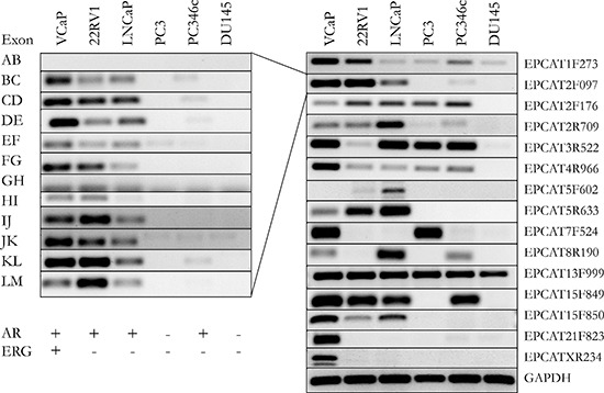
Figure 4. Validation of 15 EPCATs in 6 prostate cancer cell lines.

Intron-spanning primers were designed for each EPCAT. Exons of one transcript followed similar expression patterns (left side). Only the most representative and optimal primer set for an EPCAT is shown in the right panel. These primers were also used to design Taqman probes (see Supplementary Tables 9–10). AR and TMPRSS2-ERG status for each cell line are indicated as present (+) or absent (−).
