Abstract
Background
Vernal keratoconjunctivitis (VKC) is a severe form of allergic conjunctivitis, in which inflammatory infiltrates of the conjunctiva are characterized by CD3+ and CD30+ cells. Until today, the functional involvement of CD30+ T cells in VKC was unclear. Our aim was to evaluate the functional characteristics of CD30+ T cells after allergen stimulation in peripheral blood mononuclear cells obtained from patients with VKC.
Methods
Seventeen consecutive patients at the Institute of Ophthalmology with active forms of VKC were included.
Results
After allergen stimulation, we observed the frequency of CD30+ T cells increased compared with non-stimulated cells (p<0.0001). The CD30+ T cells responded to the specific allergen-inducing expression of intracellular interleukin-4 (IL-4), IL-5, and interferon-gamma (IFN-γ) compared with the CD30- T cells (p<0.0001). Increased early secretion of soluble CD30 was observed in the supernatant of the cultured cells from patients with keratoconjunctivitis, compared with healthy controls (p=0.03). Blockage with IL-4 significantly diminished CD30 frequency in the allergen-stimulated cells.
Conclusions
Our results suggest that after allergenic stimulation, CD4+CD30+ cells are the most important source of IL-4, IL-5, and IFN-γ. IL-4 acts as an activation loop that increases CD30 expression on T cells after specific stimulation. These findings suggest that CD4+CD30+ T cells are effector cells and play a significant role in the immune pathogenic response in patients with vernal keratoconjunctivitis.
Introduction
Allergic conjunctivitis is one of the most common ocular diseases in ophthalmologic clinical practice. Vernal keratoconjunctivitis (VKC) is a chronic form of allergic conjunctivitis with seasonal exacerbations that can lead to permanent visual impairment due to persistent inflammation. Intense itching, photophobia, tearing, and mucous discharge clinically characterize VKC [1]. In conjunctival biopsies of patients with VKC, an inflammatory infiltrate, predominantly in the epithelium and the substantia propria of the conjunctiva, characterized by CD3+ T cells expressing CD30, has been observed [2]. CD30 is a member of the tumor necrosis factor receptor (TNFR) superfamily. TNFRs have distinctive cytoplasmic death domains, related to apoptotic signaling. CD30 lacks this domain and functions as a costimulator molecule in T-cell activation [3]. CD30 is mainly expressed on TH2 cells but also identifies a subset of T cells that comprise the major cells that produce interferon-gamma (IFN-γ) and interleukin-5 (IL-5) in the T-cell compartment [4]. In patients with asthma, peripheral blood CD4+ T cells, following in vitro allergen-specific stimulation, express CD30 and IL-5 on the cell surface, which suggests that CD30 expression is related to long-term clinical manifestations [5]. Although CD30 expression has been associated with asthma and rhinitis, the role of CD30 in VKC remains unclear; thus, the aim of this study was to evaluate the functional involvement of CD30+ T cells in patients with vernal keratoconjunctivitis.
Methods
Patients
Seventeen consecutive patients from the Department of Immunology at Institute of Ophthalmology (9 males and 8 females, mean age 13.11, range 8–25 years) with active forms of VKC were included in the study. VKC diagnosis was based on clinical ophthalmological history and eye examination. All patients were classified as having active forms of vernal keratoconjunctivitis characterized by limbal, tarsal, or mixed varieties of VKC. The clinical ophthalmological characteristics of the patients were described according to [6], and are depicted in Appendix 1. The specific allergic reaction to Dermatophagoides pteronyssinus (D. pteronyssinus) was confirmed with a skin-prick test positive for D. pteronyssinus (wheal, >3 mm diameter). Healthy age- and sex-matched volunteers were used as controls. All participants gave informed consent or their assent consent for blood sampling after written information was provided, and patient anonymity was preserved during the study. The study adhered to the ethical principles of the Declaration of Helsinki and the E11 Statements of International Conference of Harmonization (E11-ICH). The Institutional Ethics Committee Board of the Institute of Ophthalmology Fundación Conde de Valenciana, Mexico City, approved this study.
Monoclonal antibodies and reagents
Phycoerythrin (PE) labeled mouse monoclonal antibodies (mAbs) against human CD30, IL-5, and IL-4; PECy5-labeled mAbs anti-human CD4 and CD8; and fluorescein isothiocyanate (FITC)-labeled antibodies against human IL-4, IFN-γ, and CD30 were purchased from BD Biosciences (San Jose, CA). Lymphoprep (Ficoll 1.077 density) was obtained from Nycomed Pharma (Nyegaard, Oslo, Norway). RPMI-1640 culture medium, Concanavalin A (Con A), Phorbol myristate acetate (PMA), ionomycin, saponin, brefeldin-A, and salts were from Sigma Chemical Co. (St. Louis, MO). Sodium pyruvate, L-glutamine, and 2-mercaptoethanol were purchased from Gibco BRL (Rockville, MD). Fetal calf serum was from HyClone Labs (Logan, UT). D. pteronyssinus was purchased from Allerstand Co. (Mexico City, Mexico).
Peripheral blood mononuclear cells
Blood samples were collected by venipuncture. Heparinized peripheral blood was diluted 1:2 in PBS (1X; 137 mM NaCl, 2.7 mM KCl, 10 mM Na2HPO4, 2 mM KH2P04, pH 7.4). Peripheral blood mononuclear cells (PBMCs) were separated on a Ficoll density gradient with centrifugation at 500 ×g for 30 min at room temperature. Then, cells in the interface were collected, washed twice, and counted using a handheld automated cell counter (Millipore Co., Billerica, MA); viability was assessed with eosin dye exclusion.
Cell cultures
The PBMCs were cultured in 96-well flat-bottomed cell culture plates (Costar, Cambridge, MA) at 2×10⁵ cells/well in RPMI-1640 medium supplemented with 1 mM sodium pyruvate, 2 mM L-glutamine, 50 μg/ml gentamicin, and 0.5% heat-inactivated fetal calf serum and incubated at 37 °C in a 5% CO2 humidified chamber. After 24 h, the culture medium was removed. Fresh culture medium supplemented with 10% heat-inactivated fetal calf serum and D. pteronyssinus (7.5 μg/ml) was added. After 7 days of culture, the cells were harvested and processed to measure intracellular cytokine expression and CD30 on the cell surface with flow cytometry. Con A mitogen (2 μg/ml) and/or PMA/ionomycin (5 ng/ml and 0.2 μg/ml, respectively) was used as the cell stimulation positive control. Supernatants were collected from the culture and stored at −70 °C to determine soluble CD30. To assess intracellular IL-4, IL-5, and IFN-γ synthesis, 4 h before the antigen or polyclonal cultures ended, brefeldin-A was added (10 µg/ml). At the end of the incubation period, the cells were harvested and then processed for immunofluorescence staining.
Immunofluorescence staining of cell surface markers
Tri- or four-color staining was performed on the PBMCs with direct immunofluorescence. Briefly, 2×10⁵ cells were suspended in 20 μl PBS supplemented with 0.2% BSA and 0.2% sodium azide (PBA), and incubated with fluorochrome-labeled mAb for 30 min at 4 °C. After incubation, the cells were washed twice with PBA, fixed with 1% p-formaldehyde, and analyzed with flow cytometry.
Immunofluorescence staining of intracellular markers
Stimulated or non-stimulated PBMCs were washed with PBA and stained with PECy5-labeled mAbs against CD4 or FITC for 30 min. After washing, the cells were fixed with 4% p-formaldehyde in PBS for 10 min at 4 °C. The cells were washed twice with PBS and permeabilized with the saponin buffer (0.1% saponin and 10% BSA in PBS) by shaking gently for 10 min at room temperature. Then, the cells were incubated with FITC-labeled anti-human IFN-γ or IL-4 and/or PE-labeled anti-human IL-5 or IL-4. In all cases, isotype-matched controls were used.
Blockade of cytokines
Capture mAbs of anti-human IL-4 (10 μg/ml) were added at the beginning of the cell culture according to the manufacturer’s protocol (R&D Systems, Minneapolis, MN). Then, 5 μg/ml of anti-IL-4 were added at day 3, and on day 5 as a blocking maintenance dose. Isotype-matched negative antibodies were used as controls at the same concentrations. After 7 days of culture, cells were harvested and then processed for immunofluorescence staining as described.
Flow cytometric analysis
All cells were analyzed for the expression of phenotypic markers on a FACScan flow cytometer (Becton Dickinson, San Jose, CA) using CellQuest software, and 10,000 events were counted. To analyze the staining of the cell-surface markers, the lymphocytes were first gated by their physical properties (forward and side scatter). Then a second gate was drawn based on the immunofluorescence characteristics of the gated cells, and fluorescence intensity was assessed with histograms. To determine CD30+ T cells, the cells were first gated on an Forward Scattered (FSC)-Side Scattered (SSC) dot plot. Then the lymphocytes were gated on CD4+ or CD8+ T cells in an SSC-CD4 or SSC-CD8 dot plot, CD4+ or CD8+ cells were selected, and a dot plot was created to select CD30+ or CD30- on CD4+ and CD8+ T cells. Finally, to analyze the intracellular cytokine staining on helper and cytotoxic CD30+ or CD30- T cells, a histogram was created to analyze the percentage of cytokine positive cells. Data are presented as dot plots or histograms. Control stains were performed using isotype-matched mAbs of unrelated specificity. Background staining was <1% and was subtracted from the experimental values.
Determination of sCD30
Soluble CD30 (human sCD30, Kamiya Biomedical Company, Seattle, WA) was measured with enzyme-linked immunosorbent assay (ELISA) in the supernatants of the cultures according to the manufacturer’s instructions and read at 620 nm in a microplate reader (ThermoLabsystems, Multiskan Ascent, Helsinki, Finland).
Statistical analysis
Results are described in this work using Media and standard deviation (SD), or Median (MD) and interquartile ranges (IQR), according with data distribution. The Mann–Whitney U test and the Wilcoxon signed-rank test were used to detect significant differences. The analysis was performed with GraphPad Prism software v.6.0. Differences were considered statistically significant when p value was less than 0.05.
Results
CD30 is expressed after D. pteronyssinus stimulation on CD4+ and CD8+ T cells from patients with VKC
We began by determining the percentage of CD4+CD30+ T cells and CD8+CD30+ T cells in the peripheral blood of patients with vernal keratoconjunctivitis and healthy controls. No significant differences were observed in the frequency of circulating CD4+CD30+ T cells between patients with VKC and healthy controls (HCs; MD 3%, IQR 1.3–3.3 versus MD 1.7%, IQR 1–1.7, respectively; p=0.08). Similarly, we did not find differences in CD8+CD30+ T cells between patients with VKC and the HCs (MD 1.5%, IQR 0.5–1.8 versus MD 1% IQR 0.4–1.8, p=0.1). However, after allergen stimulation, we observed a significant increase in the percentage of CD4+CD30+ T cells from patients with VKC compared with the HCs (MD 22.2%, IQR 14.6–41.7 versus MD 9.3%, IQR 6–11, respectively; p=0.004). Likewise, when we compared the frequency of the CD8+CD30+ T cells after the D. pteronyssinus stimulation, we observed a significant increase cytotoxic CD30+ cells in patients with VKC compared with the HCs (MD 19.9%, IQR 8–7-30.8 versus MD 7.2%, IQR 6.4–8.2, p=0.03).
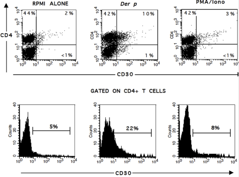
As shown in Table 1, the frequency of CD4+CD30+ T cells from patients with VKC increased 16.8-fold compared with non-stimulated cells (p<0.0001), and was 2.6 times increased compared with PMA/ionomycin (p<0.0001; Figure 1). We also observed that the helper CD30+ T cells increased 1.18-fold compared with the frequency of the CD8+CD30+ T cells after D. pteronyssinus stimulation (p=0.03; Table 1).
Table 1. Frequency of helper and cytotoxic CD30+ T cells, after Dermatophagoides pteronyssinus (Der p) stimulation in patients with VKC.
| Cell subset | RPMI MD (IQR-Range) | Der p MD (IQR-Range) | PMA/Iono MD (IQR-Range) |
P |
|
|---|---|---|---|---|---|
| RPMI versus Der p | Der p versus PMA/Iono | ||||
| CD4+CD30+ |
1.4% (0.5–2.2) |
23.6% (15–43.2) |
8.8% (4.6–11.6) |
<0.0001 |
<0.0001 |
| CD8+CD30+ | 1.0% (0.4–1.8) | 19.9% (8.7–30.8) | 1.7% (0.9–9.3) | <0.0001 | <0.0001 |
MD- Median; IQR- Interquartile range.
Figure 1.
Frequency of CD4+CD30+ T cells after stimulation. A: Representative dot plots of peripheral blood mononuclear cells (PBMCs) stimulated with Dermatophagoides pteronyssinus (Der p) or Phorbol myristate acetate (PMA) in patients with vernal keratoconjunctivitis (VKC). B: Comparative histograms of CD30 frequency on CD4+ gated cells. Dot plots and histograms are representative from 17 patients with VKC.
CD30+ helper T cells express IL-4, IL-5, and IFN-γ after D. pteronyssinus stimulation
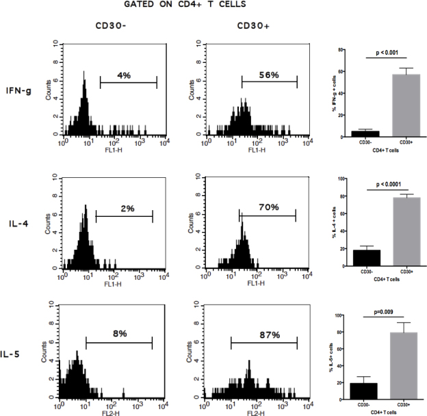
To establish the potential involvement of the specific antigenic-stimulation in the expression of Th1/Th2 cytokines in helper CD30+ or CD30-T helper cells, we assessed the percentage of IL-4, IL-5, and IFN-γ after D. pteronyssinus stimulation in patients with active VKC. Upon allergen stimulation, IFN-γ was expressed 11.4-fold more in CD4+CD30+ T cells than in CD4+CD30- T cells (p<0.0001). Similarly, IL-4 was expressed 4.3-fold more in CD4+CD30+ T cells than in CD4+CD30- T cells (p<0.0001). Likewise, IL-5 was expressed 4.1-fold more in CD30+ helper T cells than in CD30- helper T cells (p=0.009; Figure 2).
Figure 2.

Comparative histograms of intracellular cytokines on helper CD30+ and CD30- T cell subsets. Peripheral blood mononuclear cells (PBMCs) were Dermatophagoides pteronyssinus–stimulated for 7 d and stained with CD4, CD30, and intracellular interferon-gamma (IFN-γ), interleukin-4 (IL-4), or IL-5 as described in the Materials and Methods section. Left panel, CD4+CD30- T cells; central panel, CD4+CD30+ T cells; right panel, comparison of the frequency of cells positive to intracellular cytokines in either CD30+ or CD30- cell subsets. Data are expressed as mean ± standard deviation (SD).
sCD30 increased after polyclonal stimulation in patients with VKC
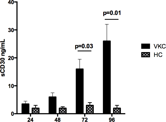
To understand whether polyclonal stimulation (Con A) induces secretion of sCD30, we evaluated soluble CD30 at 24, 48, 72, and 96 h in the supernatant of cells from patients with VKC and the HCs. We observed 4.5 times more sCD30 in the supernatant of the cultures from patients with VKC since 72 h after Con A stimulation, compared with the HCs (p=0.03), and 7.4 times more sCD30 after Con A stimulation at 96 h in the supernatant of the cultures from patients with VKC compared with the HCs (p=0.01; Figure 3).
Figure 3.

Soluble CD30 is secreted early after Concanavalin A (Con A) stimulation. Peripheral blood mononuclear cells (PBMCs) were stimulated at 24, 48, 72, and 96 h, and the supernatant of the cultures was collected to determine soluble CD30. An increased significant concentration of soluble CD30 was observed at 72 and 96 h after Con A stimulation. Results are representative of eight patients with vernal keratoconjunctivitis (VKC) and matched controls.
Blockage of IL-4 decreased CD30 expression on CD4+ T cells
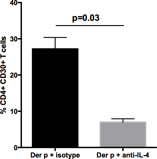
To see whether IL-4 is involved in CD30 expression on helper T cells from patients with VKC, we blockaded IL-4 function by adding neutralizing mAbs against IL-4 during the D. pteronyssinus stimulation. We observed 3.9-fold less frequency of CD4+CD30+ T cells when we added anti-IL-4 (p=0.03) (Figure 4).
Figure 4.

Effect of blockade of IL-4 on frequency of CD4+CD30+ T cells after D. pteronyssinus (Der p) stimulation. Peripheral blood mononuclear cells (PBMCs) from three patients with vernal keratoconjunctivitis (VKC) were stimulated with Dermatophagoides pteronyssinus for 7 days, and mAb anti-interleukin-4 (IL-4) was added at baseline, 3 d, and 5 d as described in the Materials and Methods section. The plot shows the significant diminished frequency of CD4+CD30+ T cells when IL-4 was blocked.
Discussion
The current study evaluated the functional involvement of CD30+ T cells in patients with vernal keratoconjunctivitis. We demonstrated that upon D. pteronyssinus stimulation, CD30 expression increased in helper and cytotoxic T cells, and that CD4+CD30+ T cells are the main source of IL-4, IL-5, and IFN-γ. Consistent with our findings, Rojas-Ramos et al. [7] demonstrated that CD30 expression on CD4+ T cells was correlated with the production of IL-4 after stimulation of CD4+ T cells isolated from patients with asthma. Other authors have suggested that IL-4 could induce CD30 on the helper T-cell surface [5] and on cytotoxic T cells after repeated stimulation [8]. In addition to Th2 cells, CD30 expression on CD4+ T cells has been associated with the maintenance of memory cells in the animal model [9]. In our work, we observed that following in vitro allergen stimulation, the frequency of CD30+ T cells from patients with VKC were increased. However, whether these CD4+CD30+ T cells have an immunophenotype related to memory in patients with VKC is unknown and requires further investigation. Likewise, interaction CD30/CD30L on cytotoxic cells is related to the generation of long-lived memory cells [10]. CD8+CD30+ T cells are an important source of IL-4 and IL-5 in patients with asthma, contributing to a Th2 microenvironment in the lung, and are a cell subset associated with worse clinical outcome [8]. In this context, the role of CD8+CD30+ Tc2 cells in corneal damage must be further evaluated, since CD8+ T cells are involved in the effector phase of allergic conjunctivitis in the mouse model [11].
The corneal damage in patients with VKC is associated with multiple Th1 and Th2 cytokines that are overexpressed on the ocular surface. IFN-γ, a Th1-derived cytokine, could influence delayed hypersensitivity ocular damage and activate the transforming growth factor-beta (TGF-β) signaling pathway favoring tissue remodeling by conjunctival fibroblasts [12,13]. In contrast, IL-4 and IL-5 cytokines are characteristically induced during the allergic response, and both cytokines are involved with chronic Th2 responses [5]. Our results showed that CD4+CD30+ T cells are the most important source of IL-4, IL-5, and IFN-γ after allergenic specific stimulation, suggesting that the CD4+CD30+ T cell subset could be the major source of Th1/Th2 cytokines during the pathogenic immune response in patients with VKC. IL-4 is also a cytokine directly involved with the increased expression of CD30 on CD4+ T cells [5,7]. In this work, we observed that blocking IL-4 is not enough to completely abrogate CD30 expression on helper T cells. Thus, IL-4 acts as an activation loop to increase CD30 expression on CD4+ T cells after specific stimulation. Investigation of other type 2 cytokines that could be involved in the upregulation of CD30 on CD4+ T cells in patients with VKC is needed to fully understand the immune pathophysiological role of CD4+CD30+ T cells in ocular allergy.
After T cell activation, CD30 is cleaved by the membrane-anchored metalloproteinase TNF-α converting enzyme (TACE), and releases a soluble ectodomain of CD30 (sCD30) [14]. In our work, we observed increased levels of sCD30 in patients with VKC after Con-A stimulation compared with the HCs. Determination of sCD30 in serum has been proposed as a disease biomarker in pediatric asthma; that is, high levels of sCD30 are related to activation of the immune response [14,15], while low levels are associated with an immune regulatory environment [16,17]. The determination of sCD30 in patients with allergic conjunctivitis could be used to assess clinical activity, to modify with opportunity medical-ophthalmological treatments, and to predict clinical outcomes. Nevertheless, further studies are needed to test this hypothesis.
Taken together, our results suggest that CD4+CD30+ T cells function as effector T cells in response to specific allergens. This cell subset produces the intracellular IL-4, IL-5, and IFN-γ cytokines provoking a Th1/Th2 microenvironment related to the immune pathogenic response in patients with vernal keratoconjunctivitis.
Acknowledgments
Authors wish to thank Veronica Romero Martinez for their technical assistance. Maria C. Jiménez-Martínez and Ricardo Lascurain (rlascurain@yahoo.com.mx) contributed equally to this work. This work was supported in part by CONACYT 71,291 and Fundación Conde de Valenciana. Diana Magaña received a Ph.D. scholarship from CONACyT number 186,211 and CVU 209,062. This article is part of the requirements for the doctoral degree of Diana Magaña. Ph.D. Program in Biomedical Sciences. Faculty of Medicine. UNAM. AUTHOR CONTRIBUTIONS: D.M., G.A., M.L.- Performed immunological evaluation; D.M.- Analyzed data, and wrote the paper; C.S. Performed clinical ophthalmological evaluation of patients, and analyzed data; J. A-B Performed clinical allergo-immunological evaluation of patients, and analyzed data; R.C., S.E-P, Y.G.- Analyzed and interpreted immunological data; R.L. and M.C.J-M.- designed the study, conducted research, and wrote the paper.
Appendix 1. Clinical-ophthalmological characteristics of patients with VKC.
To access the data, click or select the words “Appendix 1”.
References
- 1.Bonini S, Bonini S, Lambiase A, Marchi S, Pasqualetti P, Zuccaro O, Rama P, Magrini L, Juhas T, Bucci MG. Vernal keratoconjunctivitis revisited: a case series of 195 patients with long-term followup. Ophthalmology. 2000;107:1157–63. doi: 10.1016/s0161-6420(00)00092-0. [DOI] [PubMed] [Google Scholar]
- 2.El-Asrar AM, Struyf S, Al-Kharashi SA, Missotten L, Van Damme J, Geboes K. Expression of T lymphocyte chemoattractants and activation markers in vernal keratoconjunctivitis. Br J Ophthalmol. 2002;86:1175–80. doi: 10.1136/bjo.86.10.1175. [DOI] [PMC free article] [PubMed] [Google Scholar]
- 3.Lombardi V, Singh AK, Akbari O. The role of costimulatory molecules in allergic disease and asthma. Int Arch Allergy Immunol. 2010;151:179–89. doi: 10.1159/000242355. [DOI] [PMC free article] [PubMed] [Google Scholar]
- 4.Alzona M, Jäck HM, Fisher RI, Ellis TM. CD30 defines a subset of activated human T cells that produce IFN-gamma and IL-5 and exhibit enhanced B cell helper activity. J Immunol. 1994;153:2861–7. [PubMed] [Google Scholar]
- 5.Garfias Y, Ortiz B, Hernández J, Magaña D, Becerril-Angeles M, Zenteno E, Lascurain R. CD4+ CD30+ T cells perpetuate IL-5 production in Dermatophagoides pteronyssinus allergic patients. Allergy. 2006;61:27–34. doi: 10.1111/j.1398-9995.2005.00951.x. [DOI] [PubMed] [Google Scholar]
- 6.Robles-Contreras A, Santacruz A, Ayala J, Bracamontes E, Godinez V, Estrada-García I, Estrada-Parra S, Chávez R, Perez-Tapia M, Bautista-De Lucio MV, Jiménez-Martínez MC. (2011). Allergic Conjunctivitis: An Immunological Point of View, Conjunctivitis - A Complex and Multifaceted Disorder, Prof. Zdenek Pelikan (Ed.), ISBN: 978–953–307–750–5 [Google Scholar]
- 7.Rojas-Ramos E, Garfias Y. Jiménez-Martínez Mdel C, Martínez-Jiménez N, Zenteno E, Gorocica P, Lascurain R. Increased expression of CD30 and CD57 molecules on CD4(+) T cells from children with atopic asthma: a preliminary report. Allergy Asthma Proc. 2007;28:659–66. doi: 10.2500/aap.2007.28.3057. [DOI] [PubMed] [Google Scholar]
- 8.Stanciu LA, Roberts K, Lau LC, Coyle AJ, Johnston SL. Induction of type 2 activity in adult human CD8(+) T cells by repeated stimulation and IL-4. Int Immunol. 2001;13:341–8. doi: 10.1093/intimm/13.3.341. [DOI] [PubMed] [Google Scholar]
- 9.Withers DR, Gaspal FM, Bekiaris V, McConnell FM, Kim M, Anderson G, Lane PJ. OX40 and CD30 signals in CD4(+) T-cell effector and memory function: a distinct role for lymphoid tissue inducer cells in maintaining CD4(+) T-cell memory but not effector function. Immunol Rev. 2011;244:134–48. doi: 10.1111/j.1600-065X.2011.01057.x. [DOI] [PubMed] [Google Scholar]
- 10.Nishimura H. Yajima T, Muta H, Podack ER, Tani K, Yoshikai Y. A novel role of CD30/CD30 ligand signaling in the generation of long-lived memory CD8+ T cells. J Immunol. 2005;175:4627–34. doi: 10.4049/jimmunol.175.7.4627. [DOI] [PubMed] [Google Scholar]
- 11.Fukushima A, Yamaguchi T, Fukuda K, Sumi T, Kumagai N, Nishida T, Imai S, Ueno H. CD8+ T cells play disparate roles in the induction and the effector phases of murine experimental allergic conjunctivitis. Microbiol Immunol. 2006;50:719–28. doi: 10.1111/j.1348-0421.2006.tb03845.x. [DOI] [PubMed] [Google Scholar]
- 12.Leonardi A, Fregona IA, Plebani M, Secchi AG, Calder VL. Th1- and Th2-type cytokines in chronic ocular allergy. Graefes Arch Clin Exp Ophthalmol. 2006;244:1240–5. doi: 10.1007/s00417-006-0285-7. [DOI] [PubMed] [Google Scholar]
- 13.Leonardi A, Di Stefano A, Motterle L, Zavan B, Abatangelo G, Brun P. Transforming growth factor-β/Smad - signalling pathway and conjunctival remodelling in vernal keratoconjunctivitis. Clin Exp Allergy. 2011;41:52–60. doi: 10.1111/j.1365-2222.2010.03626.x. [DOI] [PubMed] [Google Scholar]
- 14.Hansen HP, Dietrich S, Kisseleva T, Mokros T, Mentlein R, Lange HH, Murphy G, Lemke H. CD30 shedding from Karpas 299 lymphoma cells is mediated by TNF-alpha-converting enzyme. J Immunol. 2000;165:6703–9. doi: 10.4049/jimmunol.165.12.6703. [DOI] [PubMed] [Google Scholar]
- 15.Remes ST, Delezuch W, Pulkki K, Pekkanen J, Korppi M, Matinlauri IH. Association of serum-soluble CD26 and CD30 levels with asthma, lung function and bronchial hyper-responsiveness at school age. Acta Paediatr. 2011;100:e106–11. doi: 10.1111/j.1651-2227.2011.02264.x. [DOI] [PubMed] [Google Scholar]
- 16.Kotaniemi-Syrjänen A, Delezuch W, Pelkonen AS, Malmström K, Malmberg LP, Punnonen K, Matinlauri IH, Mäkelä MJ. Improvement in lung function is associated with a decrease in serum soluble CD30 in atopic infants. Ann Allergy Asthma Immunol. 2015;114:156–7. doi: 10.1016/j.anai.2014.11.011. [DOI] [PubMed] [Google Scholar]
- 17.Foschi FG, Emiliani F, Savini S, Quercia O, Stefanini GF. CD30 serum levels and response to hymenoptera venom immunotherapy. J Investig Allergol Clin Immunol. 2008;18:279–83. [PubMed] [Google Scholar]


